


Imaging diagnosis of fiant cell tumor of tendon sheath
-
摘要:
目的分析腱鞘巨细胞瘤的影像学表现,以提高对该病的认识及影像诊断的准确率。 方法回顾性分析14例经手术病理证实的腱鞘巨细胞瘤患者的X线平片及MRI表现,其中6例行X线平片检查,13例行MRI平扫及增强扫描。 结果X线平片显示局部稍高密度软组织肿块影,邻近骨质未见明显异常或局部侵蚀破坏;MRI表现为相应部位软组织信号肿块影,在T1WI多呈较低信号,内可见条片状更低信号影,T2WI呈高低混杂信号影,增强扫描强化明显,病灶与邻近肌腱或关节关系密切,局部骨皮质可受侵。 结论腱鞘巨细胞瘤的影像学表现具有一定的特征性。 Abstract:ObjectiveTo study the imaging features of Giant cell tumor of tendon sheath (GCTTS), in order to improve the recognition and the accuracy of imaging in the diagnosis of the disease. Methods14 cases with surgery and pathology proved GCTTS were retrospectively reviewed. 6 patients were carried out X-ray plain film, and 13 had plain and contrast-enhanced MR imaging. ResultsX-ray plain film findings were soft-tissue swelling without osseous involvement or slight bone erosions. At MR imaging, the lesions were low signal and can be seen in the strip lower signal intensity on T1 weighted images, and the mixed signal intensity on T2 weighted images. The signal intensities tended to be moderately and heterogeneously enhanced following Gd-DTPA administration. The lesions were typically located in relation to a tendon or joint closely, local bone cortex involvement. ConclusionsThe imaging findings of GCTTS have some certain characteristics. -
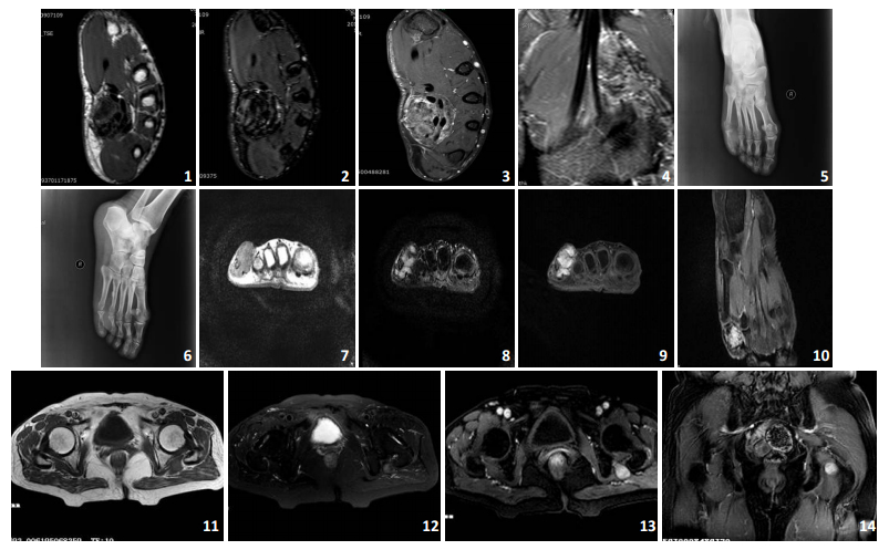
图 1~4 13岁男孩,发现左手掌及第5指肿块2年。左手掌肌腱区及第5指掌侧肌腱旁软组织肿块,边界欠清,信号不均匀,T1WI等信号肿块内见更低信号影,T2WI抑脂呈稍高及低混杂信号影,强后不均匀中等强化,邻近骨质信号未见明显异常。图 5~10 46岁女性,图 5、6 示右足近节趾骨周围见稍高密度肿块影,边界欠清,邻近近节趾骨内见囊状低密度影;7~10 MRI示右足第5近节趾骨周围软组织肿块影,边缘欠光整,T1WI呈等信号,内见点条状稍低信号影;T2WI抑脂呈稍高及低混杂信号影,强后明显欠均匀强化,邻近骨质被侵蚀。图 11~14 62岁女性,左臀部髋关节旁见卵圆形软组织肿块影,边缘光整,T1WI呈等信号,T2WI抑脂呈稍高及低混杂信号影,强后明显欠均匀强化。
-
[1] 武中弼, 杨光华.中华外科病理学[M].北京:人民卫生出版社, 2002, 2495-7. [2] Gholve PA, Hosalkar HS, Kreiger PA, et al.Giant cell tumor of tendon sheath largest signle series in children[J]. Pediatr Orthon, 2007, 27(1): 67-74. doi: 10.1097/01.bpo.0000242380.95348.8b [3] 刘子君.骨关节病理[M].北京:人民卫生出版社, 1992, 427-8. [4] Kitagawa Y, Ito H, Amano Y, et al. MR imaging for preoperative diagnosis and assessment of local tumor exrent on localized giant cell tumor of tendon sheath[J].Skeletal Radio, 2003, 32(11): 633-8. doi: 10.1007/s00256-003-0689-y [5] Chassaignac M. Cancer de la gaine des tendons[J]. Gas Hosp Civ Milit, 1852, 47(4): 185-190 [6] Jaffe HL, Lichtenstein L, Sutro CJ. Pigmented villonodular synovitis, bursitis and tenosynovitis[J]. Arch Pathol, 1941, 31(3):731-765 [7] Somerhausen NS, Cin P. Giant cell tumour of tendon sheath and dif-fuse-type giant cell tumour. In: Pathology and genetics of tumours of soft tissue and bone[M]. Lyon, France: IARC, 2002:110-4 [8] Anazawa U, Hanaoka H, Shiraishi T, et al.Similarities between giant cell tumor of bone, giant cell tumor of tendon sheath, and pigmented villonodular synovitis concerning ultrastructural cytochemical fea-tures of multinucleated giant cells and mononuclear stromal cells[J]. Ultrastruct Pathol, 2006, 30(3): 151-8 doi: 10.1080/01913120600689707 [9] 时惠平, 张新合.恶性腱鞘巨细胞瘤1例[J].中国医学影像学杂志, 2008, 16(1): 76 http://d.wanfangdata.com.cn/Periodical/zgyxyxxzz200801030 [10] 徐胜生, 肖家和, 周翔平, 等.色素沉着绒毛结节性滑膜炎、腱鞘巨细胞瘤的MRI表现及其应用价值[J].实用放射学杂志, 2005, 21(8) : 850-3. http://www.cnki.com.cn/Article/CJFDTOTAL-SYFS200508023.htm [11] Wan JM, Magarelli N, Peh WC, et al. Imaging of giant cell tumour of the tendon sheath[J]. Radiol med, 2010, 115(1):141-51 doi: 10.1007/s11547-010-0515-2 [12] Lauger J, Palmer J, Rosón N, et al. Pigmented villonodular synovitis and giant cell tumors of the tendon sheath: radiologic and pathologic features[J]. AJR, 1999, 172(4):1087-91. doi: 10.2214/ajr.172.4.10587152 [13] Mark DM, John HR, Rachel BL, et al. Pigmented Villonodular Synovi-tis: Radiologic-Pathologic Correlation[J]. RadioGraphics, 2008, 28(5):1493-1518. doi: 10.1148/rg.285085134 [14] Kransdorf MJ, Murphey MD. Synovial tumors. In: Imaging of soft tissue tumors[M]. Philadelphia, Pa: Lippincott Williams & Wilkins, 2006: 381-436. [15] 方必东, 许崇永, 章巍, 等.腱鞘巨细胞瘤MRI表现[J].中国医学影像学杂志, 2007, 15(6): 447-9 http://d.wanfangdata.com.cn/Periodical/zgyxyxxzz200706017 [16] De Schepper AM, Hogendoom PC, Bloem JL.Giant cell tumors of the tendon sheath may present radiologically as intrinsic osseous lesions[J]. Eur Radio, 2007, 17(2): 499-502. doi: 10.1007/s00330-006-0320-4 -

 下载:
下载:
 点击查看大图
图(1)
计量
- 文章访问数: 577
- HTML全文浏览量: 230
- PDF下载量: 1
- 被引次数: 0