


Evaluation the correlation between the risk of malignancy index and Doppler ultrasound examination for discrimination between benign and malignant adnexal masses
-
摘要:
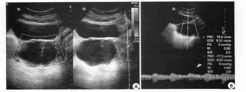
目的探讨在判断卵巢肿瘤良、恶性的诊断中,恶性危险指数(RMI4)分别与彩色多普勒超声血流阻力指数(RI)、搏动指数(PI)、时间平均最大速度(TAMXV)联合应用,判断其中最佳联合方式,并分析其诊断价值。 方法回顾150例卵巢肿瘤患者(92例良性病例,58例恶性病例)术前超声检查结果,包括二维声像图特征和频谱多普勒RI、PI、TAMXV测值;根据患者超声二维声像图像特征、绝经状态、血清CA125进行RMI4评分。记录单一变量以及双变量(RMI4分别与RI、PI、TAMXV以两种诊断标准结合)判断肿瘤为良恶性的结果与术后病理进行对照,计算各组的敏感度、假阳性率、阳性预测值、阴性预测值。 结果RMI4单独诊断时具有91%敏感性及22%假阳性率,多普勒变量中,TAMXV具有100%敏感性与49%假阳性率。而当RMI4与TAMXV的组合(同时符合二者恶性标准既判断肿瘤为恶性)具有敏感性91%与假阳性率11%。 结论彩色多普勒超声RMI4评分与TAMXV同时符合恶性标准时诊断卵巢肿瘤为恶性的判断方法,优于单一变量及其他双变量联合方式。 Abstract:ObjectiveTo investigate the best combination and the diagnosis value of combining risk of malignancy indexes (RMI4), color Doppler ultrasound resistance index (RI), pulsatility index (PI), and time-averaged maximum velocity (TAMXV) in better discrimination of benign and malignant adnexal masses. Materials and MethodsPreoperative ultrasound examination of one hundred and fifty women (ninety-two tumors were benign and fifty-eight malignant) with ovarian tumors judged by pathological were retrospectively analyzed, including spectral Doppler techniques and measurement of RI, RI, TAMXV. Calculate RMI4 score according to the two-dimensional ultrasonographic characteristics, menopausal status and CA125. The judgment results of single argument and double variables (Each one of RI, RI, TAMXV combined with RMI4 in two ways) were compared with pathological results, then, their sensitivity, false-positive rate, positive predictive value and negative predictive value with regard to malignancy were calculated. ResultsRMI4 had a sensitivity of 91% and a false-positive rate of 22%. The Doppler variable-TAMXV with a sensitivity of 100% had a false-positive rate of 49%. Combining RMI4 with TAMXV (requiring both RMI4 and TAXMV to indicate malignancy for a malignant diagnosis to be made) had a sensitivity of 91% and a false-positive rate of 11%. ConclusionThe combined use of RMI4 score and measurement of TAMXV has a higher accuracy for discrimination of benign and malignant adnexal masses than others. -
Key words:
- Ovarian neoplasm /
- risk of malignancy index (RMI) Ultrasonography /
- Doppler /
-
表 1 病理学诊断结果
病理学诊断 n 良性肿瘤 浆液性囊腺瘤 23 粘液性囊腺瘤 19 子宫内膜异位囊肿 18 成熟畸胎瘤 13 卵泡膜细胞瘤 7 纤维瘤 4 单纯甲状腺瘤 4 黄体囊肿 3 滤泡囊肿 1 恶性肿瘤 浆液性囊腺癌 18 黏液性囊腺癌 14 成熟畸胎瘤癌变 9 内胚窦瘤 6 卵巢颗粒细胞 5 卵巢透明细胞癌 4 子宫内膜样腺癌 1 转移瘤 1 总计 150  下载: 导出CSV
表 2 各变量与病理检查结果关系
变量 病理 P 恶性 良性 RMI 0.00 ≥450 53 20 < 450 5 72 RI 0.00 < 0.4 41 15 ≥0.4 13 77 PI 0.00 < 1.0 48 37 ≥1.0 10 55 TAMXV 0.00 ≥7.2 cm/s 58 45 < 7.2 cm/s 0 47 Both RMI and TAMXV 0.00 恶性 53 10 良性 5 82 Both RMI and RI 0.00 恶性 37 7 良性 15 85 Both RMI and PI 0.00 恶性 46 17 良性 12 75 Either RMI or TAMXV 0.00 恶性 58 55 良性 0 37 Either RMI or RI 0.00 恶性 57 29 良性 2 63 Either RMI or PI 0.00 恶性 55 40 良性 3 52
下载: 导出CSV
表 3 不同超声评价方法对肿瘤诊断的敏感性、假阳性率、阳性预测值及阴性预测值
方法 敏感性(%) 假阳性率(%) 阳性预测值(%) 阴性预测值(%) 单变量 RMI 91(53/58) 22(20/92) 73(53/73) 94(72/77) TAMXV 100(58/58) 49(45/92) 56(58/103) 100(47/47) PI 83(48/58) 40(37/92) 56(48/85) 85(55/65) RI 71(41/58) 16(15/92) 73(41/56) 82(77/94) 组合变量 Both RMI and TAMXV 91(53/58) 11(10/92) 84(53/63) 94(82/87) PI 79(46/58) 18(17/92) 73(46/63) 86(75/87) RI 64(37/58) 8(7/92) 82(37/45) 81(85/105) Either RMI or TAMXV 100(58/58) 60(55/92) 51(58/113) 100(37/37) PI 95(55/58) 43(40/92) 58(55/95) 95(52/55) RI 98(57/58) 32(29/92) 66(57/86) 98(63/64)
下载: 导出CSV
-
[1] Chan JK, Tian C, Monk BJ, et al. Prognostic factors for high-risk early-stage epithelial ovarian Cancer: a Gynecologic Oncology Group study[J]. Cancer, 2008, 112(10): 2202-10. doi: 10.1002/cncr.23390 [2] 刘翔宇, 王义成.卵巢肿瘤的彩色多普勒超声诊断进展[J].河北医药, 2013, 35(4): 590-1. http://www.cnki.com.cn/Article/CJFDTOTAL-HBYZ201304070.htm [3] 景履新, 王新燕. B超诊断卵巢肿瘤130例的体会[J].中国卫生产业, 2012, 29(1): 154. http://www.cnki.com.cn/Article/CJFDTOTAL-WSCY201228146.htm [4] Jacobs I, Oram D, Fairbanks J, et al. A risk of malignancy index incorporating CA 125, ultrasound and menopausal status for the accurate preoperative diagnosis of ovarian Cancer[J]. Br J Obstet Gynaecol, 1990, 97(10): 922-9. doi: 10.1111/bjo.1990.97.issue-10 [5] Kurjak A, Predanić M. New scoring system for prediction of ovarian malignancy based on transvaginal color Doppler sonography[J]. J Ultrasound Med, 1992, 11(12): 631-8. doi: 10.7863/jum.1992.11.12.631 [6] Weiner Z, Thaler I, Beck D, et al. Differentiating malignant from benign ovarian tumors with transvaginal color flow imaging[J]. Obstet Gynecol, 1992, 79(2): 159-62. [7] Valentin L. Ovarian masses, color Doppler. In: Boume[C]//TB, 1998. [8] Jacobs I, Oram D, Fairbanks J, et al. A risk of malignancy index incorporating CA 125, ultrasound and menopausal status for the accurate preoperative diagnosis of ovarian Cancer[J]. Br J Obstet Gynaecol, 1990, 97(10): 922-9. doi: 10.1111/bjo.1990.97.issue-10 [9] Tingulstad S, Hagen B, Skjeldestad FE, et al. The risk-ofmalignancy index to evaluate potential ovarian cancers in local hospitals[J]. Obstet Gynecol, 1999, 93(3): 448-52. https://www.researchgate.net/publication/13215892_The_Risk_of_Malignancy_Index_to_evaluate_potential_cancers_in_local_hospitals [10] Yamamoto Y, Yamada R, Oguri H, et al. Comparison of four malignancy risk indices in the preoperative evaluation of patients with pelvic masses[J]. Eur J Obstet Gynecol Reprod Biol, 2009, 144(2): 163-7. doi: 10.1016/j.ejogrb.2009.02.048 [11] Anton C, Carvalho FM, Oliveira EI, et al. A comparison of CA125, HE4, risk ovarian malignancy algorithm (ROMA), and risk malignancy index (RMI) for the classification of ovarian masses[J]. Clinics (São Paulo, Brazil), 2012, 67(5): 437-41. doi: 10.6061/clinics [12] Moore RG, Jabre-Raughley M, Brown AK, et al. Comparison of a novel multiple marker assay vs the Risk of Malignancy Index for the prediction of epithelial ovarian Cancer in patients with a pelvic mass[J]. Am J Obstet Gynecol, 2010, 203(3): 228.e1-6. [13] Shy K, Dubinsky T. Is color Doppler ultrasound useful in diagnosing ovarian Cancer[J]. Clin Obstet Gynecol, 1999, 42(4):902-15. doi: 10.1097/00003081-199912000-00018 [14] Costantini M, Belli P, Ierardi C, et al. Solid breast mass characterisation: use of the sonographic BI-RADS classification[J]. Radiol Med, 2007, 112(6): 877-94. doi: 10.1007/s11547-007-0189-6 [15] Valentin L. Gray scale sonography, subjective evaluation of the color Doppler image and measurement of blood flow velocity for distinguishing benign and malignant tumors of suspected adnexal origin[J]. Eur J Obstet Gynecol Reprod Biol, 1997, 72(1): 63-72. doi: 10.1016/S0301-2115(96)02661-9 [16] Valentin L. Comparison of lerner score, doppler ultrasound examination, and their combination for discrimination between benign and malignant adnexal masses[J]. Ultrasound Obstet Gynecol, 2000, 15(2): 143-7. doi: 10.1046/j.1469-0705.2000.00028.x [17] Valentin L. Prospective cross-validation of Doppler ultrasound examination and gray-scale ultrasound imaging for discrimination of benign and malignant pelvic masses[J]. Ultrasound Obstet Gynecol, 1999, 14(4): 273-83. doi: 10.1046/j.1469-0705.1999.14040273.x -

 点击查看大图
图(5) / 表(3)
计量
- 文章访问数: 629
- HTML全文浏览量: 228
- PDF下载量: 6
- 被引次数: 0