


Value of MRI texture analysis combined with ZOOMit IVIM sequence for differential diagnosis of benign and malignant prostate nodules
-
摘要:
目的 探究MRI纹理分析和基于并行发射平台选择性激发成像(ZOOMit)的体素内不相干运动(IVIM)序列对于前列腺结节良性和恶性的鉴别诊断价值。 方法 选取我院2021年3月~2022年8月收治的95例前列腺结节患者作为研究对象,共112个结节,根据结节良恶性将其分为良性组(n=59)和恶性组(n=53)。所有患者均行均MRI T2WI和ZOOMit IVIM序列扫描,形成平扫T2WI图像、表观扩散系数(ADC)、纯扩散系数(D)、伪扩散系数(D*)、灌注分数(f)伪彩图,获取纹理分析参数ADC、D、D*、f值,比较两种扫描序列的误诊率,采用ROC曲线评估个参数对于前列腺良恶性结节的诊断价值。 结果 MRI纹理分析、ZOOMit IVIM序列扫描单独及联合诊断前列腺结节的良恶性情况的误判率差异无统计学意义(P > 0.05)。与良性组相比,恶性组结节的平均ADC值、方差、D值和f值降低(P < 0.05),偏度、峰度和熵上升(P < 0.05),D*值的差异无统计学意义(P > 0.05)。ROC曲线分析可知,ADC值鉴别诊断前列腺良恶性结节的敏感度较高,D值鉴别诊断前列腺良恶性结节的特异性较高,ADC值、D值和f值联合诊断的整体效能最高。 结论 MRI纹理分析和ZOOMit IVIM序列对于鉴别前列腺结节的良恶性有较高的价值。 Abstract:Objective To investigate the value of MRI texture analysis and intravoxel incoherent motion (IVIM) sequences based on zoomed imaging with parallel transmission technique (ZOOMit) for the differential diagnosis of benign and malignant prostate nodules. Methods Ninety-five patients with prostate nodules in our hospital between March 2021 and August 2022 were selected. A total of 112 nodules were divided into benign group (n=59) and malignant group (n=53). All patients underwent MRI including T2* mapping and ZOOMit IVIM sequence scans before surgery, then the MRI plain scanning images, diffusion weighted imaging (DWI) images, parameters including apparent diffusion coefficient (ADC), diffusion coefficient of pure diffusion (D), pseudo-diffusion coefficient (D*) and perfusion fraction (f) were obtained. The misjudgment rates of the two scanning sequences were compared. ROC curve was plotted to evaluate the diagnostic value of each parameter for benign and malignant prostate nodules. Results There was no significant difference in the misjudgment rate between benign and malignant prostate nodules diagnosed by MRI texture analysis and ZOOMit IVIM sequence scan alone and in combination (P > 0.05). Compared with benign group, malignant group had remarkably decreased ADCMean, variance, D value and f value (P < 0.05), notably increased skewness, kurtosis and entropy (P < 0.05), and slightly increased D* value (P > 0.05). Among the parameters in the differential diagnosis of benign and malignant prostate nodules, ADC value had high sensitivity and D value had high specificity, while the diagnostic efficacy of ADC combined with D and f values was the highest. Conclusion MRI texture analysis and ZOOMit IVIM sequence have high value in differentiating benign and malignant prostate nodules. -
Key words:
- MRI texture analysis /
- intravoxel incoherent motion /
- prostate /
- benign nodule /
- malignant nodule
-

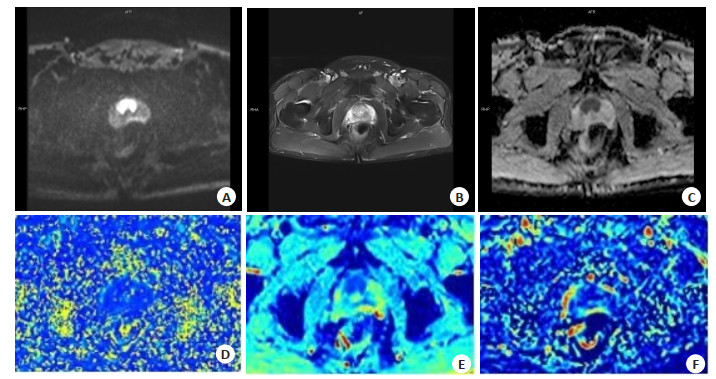
图 2 不同扫描模式所得前列腺图像比较
Figure 2. Comparison of prostate images obtained by different scanning modes. A: Image of nodules with slightly low signal intensity on T2WI; B: Image of nodules with high signal intensity on DWI; C: Image of nodules with low signal intensity on ADC map; D: Pseudo-colour map of pure diffusion coefficient D of IVIM; E: Pseudo-colour map of pseudo-diffusion coefficient D* of IVIM; F: Pseudo-colour map of perfusion fraction f of IVIM.
表 1 两种方法对于前列腺良恶性结节的误判情况比较
Table 1. Comparison of misjudgment rates of two scanning sequences for differential diagnosis of benign and malignant prostate nodules
Group Texture analysis ZOOMit IVIM sequence Combined diagnosis χ2 P Benign group (n=59) 15(25.42) 14(23.73) 9(15.25) 2.0776 0.3539 Malignant group (n=53) 20(37.74) 17(32.06) 12(22.64) 2.8909 0.2356 IVIM: Intravoxel incoherent motion.  下载: 导出CSV
表 2 两组结节的MRI纹理分析参数比较
Table 2. Comparison of MRI texture parameters between two groups (Mean±SD)
Group ADCMean (mm2/s) Variance Skewness Kurtosis Entropy Benign group (n=59) 1.12±0.16 3472.63±1020.41 0.30±0.11 -0.23±0.07 7.06±2.10 Malignant group (n=53) 0.91±0.19 2170.87±1019.54 0.51±0.15 0.56±0.11 10.49±2.27 t 6.3470 6.7435 8.5063 45.8087 8.3060 P < 0.001 < 0.001 < 0.001 < 0.001 < 0.001
下载: 导出CSV
表 3 两组结节的ZOOMit IVIM序列参数比较
Table 3. Comparison of ZOOMit IVIM sequence parameters between two groups (Mean±SD)
Group D (mm2/s) D* (mm2/s) f (%) Benign group (n=59) 1.15±0.26 10.38±3.58 15.59±3.46 Malignant group (n=53) 0.81±0.11 11.57±4.04 11.37±2.15 t 8.8333 1.6528 7.6493 P < 0.001 1.1012 < 0.001 D: Diffusion coefficient of pure diffusion; D*: Pseudo-diffusion coefficient; f: Perfusion fraction.
下载: 导出CSV
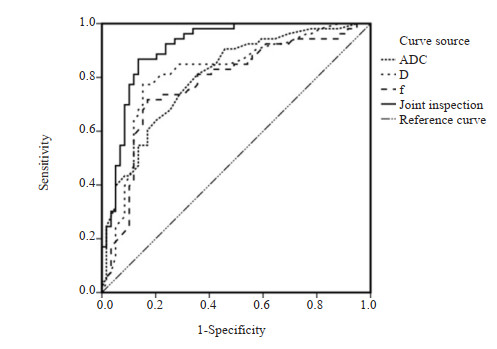
表 4 MRI纹理分析和ZOOMit IVIM序列参数诊断前列腺良恶性结节ROC曲线分析
Table 4. ROC curve analysis of MRI texture analysis and ZOOMit IVIM sequence parameters for differential diagnosis of benign and malignant prostate nodules.
Parameters Cut-off value AUC 95% CI P Sensitivity (%) Specificity(%) ADC 0.99 0.804 0.723-0.884 < 0.001 81.1 64.4 D 0.92 0.814 0.730-0.897 < 0.001 77.4 84.7 f 13.57 0.778 0.689-0.867 < 0.001 71.7 83.1 Combined test - 0.911 0.856-0.966 < 0.001 86.8 86.4
下载: 导出CSV
-
[1] 崔亚东, 李春媚, 韩思圆, 等. 合成MRI定量参数对前列腺癌的诊断价值[J]. 中华放射学杂志, 2021, 55(9): 975-80. doi: 10.3760/cma.j.cn112149-20200721-00935 [2] Hayee A, Lugo I, Iakymenko OA, et al. Anterior or posterior prostate cancer tumor nodule location predicts likelihood of certain adverse outcomes at radical prostatectomy[J]. Arch Pathol Lab Med, 2022, 146(7): 833-9. doi: 10.5858/arpa.2021-0104-OA [3] 孟祥, 王庆国, 邵福明, 等. MR扩散加权成像在小脑良、恶性占位病变鉴别诊断中的价值[J]. 实用放射学杂志, 2021, 37(4): 535-8. doi: 10.3969/j.issn.1002-1671.2021.04.005 [4] 李振凯, 杜红娣, 王莺, 等. 基于磁共振T2WI的影像组学在前列腺癌诊断中的应用研究[J]. 重庆医学, 2021, 50(22): 3892-5, 3899. doi: 10.3969/j.issn.1671-8348.2021.22.026 [5] 楚蕾, 斯艺, 刘荣波. MRI纹理分析在识别前列腺导管内癌成分中的价值[J]. 四川大学学报: 医学版, 2020, 51(1): 42-8. https://www.cnki.com.cn/Article/CJFDTOTAL-HXYK202001008.htm [6] Merisaari H, Federau C. Signal to noise and b-value analysis for optimal intra‑voxel incoherent motion imaging in the brain[J]. PLoS One, 2021, 16(9): e0257545. doi: 10.1371/journal.pone.0257545 [7] Stabinska J, Ljimani A, Zöllner HJ, et al. Spectral diffusion analysis of kidney intravoxel incoherent motion MRI in healthy volunteers and patients with renal pathologies[J]. Magn Reson Med, 2021, 85(6): 3085-95. doi: 10.1002/mrm.28631 [8] 刘健萍, 金亚彬, 成东亮, 等. 基于FireVoxel软件MR-T2WI纹理分析在前列腺癌诊断中的初步应用[J]. 临床放射学杂志, 2021, 40(7): 1426-30. https://www.cnki.com.cn/Article/CJFDTOTAL-LCFS202107040.htm [9] 肖建明, 彭涛, 张仕慧, 等. 基于磁共振纹理及定量分析提高前列腺癌诊断效能的研究[J]. 实用放射学杂志, 2020, 36(5): 764-7. doi: 10.3969/j.issn.1002-1671.2020.05.019 [10] Taher A, Jensen CT, Yedururi S, et al. Imaging of neuroendocrine prostatic carcinoma[J]. Cancers, 2021, 13(22): 5765. doi: 10.3390/cancers13225765 [11] 肖建明, 牛翔科, 王娜, 等. 双参数纹理分析结合机器学习在高级别前列腺癌中的诊断价值[J]. 中国医学影像学杂志, 2021, 29(2): 177-80. https://www.cnki.com.cn/Article/CJFDTOTAL-ZYYZ202102019.htm [12] 沈力, 徐圆, 叶靖, 等. 基于MRI T2WI图像的游程矩阵纹理分析联合ADC值对前列腺癌分化程度的评估[J]. 临床放射学杂志, 2020, 39(1): 102-6. https://www.cnki.com.cn/Article/CJFDTOTAL-LCFS202001024.htm [13] 郭丹, 刘爱连, 孙美玉. 体素内不相干运动与动态对比增强磁共振成像纹理分析鉴别前列腺癌与前列腺增生的价值[J]. 实用放射学杂志, 2020, 36(12): 1980-4. doi: 10.3969/j.issn.1002-1671.2020.12.024 [14] 王荣甲, 任克, 王永芳. 体素内不相干运动扩散成像及血氧水平依赖成像评估碘对比剂致兔急性肾损伤[J]. 中国医学影像技术, 2020, 36(5): 641-7. https://www.cnki.com.cn/Article/CJFDTOTAL-ZYXX202005001.htm [15] 汪林, 陈向荣, 许淑惠, 等. IVIM定量分析在不同分子分型乳腺癌鉴别诊断中的效果分析[J]. 影像科学与光化学, 2020, 38(2): 368-75. https://www.cnki.com.cn/Article/CJFDTOTAL-GKGH202002035.htm [16] 何珍珍, 周清清, 余玉盛, 等. 基于常规DWI和ZOOMit DWI技术对甲状腺图像质量的对比评估[J]. 中国医学计算机成像杂志, 2020, 26(4): 324-8. https://www.cnki.com.cn/Article/CJFDTOTAL-YJTY202004006.htm [17] 尹希, 吴慧, 高阳, 等. 不同弥散模型对子宫内膜癌诊断及分级的价值[J]. 临床放射学杂志, 2019, 38(10): 1904-8. https://www.cnki.com.cn/Article/CJFDTOTAL-LCFS201910025.htm [18] 罗潇, 何永胜, 戚轩, 等. T 2* mapping和ZOOMit IVIM序列鉴别诊断甲状腺良恶性结节的价值[J]. 中华放射学杂志, 2021, 55(7): 729-33. https://www.cnki.com.cn/Article/CJFDTOTAL-PPXZ202302001.htm [19] 但汉丽, 谭钰川, 杨露, 等. MR不同弥散加权序列前列腺图像质量评价研究[J]. 磁共振成像, 2021, 12(3): 54-8. https://www.cnki.com.cn/Article/CJFDTOTAL-CGZC202103013.htm [20] Markiet K, Glinska A, Nowicki T, et al. Feasibility of intravoxel incoherent motion (IVIM) and dynamic contrast‑enhanced magnetic resonance imaging (DCE-MRI) in differentiation of benign parotid gland tumors[J]. Biology, 2022, 11(3): 399. [21] 张丽君, 邢伟, 邢士军. 3.0T磁共振扩散峰度成像联合扩散加权成像评估侵袭性前列腺癌[J]. 分子影像学杂志, 2020, 43(1): 76-81. doi: 10.12122/j.issn.1674-4500.2020.01.16 [22] Shor N, Sené T, Zuber K, et al. Discriminating between IgG4-related orbital disease and other causes of orbital inflammation with intra voxel incoherent motion (IVIM) MR imaging at 3T[J]. Diagn Interv Imaging, 2021, 102(12): 727-34. [23] 冷晓明, 韩晓蕊, 徐嬿, 等. IVIM-DWI和定量DCE-MRI鉴别前列腺癌和前列腺增生: 灌注系数的相关性研究[J]. 影像诊断与介入放射学, 2016, 25(5): 390-5. https://www.cnki.com.cn/Article/CJFDTOTAL-YXZD201605008.htm [24] 王睿, 任静, 杨如武, 等. IVIM在前列腺癌诊断中的价值及其与第八版AJCC临床病理分级的相关性研究[J]. 临床放射学杂志, 2020, 39(1): 86-90. https://www.cnki.com.cn/Article/CJFDTOTAL-LCFS202001021.htm [25] 谭慧, 陈军, 许启仲, 等. 体素内不相干运动在甲状腺良恶性结节中的诊断价值[J]. 中国医学影像学杂志, 2016, 24(3): 166-9, 174. https://www.cnki.com.cn/Article/CJFDTOTAL-ZYYZ201603003.htm -

 点击查看大图
计量
- 文章访问数: 93
- HTML全文浏览量: 33
- PDF下载量: 7
- 被引次数: 0