


Machine learning model based on Gd-EOB-DTPA-enhanced MRI in predicting microvascular invasion of hepatocellular carcinoma
-
摘要:
目的 探讨基于钆塞酸二钠增强MRI的机器学习模型预测肝细胞癌微血管侵犯(MVI)的价值。 方法 回顾性分析2017年1月~2020年12月接受钆塞酸二钠增强MR扫描的59例经病理证实为肝细胞癌患者的MRI图像资料及临床资料,依据术后病理结果分为MVI阴性组(n=34)及阳性组(n=25)。分别在肝胆特异期及表观弥散系数图像上测量得到信噪比及对比噪声比。采用主成分分析法对特征进行降维并构建支持向量机模型,采用ROC曲线及混淆矩阵评价模型的诊断效能。 结果 构建的支持向量机预测模型诊断MVI的曲线下面积为0.92(95% CI: 0.83, 0.95),准确率为0.80,敏感度为0.64,特异性为0.91。 结论 基于钆塞酸二钠增强MRI构建的机器学习模型在肝细胞癌术前诊断MVI具有较好的应用价值。 Abstract:Objective To explore the effectiveness of a machine learning model based on Gd-EOB-DTPA enhanced MRI in predicting microvascular invasion (MVI) of hepatocellular carcinoma. Methods We retrospectively analyzed MRI images and clinical data from 59 patients who underwent Gd-EOB-DTPA enhanced imaging and were pathologically confirmed with hepatocellular carcinoma from January 2017 to December 2020. Based on histopathological results, the patients were divided into MVI-positive group and MVI-negative group. The signal noise ratio and contrast to noise ratio in the hepatobiliary-specific phase and apparent dispersion coefficient images were measured. Principal component analysis was used for feature dimension reduction, and a support vector machine model was developed. The diagnostic performance of the model was evaluated using ROC curves and a confusion matrix. Results The AUC value of the support vector machine model was 0.86 (95% CI: 0.83-0.95). The accuracy, sensitivity and specificity were 0.80, 0.64 and 0.91, respectively. Conclusion The machine learning model based on Gd-EOB-DTPA enhanced MRI has potential in predicting MVI before hepatocellular carcinoma surgery. -

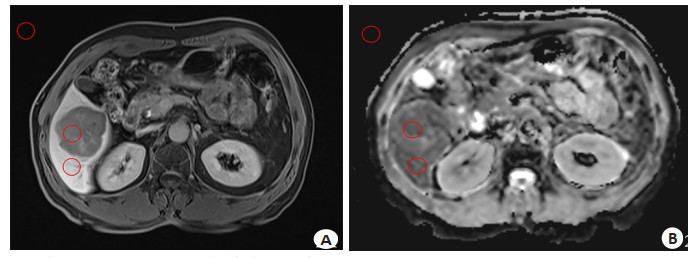
图 1 术后病理为HCC MVI阳性患者的MRI图像
Figure 1. MRI images of a patient with postoperative pathology confirmed as HCC with MVI-positive. A: Liver and gallbladder specific phase image measurement schematic diagram; B: ADC map measurement schematic diagram.

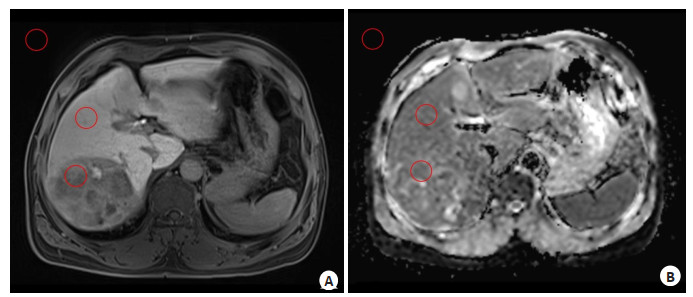
图 2 术后病理为HCC MVI阴性患者的MR图像
Figure 2. MRI images of a patient with postoperative pathology confirmed as HCC with MVI-negative. A: Liver and gallbladder specific phase image measurement schematic diagram; B: ADC map measurement schematic diagram.

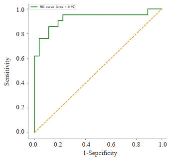
图 3 基于Gd-EOB-DTPA增强MRI构建的SVM模型预测MVI的ROC曲线
Figure 3. ROC curve for predicting MVI using the SVM model constructed based on Gd-EOB-DTPA enhanced MRI.

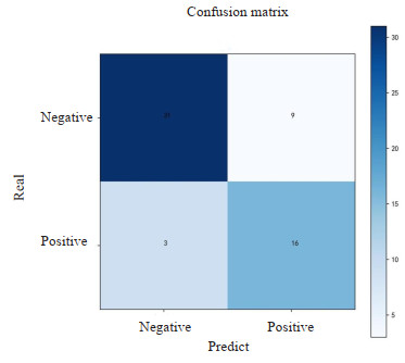
图 4 基于Gd-EOB-DTPA增强MRI构建的SVM模型的混淆矩阵
Figure 4. Confusion matrix of the SVM model constructed based on Gd-EOB-DTPA enhanced MRI.
表 1 MVI阴性组与MVI阳性组临床资料结果比较
Table 1. Comparison of clinical data between the MVI-negative group and the MVI-positive group.
Index MVI-negative group(n=34) MVI-positive group(n=25) t/U/χ2 P Age (years, Mean±SD) 59.7±10.3 61.2±9.7 -0.55 0.58 Gender [n(%)] 1.61 0.39 Male 32 21 Female 2 4 AFP [ng/mL, n(%)] 8.64 < 0.01 ≤20 24 8 > 20 10 17 Child-Pugh classification [n(%)] 0.13 0.91 A 31 23 B 3 2 C 0 0 Tumor diameter (cm, Mean±SD) 5.25±2.97 8.27±3.92 -3.20 < 0.01 AFP: Alpha-fetoprotein; MVI: Microvascular invasion.  下载: 导出CSV
-
[1] Sung H, Ferlay J, Siegel RL, et al. Global cancer statistics 2020: GLOBOCAN estimates of incidence and mortality worldwide for 36 cancers in 185 countries[J]. CA A Cancer J Clin, 2021, 71(3): 209-49. doi: 10.3322/caac.21660 [2] Kudo M, Trevisani F, Abou-Alfa GK, et al. Hepatocellular carcinoma: therapeutic guidelines and medical treatment[J]. Liver Cancer, 2017, 6(1): 16-26. doi: 10.1159/000449343 [3] Hirokawa F, Hayashi M, Asakuma M, et al. Risk factors and patterns of early recurrence after curative hepatectomy for hepatocellular carcinoma[J]. Surg Oncol, 2016, 25(1): 24-9. doi: 10.1016/j.suronc.2015.12.002 [4] Sun SW, Liu QP, Xu X, et al. Direct comparison of four presurgical stratifying schemes for prediction of microvascular invasion in hepatocellular carcinoma by gadoxetic acid-enhanced MRI[J]. J Magn Reson Imaging, 2020, 52(2): 433-47. doi: 10.1002/jmri.27043 [5] Pawlik TM, Gleisner AL, Anders RA, et al. Preoperative assessment of hepatocellular carcinoma tumor grade using needle biopsy: implications for transplant eligibility[J]. Ann Surg, 2007, 245(3): 435-42. doi: 10.1097/01.sla.0000250420.73854.ad [6] An C, Kim MJ. Imaging features related with prognosis of hepatocellular carcinoma[J]. Abdom Radiol, 2019, 44(2): 509-16. doi: 10.1007/s00261-018-1758-y [7] Witjes CDM, Willemssen FEJA, Verheij J, et al. Histological differentiation grade and microvascular invasion of hepatocellular carcinoma predicted by dynamic contrast-enhanced MRI[J]. J Magn Reson Imaging, 2012, 36(3): 641-7. doi: 10.1002/jmri.23681 [8] Ariizumi SI, Kitagawa K, Kotera Y, et al. A non-smooth tumor margin in the hepatobiliary phase of gadoxetic acid disodium (Gd-EOB-DTPA)-enhanced magnetic resonance imaging predicts microscopic portal vein invasion, intrahepatic metastasis, and early recurrence after hepatectomy in patients with hepatocellular carcinoma[J]. J Hepatobiliary Pancreat Sci, 2011, 18(4): 575-85. doi: 10.1007/s00534-010-0369-y [9] 焦琳琳, 段崇锋, 于海洋, 等. 钆塞酸二钠增强MRI定量及定性评价肝癌微血管侵犯的价值[J]. 放射学实践, 2021, 36(8): 1026-31. https://www.cnki.com.cn/Article/CJFDTOTAL-FSXS202108021.htm [10] Chong CC, Lee KF, Ip PC, et al. Pre-operative predictors of post-hepatectomy recurrence of hepatocellular carcinoma: can we predict earlier?[J]. Surgeon, 2012, 10(5): 260-6. doi: 10.1016/j.surge.2011.07.004 [11] Park UJ, Kim YH, Kang KJ, et al. Risk factors for early recurrence after surgical resection for hepatocellular carcinoma[J]. Korean J Hepatol, 2008, 14(3): 371. doi: 10.3350/kjhep.2008.14.3.371 [12] Sumie SJ, Kuromatsu R, Okuda K, et al. Microvascular invasion in patients with hepatocellular carcinoma and its predictable clinicopathological factors[J]. Ann Surg Oncol, 2008, 15(5): 1375-82. doi: 10.1245/s10434-008-9846-9 [13] 李洁, 曹吉平, 李莉, 等. 肝细胞癌MRI钆贝葡胺增强肝胆期与病理学对照分析[J]. 中国医学影像技术, 2018, 34(12): 1825-9. https://www.cnki.com.cn/Article/CJFDTOTAL-ZYXX201812021.htm [14] 刘曦娇, 唐鹤菡, 张笑, 等. 钆塞酸二钠增强MRI肝胆期信号与肝细胞肝癌分化程度的关系[J]. 中国普外基础与临床杂志, 2014, 21(12): 1583-6. doi: 10.7507/1007-9424.20140377 [15] 金玉梅, 王叶武, 张军, 等. 影像组学在混合型肝癌与肝内胆管细胞癌鉴别诊断中的价值[J]. 中国CT和MRI杂志, 2021, 19(11): 118-22, 126. https://www.cnki.com.cn/Article/CJFDTOTAL-CTMR202111039.htm [16] 张泳欣, 张水兴, 肖学红, 等. 基于影像学和血清学特征构建的列线图模型可较好预测原发性肝细胞癌的微血管浸润[J]. 分子影像学杂志, 2022, 45(4): 518-25. doi: 10.12122/j.issn.1674-4500.2022.04.10 [17] Zhou W, Jian W, Cen XP, et al. Prediction of microvascular invasion of hepatocellular carcinoma based on contrast-enhanced MR and 3D convolutional neural networks[J]. Front Oncol, 2021, 11: 588010. doi: 10.3389/fonc.2021.588010 -

 点击查看大图
计量
- 文章访问数: 112
- HTML全文浏览量: 26
- PDF下载量: 7
- 被引次数: 0