


Clinical manifestations and ultrasonic characteristics of uterine lipoleiomyomas at different sites
-
摘要:
目的 分析不同部位子宫脂肪平滑肌瘤(LL)的临床表现、超声特征。 方法 回顾性分析2019年9月~2022年9月在我院经手术病理证实为LL的60例患者的临床信息、实验室检查、超声图像特征、病理结果等临床资料。 结果 60例LL患者,无临床表现41例,下腹胀痛7例,月经量增多6例,绝经后阴道流血3例,同房痛1例,肛门坠胀感1例,尿频尿急1例,不同部位LL临床表现差异无统计学意义(P > 0.05)。41例病灶位于子宫体肌壁间或浆膜下,其中5例发生在阔韧带;13例位于子宫颈,其中1例位于宫颈管粘膜下;6例位于宫腔粘膜下。LL在超声多为圆形或类圆形,有假包膜样回声,多边界清楚,均表现为实性稍高回声团块,内部回声较均质,肿物周边均可探及血流信号。术前超声误诊30例,误诊率50%,不同部位LL误诊率差异无统计学意义(P > 0.05)。 结论 LL超声表现具有一定特征,以圆形或类圆形为主,均表现为实性稍高回声团块,能够通过术前超声进行初步诊断,但误诊率较高。 Abstract:Objective To analyze the clinical manifestations and ultrasonic characteristics of uterine lipoleiomyomas (LL) at different sites. Methods A retrospective analysis was performed on the clinical data of 60 patients with LL confirmed by surgical pathology in the hospital from September 2019 to September 2022. The clinical information, laboratory examinations, characteristics of ultrasonic images and pathological results were analyzed. Results In the 60 patients with LL, there were 41 cases without clinical manifestations, 7 cases with abdominal distension, 6 cases with increased menstruation, 3 cases with postmenopausal vaginal bleeding, 1 case with dyspareunia, 1 case with anal distension and 1 case with frequent and urgent urination. There was no significant difference in clinical manifestations among patients with LL at different sites (P > 0.05). There were 41 cases with lesions located in the intermuscular wall of uterine body or subserous membrane (5 cases in broad ligament), 13 cases in the cervix (1 case in submucosa of cervical canal) and 6 cases in uterine submucosa. In ultrasound detection, LL were mostly round or quasi-round, there were false envelope-like echoes, most boundaries were clear. There were solid and slightly hyperechoic lumps, relatively homogeneous internal echoes and blood flow signals around tumors. Preoperative ultrasound showed that there were 30 misdiagnosed cases, with misdiagnosis rate of 50%. There was no significant difference in misdiagnosis rate among patients with LL at different sites (P > 0.05). Conclusion There are certain characteristics of ultrasonic manifestations in LL. Most of them are round or quasi-round, with solid and slightly hyperechoic lumps. Preoperative ultrasound can preliminarily determine LL, but the misdiagnosis rate is high. -
Key words:
- uterus /
- lipoleiomyoma /
- clinical manifestation /
- ultrasonic characteristic /
- diagnostic value
-

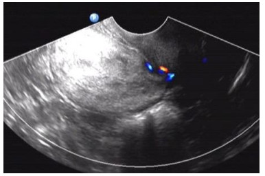
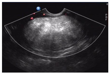
图 1 子宫体肌壁间LL
Figure 1. LL in intermuscular wall of uterine body. There was solid echo mass with homogeneous, diffuse and slightly high echo, with clear boundaries, regular morphology, peripheral blood flow signals, pseudocapsule echoes and a few peripheral muscular tissues. It was easily misdiagnosed as fibroid calcification or steatosis.

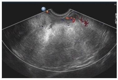
图 2 子宫颈前壁浆膜下LL
Figure 2. LL in anterior cervix wall under subserous membrane. There were large lumps protruding toward the bladder, which resulted in frequent and urgent urination and other compression symptoms.

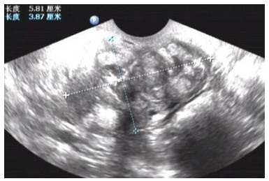
图 3 巨大的右侧阔韧带LL
Figure 3. Large LL in right broad ligament. They were misdiagnosed as ovarian teratoma by ultrasound and were difficult to distinguish from solid teratoma.

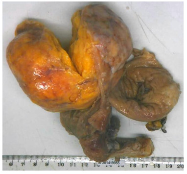
图 5 切除的子宫及子宫浆膜下LL
Figure 5. The resected LL in cervix and uterine submucosa. There was complete pseudocapsule around them, profile was light yellow, with soft and tough characters.

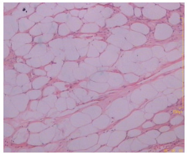
图 6 LL病理检查
Figure 6. Pathological examination of LL. There were adipose cells with diffuse distribution, smooth muscle cells and fibrous tissues (Hematoxylin-eosin staining, ×400).
表 1 60例不同部位的子宫LL的临床表现
Table 1. Clinical manifestations of LL at different sites in the 60 cases (n)
Sites of LL No clinical manifestations (lumps in physical examination) Abdominal distension Increased menstruation Postmenopausal vaginal bleeding Dyspareunia Anal distension Frequent and urgent urination Intermuscular wall of uterine body or subserous membrane (n=36) 28 4 1 2 0 1 0 Cervix (n=13) 8 0 3 0 1 0 1 Uterine submucosa(n=6) 3 0 2 1 0 0 0 Broad ligament (n=5) 2 3 0 0 0 0 0 χ2 4.548 P 0.208 LL: lipoleiomyoma.  下载: 导出CSV
表 2 不同部位子宫LL的误诊疾病
Table 2. Misdiagnosed diseases of LL at different sites (n)
Sites of LL Calcification or steatosis of uterine fibroid Adnexal tumor Endometrium-derived occupation Cervical polyp Incision pregnancy In total Intermuscular wall of uterine body or subserous membrane (n=36) 12 5 1 0 0 18 Cervix (n=13) 2 1 0 1 1 5 Uterine submucosa(n=6) 0 0 2 0 0 2 Broad ligament (n=5) 0 5 0 0 0 5 χ2 6.359 P 0.095
下载: 导出CSV
-
[1] Wilke S, Benson J, Roller L. Uterine lipoleiomyoma: case report and review of the literature[J]. Radiol Case Rep, 2022, 17(3): 954-8. doi: 10.1016/j.radcr.2022.01.012 [2] Sun D, Yang P, Liu Y, et al. Fallopian tube lipoleiomyoma with degeneration: a case report and literature review[J]. Int J Clin Exp Pathol, 2020, 13(8): 2163-8. [3] Rampersad FS, Verma S, Diljohn J, et al. Uterine lipoleiomyoma presenting with pelvic pain in a post-menopausal woman[J]. Cureus, 2021, 13(5): e14929. [4] Hu J, Surti U, Tobon H. Cytogenetic analysis of a uterine lipoleiomyoma[J]. Cancer Genet Cytogenet, 1992, 62(2): 200-2. doi: 10.1016/0165-4608(92)90263-8 [5] Nazir HM, Mehta S, Seena CR, et al. Uterine lipoleiomyoma: a report of two cases[J]. J Clin Imaging Sci, 2017, 7: 26. doi: 10.4103/jcis.JCIS_13_17 [6] 程晓晓, 贺应林, 涂开家, 等. 子宫脂肪平滑肌瘤31例报道[J]. 诊断病理学杂志, 2018, 25(5): 378-9. doi: 10.3969/j.issn.1007-8096.2018.05.015 [7] 韩逢皎, 屈星, 李玉兰, 等. 子宫脂肪平滑肌瘤的发病机制及诊断治疗研究进展[J]. 山东医药, 2022, 62(26): 98-101. https://www.cnki.com.cn/Article/CJFDTOTAL-SDYY202226024.htm [8] Akbulut M, Gündoğan M, Yörükoğlu A. Clinical and pathological features of lipoleiomyoma of the uterine corpus: a review of 76 cases[J]. Balkan Med J, 2014, 31(3): 224-9. doi: 10.5152/balkanmedj.2014.13079 [9] Lin KC, Sheu BC, Huang SC. Lipoleiomyoma of the uterus[J]. Int J Gynecol Obstet, 1999, 67(1): 47-9. doi: 10.1016/S0020-7292(99)00094-6 [10] 邢肖肖, 黄亚青, 赵伟. 子宫脂肪平滑肌瘤超声表现1例[J]. 中国超声医学杂志, 2018, 34(11): 1054. doi: 10.3969/j.issn.1002-0101.2018.11.033 [11] Palicelli A, Ardighieri L, Broggi G, et al. Lipoleiomyomas of the uterine cervix: a new series including the first recurrent case and the first systematic literature review[J]. J Pers Med, 2022, 12(11): 1852. doi: 10.3390/jpm12111852 [12] 李妹庆, 张阳阳, 于楠楠, 等. 5例子宫平滑肌脂肪瘤的临床分析[J]. 中国妇产科临床杂志, 2019, 20(1): 57-8. https://www.cnki.com.cn/Article/CJFDTOTAL-FKLC201901020.htm [13] 刘婷婷, 王翔宇, 韩毅, 等. 子宫脂肪平滑肌瘤临床及病理学特征探讨(附1例报告及文献复习)[J]. 青岛大学学报: 医学版, 2020, 56(1): 109-11. https://cpfd.cnki.com.cn/Article/CPFDTOTAL-ZHYX201211001522.htm [14] 叶小剑, 徐荣全, 鄢磊, 等. 子宫脂肪平滑肌瘤超声与临床病理表现对照分析[J]. 中国介入影像与治疗学, 2016, 13(10): 627-31. doi: 10.13929/j.1672-8475.2016.10.011 [15] 王翠翠, 张晓霞, 张松灵, 等. 子宫脂肪平滑肌瘤2例误诊报告[J]. 吉林大学学报: 医学版, 2010, 36(2): 311. https://www.cnki.com.cn/Article/CJFDTOTAL-BQEB201002033.htm [16] 王靖莹, 路英丽, 谢娱新, 等. 原发性卵巢平滑肌瘤误诊为子宫平滑肌瘤1例[J]. 中国实验诊断学, 2020, 24(7): 1223-4. [17] 卫丹, 翟瑞芳, 廖平川. 彩色多普勒超声在子宫平滑肌瘤、非典型平滑肌瘤及子宫平滑肌肉瘤鉴别诊断中的差异性分析[J]. 中国计划生育和妇产科, 2020, 12(11): 48-51. https://www.cnki.com.cn/Article/CJFDTOTAL-JHFC202011015.htm [18] 韩春宏, 吴明灿, 张建丰, 等. 子宫脂肪平滑肌瘤的CT及MRI特征[J]. 医学影像学杂志, 2012, 22(8): 1370-3. https://www.cnki.com.cn/Article/CJFDTOTAL-XYXZ201208041.htm [19] 黄波涛, 区俊兴, 韩淑珍, 等. 子宫特殊类型平滑肌瘤的临床病理特征与MRI表现[J]. 实用放射学杂志, 2019, 35(7): 1103-6. [20] Chu CY, Tang YK, Chan TS A, et al. Diagnostic challenge of lipomatous uterine tumors in three patients[J]. World J Radiol, 2012, 4(2): 58-62. -

 点击查看大图
计量
- 文章访问数: 126
- HTML全文浏览量: 87
- PDF下载量: 9
- 被引次数: 0