


Analysis of the diagnostic value of CT value combined with coagulation and fibrinolysis related indexes in the occurrence of deep vein thrombosis of lower extremity
-
摘要:
目的 分析CT值联合凝血纤溶指标对下肢深静脉血栓(DTV)的诊断价值分析。 方法 选取2019年1月~2022年5月本院收治的发生下肢DTV的98例患者为研究组,另取同期本院未发生下肢DTV的98例患者为对照组。所有患者均进行CT检查和测定凝血纤溶指标[活化部分凝血酶原时间(aPTT)、凝血酶原时间(PT)、纤维蛋白原(FIB)、D-二聚体(D-D)、抗凝血酶Ⅲ(ATⅢ)],采用Logistic回归分析CT值、凝血纤溶指标与下肢DTV的相关性,并采用ROC曲线分析CT值和凝血纤溶指标对下肢DTV的诊断价值,计算曲线下面积。 结果 CT图像显示,下肢DTV的静脉管腔未见造影剂填充,呈低密度;相较于对照组,研究组CT值、FIB、D-D均较高(P < 0.05),aPTT、PT、ATⅢ均较低(P < 0.05);Logistic回归结果显示,CT值、aPTT、PT、FIB、D-D、ATⅢ与下肢DTV存在显著相关性(P < 0.05);ROC曲线结果显示,联合诊断的曲线下面积为0.946,敏感度为81.63%,特异性为95.92%。 结论 CT值、凝血纤溶指标与下肢DTV具有相关性,其联合诊断在下肢DTV患者方面具有较高的应用价值。 Abstract:Objective To analyze the diagnostic value of CT value combined with coagulation and fibrinolysis index in the diagnosis of lower extremity deep vein thrombosis (DTV). Methods A total of 98 patients with lower extremity DTV admitted to our hospital from January 2019 to May 2022 were selected as the research group, and 98 patients without lower extremity DTV in our hospital during the same period were selected as the control group. All patients underwent CT examination and determination of coagulation and fibrinolysis indexes [activated partial prothrombin time (aPTT), prothrombin time (PT), fibrinogen (FIB), D-dimer (D-D), antithrombin Ⅲ (ATⅢ), Logistic regression was used to analyze the correlation between CT value, coagulation and fibrinolysis index and lower extremity DTV, and ROC curve was used to analyze the diagnostic value of CT values and coagulation and fibrinolysis indexes for lower extremity DTV, and the area under the curve was calculated. Results CT images showed that the venous lumen of DTV in lower limbs was not filled with contrast agent, showing low density; compared with the control group, the CT value, FIB, and D-D of the study group were significantly higher (P < 0.05), aPTT, PT and ATⅢ were significantly lower (P < 0.05); Logistic regression results showed that CT value, aPTT, PT, FIB, D-D, ATⅢ were significantly correlated with lower extremity DTV (P < 0.05). ROC curve results showed that the area under the curve of the combined diagnosis was 0.946, the sensitivity was 81.63%, and the specificity was 95.92%. Conclusion CT value, coagulation and fibrinolysis index are correlated with lower extremity DTV, and their combined diagnosis has high application value in patients with lower extremity DTV. -

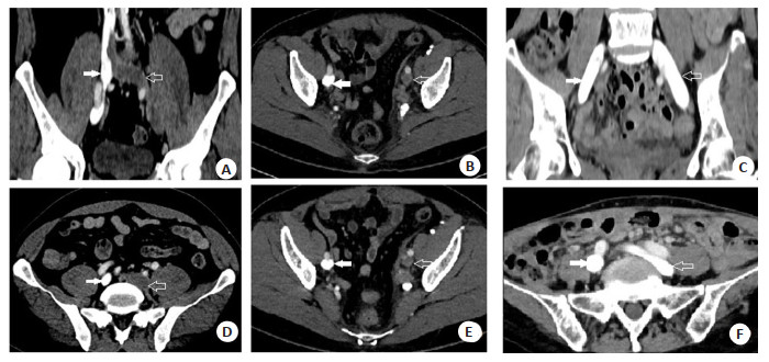
图 1 下肢DVT CT造影图
Figure 1. Lower limb DVT CT imaging. A-B: A 51-year-old male patient with left common iliac vein thrombosis as shown by the hollow arrow could be seen in both coronal bitmap (A) and transverse image (B). There was no contrast agent filling in the lumen, showing low density. The contrast agent filling in the right common iliac vein as shown by the solid arrow shows uniform high-density shadow. C-D: A 51-year-old female patient with thrombosis in the left common iliac vein as shown by the hollow arrow. Compared with the right common iliac vein as shown by the solid arrow, the lumen was not filled with contrast agent and is low density. E-F: A 54-year-old female patient, as shown by the arrow, had left and right common iliac veins filled with contrast medium, showed uniform high density.

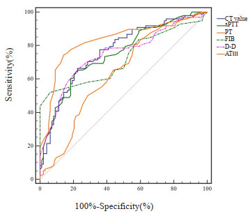
图 2 CT值、aPTT、PT、FIB、D-D、ATⅢ诊断下肢DTV的ROC曲线
Figure 2. ROC curve of CT value, aPTT, PT, FIB, D-D and ATⅢ for diagnosing DTV of lower limbs.

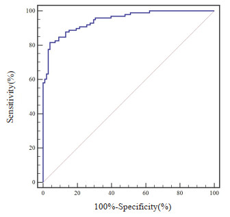
图 3 CT值联合aPTT、PT、FIB、D-D、ATⅢ诊断下肢DTV的ROC曲线
Figure 3. ROC curve of CT value combined with aPTT, PT, FIB, D-D and ATⅢ in diagnosing DTV of lower limbs.
表 1 两组一般资料比较
Table 1. Comparison of general data between the two groups (n=98)
Index Research group Control group t/χ2 P Gender (n) 0.519 0.471 Male 58 53 Female 40 45 Average age (year, Mean±SD) 42.56±10.22 43.47±11.06 0.598 0.550 Average BMI(kg/m2, Mean±SD) 22.20±2.54 22.57±2.83 0.963 0.337 Merger of underlying diseases (n) 0.822 0.364 Yes 36 30 No 62 68  下载: 导出CSV
表 2 两组CT值、凝血纤溶指标比较
Table 2. Comparison of CT values and coagulation and fibrinolysis indexes between the two groups (n=98, Mean±SD)
Index Research group Control group t P CT value(Hu) 55.42±9.81 45.96±9.60 6.823 < 0.001 Coagulation function indicators aPTT(s) 27.70±1.92 29.41±1.60 6.773 < 0.001 PT(s) 10.49±0.74 11.41±0.52 10.07 < 0.001 FIB(g/L) 3.33±0.60 2.92±0.47 5.325 < 0.001 Fibrinolytic function indicators D-D(mg/L) 0.47±0.11 0.39±0.12 4.865 < 0.001 ATⅢ(%) 95.92±10.68 101.52±12.69 3.402 0.001 aPTT: Activated partial prothrombin time; PT: Prothrombin time; FIB: Fibrinogen; D-D: D-dimer; ATⅢ: Antithrombin Ⅲ.
下载: 导出CSV
表 3 CT值、凝血纤溶指标与下肢DTV的相关性
Table 3. Correlation between CT value, coagulation and fibrinolytic indexes and DTV of lower limbs.
Target β SE Wald χ2 OR P 95% CI CT value 0.962 0.331 8.447 2.617 0.004 1.368-5.007 aPTT 0.632 0.196 10.397 1.881 0.001 1.281-2.763 PT 0.782 0.289 7.322 2.186 0.007 1.241-3.851 FIB 0.825 0.336 6.029 2.282 0.014 1.181-4.409 D-D 1.285 0.533 5.812 3.615 0.016 1.272-10.275 ATⅢ 0.692 0.325 4.534 1.998 0.034 1.057-3.777
下载: 导出CSV
表 4 CT值联合凝血纤溶指标对下肢DPV发生的诊断效能
Table 4. Diagnostic efficacy of CT value combined with coagulation and fibrinolysis indexes on DPV occurrence in lower limbs
Index Cut-off value AUC Maximum Jordan index Sensibility(%) Specificity(%) 95% CI P vs six joint ventures CT value(Hu) > 51.81 0.765 0.439 66.33 77.65 0.699-0.822 < 0.001 aPTT(s) ≤28.4 0.749 0.429 64.29 78.57 0.682-0.808 < 0.001 PT(s) ≤10.9 0.830 0.602 74.49 85.71 0.770-0.880 < 0.001 FIB(g/L) > 3.4 0.723 0.459 52.04 93.88 0.655-0.785 < 0.001 D-D(mg/L) > 0.48 0.747 0.429 66.33 76.53 0.681-0.807 < 0.001 ATⅢ(%) ≤104 0.634 0.245 80.61 43.88 0.563-0.702 < 0.001 Six joint projects - 0.946 0.776 81.63 95.92 0.904-0.973 -
下载: 导出CSV
-
[1] Braun MM, Kassop D. Cardiovascular disease: lower extremity deep venous thrombosis[J]. Fp Essentials, 2019, 479: 21-9. [2] Hattab Y, Küng S, Fasanya A, et al. Deep venous thrombosis of the upper and lower extremity[J]. Crit Care Nurs Q, 2017, 40(3): 230-6. doi: 10.1097/CNQ.0000000000000165 [3] 侯瑞鸿, 母建奎, 孙梦月, 等. MSCTV对下肢静脉血栓性疾病的诊断价值[J]. 河北医科大学学报, 2020, 41(10): 1195-9. doi: 10.3969/j.issn.1007-3205.2020.10.017 [4] 朱海荣. 急诊床旁超声联合快速床旁D-二聚体、纤维蛋白原检测对下肢深静脉血栓的诊断价值[J]. 广西医科大学学报, 2019, 36(8): 1327-30. doi: 10.16190/j.cnki.45-1211/r.2019.08.025 [5] Li HL, Chan Y, Li N, et al. Prevalence and predictor of pulmonary embolism in a cohort of Chinese patients with acute proximal deep vein thrombosis[J]. Ann Vasc Surg, 2020, 63: 293-7. doi: 10.1016/j.avsg.2019.06.042 [6] Kruger PC, Eikelboom JW, Douketis JD, et al. Deep vein thrombosis: update on diagnosis and management[J]. Med J Aust, 2019, 210(11): 516-24. doi: 10.5694/mja2.50201 [7] 胡剑, 任魁, 牟欢. 超声弹性成像参数及CT值与妇科术后下肢深静脉血栓形成临床分期的相关性及疗效预测价值[J]. 医疗卫生装备, 2021, 42(3): 51-5. doi: 10.19745/j.1003-8868.2021053 [8] 周瀚, 梁鲁, 姚碧辉, 等. Force CT评价下肢深静脉血栓CT值与血液成分的关系[J]. 临床外科杂志, 2021, 29(5): 474-7. doi: 10.3969/j.issn.1005-6483.2021.05.022 [9] 赵小杰, 谢粉霞. 间接下肢CT静脉成像联合D二聚体对股骨颈骨折术后下肢深静脉血栓形成的诊断价值[J]. 血栓与止血学, 2022, 28(3): 732-4, 737. doi: 10.3969/j.issn.1009-6213.2022.03.171 [10] 龚娜, 赵鹏飞. 间接下肢CT静脉成像与凝血指标诊断髋关节置换术后下肢深静脉血栓形成的价值[J]. 血栓与止血学, 2020, 26(2): 239-40, 245. doi: 10.3969/j.issn.1009-6213.2020.02.023 [11] Min SK, Kim YH, Joh JH, et al. Diagnosis and treatment of lower extremity deep vein thrombosis: Korean practice guidelines[J]. Vasc Specialist Int, 2016, 32(3): 77-104. doi: 10.5758/vsi.2016.32.3.77 [12] Duffy S. Understanding patient assessment and treatment in deep vein thrombosis[J]. Nurs Stand, 2022, 37(11): 71-5. doi: 10.7748/ns.2022.e12050 [13] Wenger N, Sebastian T, Engelberger RP, et al. Pulmonary embolism and deep vein thrombosis: similar but different[J]. Thromb Res, 2021, 206: 88-98. doi: 10.1016/j.thromres.2021.08.015 [14] Tamura S, Yamamoto M, Kitagawa A, et al. Deep vein thrombosis (DVT) prophylactic team activity to support DVT prevention protocol for the purpose of the prophylaxis of pulmonary thromboembolism (PTE) and operation[J]. Ann Vasc Dis, 2021, 14(2): 99-107. doi: 10.3400/avd.oa.21-00017 [15] 王春辉, 袁红维. 超声弹性成像参数及CT值与妇科术后下肢深静脉血栓形成临床分期的相关性[J]. 血栓与止血学, 2022, 28(3): 477-8, 481. https://www.cnki.com.cn/Article/CJFDTOTAL-XSZX202203052.htm [16] 吴英宁, 韦勤将, 柴梦琪, 等. 间接法CT静脉造影在诊断盆腔和下肢深静脉血栓中的价值[J]. 广西医学, 2021, 43(4): 434-7. https://www.cnki.com.cn/Article/CJFDTOTAL-GYYX202104011.htm [17] 黄莺, 刘长俊, 江雪莲, 等. 骨科特殊护理预防老年患者下肢骨折术后下肢深静脉血栓形成的效果[J]. 血栓与止血学, 2017, 23(6): 1064-5. doi: 10.3969/j.issn.1009-6213.2017.06.061 [18] Zhu SB, Song Y, Chen X, et al. Traditional Chinese and western medicine for the prevention of deep venous thrombosis after lower extremity orthopedic surgery: a meta-analysis of randomized controlled trials[J]. J Orthop Surg Res, 2018, 13(1): 79. doi: 10.1186/s13018-018-0785-2 [19] Wang C, Liu QH, Sun L, et al. Application of thrombelastography in primary total knee and total hip replacement: a prospective 87 patients study[J]. Blood Coagul Fibrinolysis, 2019, 30(6): 281-90. doi: 10.1097/MBC.0000000000000839 [20] Cheng J, Fu ZB, Zhu JX, et al. The predictive value of plasminogen activator inhibitor-1, fibrinogen, and D-dimer for deep venous thrombosis following surgery for traumatic lower limb fracture[J]. Ann Palliat Med, 2020, 9(5): 3385-92. doi: 10.21037/apm-20-1604 [21] Joraku A, Nitta S, Tanaka K, et al. Screening of deep vein thrombosis prior to surgery using D-dimer testing and venous ultrasonography of lower extremities[J]. Hinyokika Kiyo, 2017, 63(6): 229-33. [22] Ünlü B, Versteeg HH. Cancer-associated thrombosis: the search for the holy grail continues[J]. Res Pract Thromb Haemost, 2018, 2(4): 622-9. https://www.cnki.com.cn/Article/CJFDTOTAL-ZGTO202305023.htm [23] Abdalhabib EK, Jackson DE, Alzahrani B, et al. Assessment of risk factors for deep vein thrombosis associated with natural anticoagulants and fibrinolytic regulatory proteins[J]. Blood Coagul Fibrinolysis, 2022, 33(3): 149-52. doi: 10.1097/MBC.0000000000001116 [24] 李大千, 蒋云, 梅燕萍. 抗凝血酶Ⅲ、D-二聚体与纤维蛋白原在下肢静脉血栓形成中的临床应用[J]. 医学信息, 2019, 32(17): 167-9 https://www.cnki.com.cn/Article/CJFDTOTAL-YXXX201917057.htm [25] 贾托, 贺世集, 祁学强. 创伤骨科患者围术期凝血指标及纤溶活性指标的检测意义[J]. 血栓与止血学, 2020, 26(5): 835-6. https://www.cnki.com.cn/Article/CJFDTOTAL-XSZX202104048.htm -

 点击查看大图
计量
- 文章访问数: 82
- HTML全文浏览量: 60
- PDF下载量: 5
- 被引次数: 0