


Imaging differential diagnosis of giant cell tumor of bone and chondrosarcoma in the end of long bone
-
摘要:
目的 探讨长骨骨端发生的骨巨细胞瘤与软骨肉瘤的影像鉴别诊断特征。 方法 选择我院2015年5月~2022年5月经手术病理证实为长骨骨端原发的20例骨巨细胞瘤(骨巨细胞瘤组)和16例软骨肉瘤(软骨肉瘤组)患者作为研究对象,采用独立样本t检验和Fisher确切概率法对两组患者的临床及影像学特征进行回顾性分析。 结果 两组影像特征比较,差异有统计学意义(P < 0.05):长骨骨端骨巨细胞瘤患者发病年龄在20~40岁者占比更高(60%),单纯累及骨端(90%)、偏心性生长(70%)、边界清楚(100%)、骨皮质膨胀(95%)、均匀变薄(100%)、软组织肿块无分叶(100%)、软组织肿块局限性生长(100%)、T2WI以稍高信号为主(100%)、伴有囊变/坏死/出血/液-液平(60%)的比例更高,而长骨骨端软骨肉瘤患者发病年龄 > 40岁者占87.5%,病灶同时累及骨端及骨干(81.2%)、非偏心性生长(87.5%)、病灶边界不清(56.2%)、骨皮质无膨胀改变(87.5%)、皮质厚薄不均/毛糙(100%)、软组织肿块分叶状(100%)、软组织肿块包绕骨干/浸润周围组织(100%)、T2WI高信号或明显高信号(100%)、无囊变/坏死/出血/液-液平(100%)的占比更高;两组性别、有无软组织肿块、骨髓水肿、软组织渗出以及MR边缘低信号环是否完整的差异无统计学意义(P > 0.05)。 结论 长骨骨端原发骨巨细胞瘤与软骨肉瘤在发病年龄及影像学表现中有一定相似性,通过综合评估患病年龄、发病部位、偏心与否、边界是否清晰、骨皮质有无异常改变、软组织肿块特征及MR上病变内部特点有助于二者鉴别诊断。 Abstract:Objective To investigate the imaging differential diagnosis features of giant cell tumor of bone and chondrosarcoma occurring in the end of long bone. Methods Twenty patients with giant cell tumor of bone (giant cell tumor of bone group) and 16 patients with chondrosarcoma (chondrosarcoma group), which were confirmed by surgical pathology to be primary in the bone end of long bone in our hospital from May 2015 to May 2022 were selected for the study, and the clinical and imaging characteristics between the two groups were retrospectively analyzed using independent samples t-test and Fisher's precision probability test. Results Comparing the imaging features of the two groups, the differences were statistically significant (P < 0.05): A higher percentage of patients with giant cell tumor of bone in the end of long bone are 20 to 40 years old (60%), and there was a higher percentage of simple involvement of bone end (90%), eccentric growth (70%), clear boundary (100%), bone cortex expansion (95%), uniform cortical thinning (100%), soft tissue masses without lobulation (100%), localized growth of soft tissue masses (100%), slightly high signal predominates on T2WI (100%), with Cystic degeneration/necrosis /bleeding/liquid-liquid level (60%). While the percentage of patients with chondrosarcoma in the end of long bone greater than 40 years was 87.5%, and there was a higher percentage of the lesions involved both the bone end and the diaphysis (81.2%), non-eccentric growth (87.5%), unclear boundary (56.2%), without bone cortex expansion (87.5%), uneven cortical thickness/roughness (100%), lobulated soft tissue mass (100%), soft tissue mass surrounding the diaphysis/infiltrating surrounding tissue (100%), high signal or Significantly high signal on T2WI (100%), without Cystic degeneration/necrosis/bleeding/liquid-liquid level (100%). There was no statistically significant difference between the two groups of gender, presence or absence of soft tissue mass, bone marrow edema, soft tissue exudation and MR edge low signal ring (P > 0.05). Conclusion There are some similarities in age of onset and imaging manifestations between giant cell tumor of bone and chondrosarcoma occurring in the end of long bone. Comprehensive assessment of onset age, onset location, eccentric growth or not, clear boundary or not, abnormal changes of the bone cortex or not, features of the soft tissue mass, and internal signal characteristics of the lesion on MR can help in the differential diagnosis of the two deases. -
Key words:
- end of long bone /
- giant cell tumor of bone /
- chondrosarcoma /
- magnetic resonance imaging
-

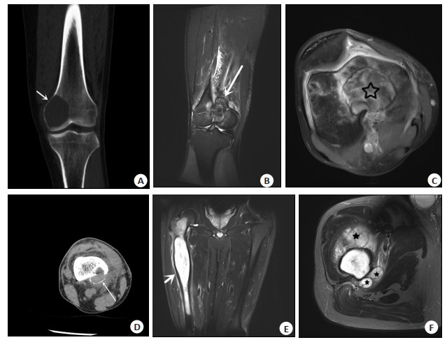
图 1 骨巨细胞瘤与软骨肉瘤影像征象对比
Figure 1. Comparison of imaging features between GCT and chondrosarcoma. A: A 31-year-old male patient presented with a GCT at the distal end of the left femur. Coronal CT showed eccentricity, clear boundary, mild distention and uniform thinning of the cortical bone (short and thin arrow). B-D: A 37-year-old male patient presented with a GCT at the distal end of the right femur. B: Coronal T2WI MR showed eccentric lesions with slightly elevated mixed signals. The soft tissue mass is localized and prominent (long and coarse arrow). Incomplete low signal ring was seen at the edge of the lesion with clear boundary, adjacent bone marrow edema and soft tissue exudation; C: Axial T1WI C+ MR showed avidly uneven enhancement of the lesion (☆); D: Axial CT showed thin bony cladding along the margin of the soft tissue mass (long thin arrow). E-F: A 56-year-old man with a chondrosarcoma of the right femur. E: Coronal T2WI MR showed that the lesion involved the proximal and upper middle medullary cavity of the femur, without eccentricity, with mild swelling of the bone cortex and uneven thickness (short and thick arrow). The main body of the lesion showed obvious high signal close to water, surrounding bone marrow edema and soft tissue exudation; F: Axial T2WI MR showed a multilobulated soft tissue mass growing around the femur (★).
表 1 长骨骨端骨巨细胞瘤与软骨肉瘤年龄分段、性别及MR影像特征比较
Table 1. Comparison of age segment, sex and MR image characteristics of giant cell tumor of bone and chondrosarcoma in the end of long bone [n(%)]
Index GCT group (n=20) Chondrosarcoma group(n=16) P Age (years) 0.003 < 20 1(5) 0(0) 20~40 12(60) 2(12.5) > 40 7(35) 14(87.5) Gender 0.515 Male 10(50) 10(62.5) Female 10(50) 6(37.5) Range of involvement < 0.001 Bony end only 18(90) 3(18.8) Bony end and diaphysis 2(10) 13(81.2) Eccentric growth 0.001 Yes 14(70) 2(12.5) No 6(30) 14(87.5) Focal boundary < 0.001 Clear 20(100) 7(43.8) Unclear 0(0) 9(56.2) Edge low signal ring 0.492 Complete 2(10) 0(0) Incomplete 18(90) 16(100) Bone cortex expansion < 0.001 Yes 19(95) 2(12.5) No 1(5) 14(87.5) Bone cortex < 0.001 Expanded, evenly thinned, smooth inner edge 20(100) 0(0) Uneven thickness, rough 0(0) 16(100) Soft tissue mass 0.176 Yes 10(50) 12(75) No 10(50) 4(25) Lobulated soft tissue mass* < 0.001 Yes 0(0) 12(100) No 10(100) 0(0) Soft tissue mass features* < 0.001 Localized mass protrusion 10(100) 0(0) Surrounds the diaphysis and/or infiltrates the surrounding 0(0) 12(100) The dominant T2WI signal intensity of the lesion < 0.001 High signal or High signal clearly approaching water 0(0) 16(100) Slightly high signal or significantly low signal 20(100) 0(0) Cystic degeneration/necrosis/bleeding/liquid-liquid level < 0.001 Yes 12(60) 0(0) No 8(40) 16(100) Bone marrow edema 0.106 Yes 8(40) 11(68.8) No 12(60) 5(31.2) Soft tissue exudation > 0.999 Yes 16(80) 12(75) No 4(20) 4(25) * A total of 10 of 20 patients in the GCT group have soft tissue masses. A total of 12 of 16 patients in chondrosarcoma group have soft tissue masses; GCT: Giant cell tumor.  下载: 导出CSV
-
[1] Ali N, Shah AA, Rakshan I. Clinical scenario and imaging with illustrations of giant cell tumor of bone: a retrospective analysis[J]. Arch Bone Jt Surg, 2022, 10(1): 60-6. [2] Jain V, Oliveira I, Chavda A, et al. MRI differentiation of low-grade and high-grade chondrosarcoma of the shoulder girdle, chest wall and pelvis: a pictorial review based on 111 consecutive cases[J]. Br J Radiol, 2021, 94(1126): 20201404. doi: 10.1259/bjr.20201404 [3] 李觅, 杨彩虹. 美国国家综合癌症网络临床实践指南: 骨肿瘤(2020V1)解读[J]. 临床外科杂志, 2021(1): 35-7. https://www.cnki.com.cn/Article/CJFDTOTAL-LCWK202101012.htm [4] Liede A, Hernandez RK, Tang ET, et al. Epidemiology of benign giant cell tumor of bone in the Chinese population[J]. J Bone Oncol, 2018, 12: 96-100. doi: 10.1016/j.jbo.2018.07.003 [5] Parmeggiani A, Miceli M, Errani C, et al. State of the art and new concepts in giant cell tumor of bone: imaging features and tumor characteristics[J]. Cancers, 2021, 13(24): 6298. doi: 10.3390/cancers13246298 [6] Roessner A, Smolle M, Haybäck J. Giant cell tumor of bone: Morphology, molecular pathogenesis, and differential diagnosis[J]. Pathologe, 2020, 41(2): 134-42. doi: 10.1007/s00292-020-00760-5 [7] Brown JM, Rakoczy K, Hart J, et al. Presenting features and overall survival of chondrosarcoma of the pelvis[J]. Cancer Treat Res Commun, 2022, 30: 100510. doi: 10.1016/j.ctarc.2022.100510 [8] Wahl S, Domson G, Greenwood A, et al. Giant cell tumor of bone: a report of two cases with metaphyseal lesions and their progression to the articular surface[J]. Int Canc Conf J, 2022, 11(1): 31-40. doi: 10.1007/s13691-021-00511-0 [9] Wang CS, Duan Q, Xue YJ, et al. Giant cell tumour of tendon sheath with bone invasion in extremities: analysis of clinical and imaging findings[J]. Radiol Med, 2015, 120(8): 745-52. doi: 10.1007/s11547-015-0520-6 [10] 刘德斌, 崔学锋, 梁文杰. 长管状骨骨巨细胞瘤的影像诊断分析[J]. 中国矫形外科杂志, 2019, 27(11): 1050-1. https://www.cnki.com.cn/Article/CJFDTOTAL-ZJXS201911024.htm [11] Ghafoor S, Hameed MR, Tap WD, et al. Mesenchymal chondrosarcoma: imaging features and clinical findings[J]. Skeletal Radiol, 2021, 50(2): 333-41. doi: 10.1007/s00256-020-03558-x [12] Wang QZ, Zhang EL, Xing XY, et al. Clinical significance of Preoperative CT and MR imaging findings in the prediction of postoperative recurrence of spinal giant cell tumor of bone[J]. Orthop Surg, 2021, 13(8): 2405-16. doi: 10.1111/os.13173 [13] Chen MF, Lai QQ. Primary intra-and extradural extramedullary mesenchymal chondrosarcoma with isolated punctate calcification: case report and literature review[J]. BMC Neurol, 2022, 22(1): 112. doi: 10.1186/s12883-022-02645-x [14] 杜联军, 丁晓毅, 颜凌, 等. 骨巨细胞瘤常见和典型的CT表现分析[J]. 实用放射学杂志, 2007, 23(8): 1070-3. https://www.cnki.com.cn/Article/CJFDTOTAL-SYFS200708026.htm [15] Rekhi B, Dave VD. Giant cell tumor of bone: an update, including spectrum of pathological features, pathogenesis, molecular profile and the differential diagnoses[J]. Histol Histopathol, 2023, 38(2): 139-53. [16] 马千里, 李晓莉, 邢杰, 等. 骨巨细胞瘤硬化边征象的分析与病理基础探讨[J]. 医学影像学杂志, 2018, 28(7): 1196-8, 1203. https://www.cnki.com.cn/Article/CJFDTOTAL-XYXZ201807043.htm [17] Chen L, Shi XL, Zhou ZM, et al. Clinical significance of MRI and pathological features of giant cell tumor of bone boundary[J]. Orthop Surg, 2019, 11(4): 628-34. doi: 10.1111/os.12510 [18] 高赛, 杨志涛, 李翔, 等. MR评价良性骨肿瘤及肿瘤样病变与恶性骨肿瘤周围水肿的不同[J]. 磁共振成像, 2019, 10(6): 430-4. https://www.cnki.com.cn/Article/CJFDTOTAL-CGZC201906010.htm [19] Sharif B, Rajakulasingam R, Sharifi S, et al. MRI features of low-grade and high-grade chondrosarcoma in enchondromatosis[J]. Skeletal Radiol, 2021, 50(8): 1637-46. [20] 曾燕妮, 宋亭, 张卜天, 等. MRI在长骨高低级别软骨肉瘤诊断中的临床价值[J]. 中国骨与关节杂志, 2018, 7(1): 66-71. https://www.cnki.com.cn/Article/CJFDTOTAL-GZGL201801019.htm [21] 陈基明, 颜秀芳, 翟建, 等. MRI评价长骨中心型低度恶性软骨肉瘤早期侵袭性[J]. 中国医学影像技术, 2019, 35(6): 895-8. https://www.cnki.com.cn/Article/CJFDTOTAL-ZYXX201906035.htm [22] Broehm CJ, Inwards CY, Al-Ibraheemi A, et al. Giant cell tumor of bone in patients 55 years and older: a study of 34 patients[J]. Am J Clin Pathol, 2018, 149(3): 222-33. [23] Chang CY, Garner HW, Ahlawat S, et al. Society of Skeletal Radiology-white paper. Guidelines for the diagnostic management of incidental solitary bone lesions on CT and MRI in adults: bone reporting and data system (Bone-RADS)[J]. Skeletal Radiol, 2022, 51(9): 1743-64. -

 点击查看大图
计量
- 文章访问数: 136
- HTML全文浏览量: 108
- PDF下载量: 9
- 被引次数: 0