


Cardiac magnetic resonance imaging can evaluate early cardiac function in patients with acute myocardial infarction
-
摘要:
目的 探讨心脏磁共振对急性心肌梗死患者早期心功能状态评估价值。 方法 选择2022年6月~2022年12月在我科住院并确诊的急性ST段抬高型心肌梗死患者24例,男性22例,女性2例,年龄55.3±11.3岁。所有患者均在入院后行冠状动脉介入手术,并在术后5~7 d行心脏磁共振检查。应用电影成像技术分析心功能状态、是否存在反向运动和室壁瘤;组织追踪技术分析心肌各节段的应变能力;延迟强化技术分析心肌梗死部位、梗死面积大小和是否存在微循环障碍。结果梗死节段心肌应变分析显示:24例患者中,21例患者径向应变下降,18例患者周向应变下降,21例患者纵向应变下降,16例患者三向应变均下降;整体心肌应变分析显示:15例患者径向应变下降,10例患者周向应变下降,20例患者纵向应变下降,9例患者三向应变均下降;梗死节段心肌平均径向应变和周向应变低于整体心肌平均径向应变和周向应变(P < 0.05),梗死节段与整体心肌纵向应变的差异无统计学意义(P > 0.05)。13例患者出现心肌反向运动;左室射血分数(LVEF)下降者10例;LVEF未下降的14例中,心肌反向运动6例,梗死节段三向心肌应变下降6例,梗死区内微循环障碍7例,NT-proBNP水平升高7例。出现反向运动组患者LVEF、梗死节段心肌和整体心肌平均周向应变、纵向应变均小于未出现反向运动组患者(P < 0.05)。相关性分析显示:LVEF与心肌整体应变、梗死节段应变和左房射血分数呈正相关关系,与心肌梗死面积、左室收缩末期容积/体表面积比值、NT-proBNP水平和Genisini评分呈负相关关系(P < 0.05)。二元Logistic回归显示,梗死节段纵向应变的降低可以独立预测患者是否出现心肌反向运动。 结论 心脏磁共振技术可以多角度评价心功能状态,对急性心肌梗死患者术后早期心功能评估、治疗和干预,改善患者的预后提供参考依据。 Abstract:Objective To explore the value of cardiac magnetic resonance imaging in evaluating early cardiac function in patients with acute myocardial infarction. Methods The study included 24 patients with acute ST-segment elevation myocardial infarction who were admitted to our department from June 2022 to December 2022, including 22 male patients and 2 female patients, with the age at 55.3±11.3 years old. All patients underwent coronary intervention followed by cardiac magnetic resonance imaging 5-7 d after the operation. Motion picture imaging was used to analyze cardiac function status, presence or absence of reverse motion and ventricular aneurysm. The strain capacity of each myocardium segment was analyzed by tissue tracking technique. The location, size and microcirculation disturbance of myocardial infarction were analyzed by delayed enhancement technique. Results Myocardial strain analysis revealed that 21 patients were found to have decreased radial strain and longitudinal strain among the 24 patients, 18 had decreased circumferential strain, and 16 showed a reduction in the three-dimensional myocardial strain. According to the overall analysis of the myocardial strain revealed that 15 patients revealed decreased radial strain, 10 had decreased circumferential strain, 20 had decreased longitudinal strain, and 9 showed a reduction in three-dimensional myocardial strain. The mean radial strain and circumferential strain of the MI segment were significantly lower than that of the average radial strain and circumferential strain of the entire myocardium (P < 0.05), but no statistically significant difference was observed in the longitudinal strain (P > 0.05). Moreover, 13 patients were observed to have myocardial reverse movement. There were 10 and 14 patients with and without decreased left ventricular ejection fraction, respectively, among which 6 had myocardial reverse movement, 6 showed a decrease in three-dimensional myocardial strain in the infarcted segment, 7 presented microcirculatory disturbance within the infarct zone, and 7 had increased level of NT-proBNP. The mean circumferential and longitudinal strains of left ventricular ejection fraction, infarct segment myocardium and global myocardium in patients with reverse motion were all smaller than those without reverse motion (P < 0.05). Correlation analysis indicated that left ventricular ejection fraction was positively correlated with the overall myocardial strain, myocardial strain in the infarcted segment and left atrial emptying fraction. It was negatively correlated with infarct size, left ventricular end-systolic volume/body surface area ratio, NT-proBNP level and Gensini score (P < 0.05). The binary Logistic regression analysis showed that the reduction in the longitudinal strain in the infarcted segments could independently predict whether patients possessed myocardial reverse movement. Conclusion Cardiac magnetic resonance imaging provides a comprehensive evaluation of the cardiac function status, which will be a reference for patients with acute myocardial infarction, offering valuable insights into the improvement of early post-operative cardiac function assessment, treatment, intervention and prognosis. -

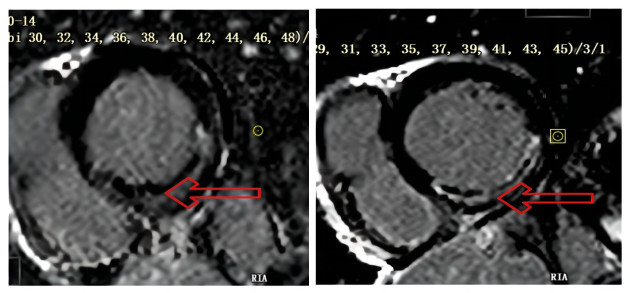
图 1 前壁心肌梗伴微循环障碍2例
Figure 1. Anterior myocardial infarction with microcirculation disturbance in 2 cases.

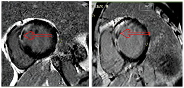
图 2 下壁心肌梗死伴微循环障碍2例
Figure 2. Inferior myocardial infarction with microcirculation disturbance in 2 cases.

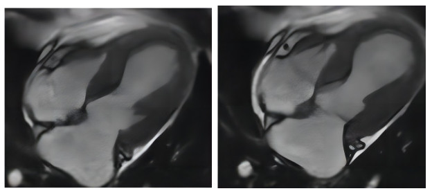
图 3 前壁心肌梗死伴反向运动电影序列图像
Figure 3. Anterior myocardial infarction with reverse motion in film sequence images.

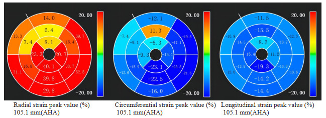
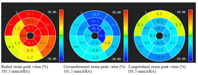
图 4 前壁心肌梗死心肌应变下降
Figure 4. Anterior myocardial infarction myocardial strain decreased.

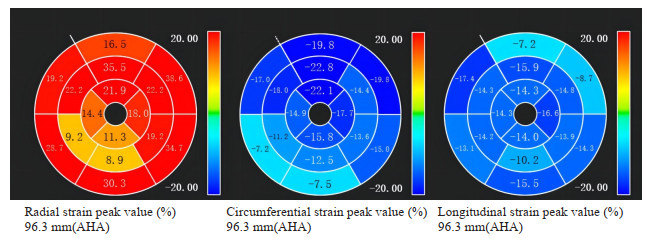
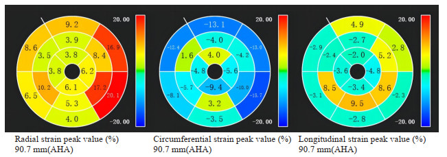
图 5 下壁心肌梗死心肌应变下降
Figure 5. Inferior myocardial infarction myocardial strain decreased.

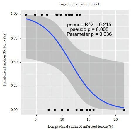
图 6 前壁心肌梗死应变下降伴反向运动
Figure 6. Anterior myocardial infarction decreased myocardial strain with reverse movement.

图 7 下壁心肌梗死应变下降伴反向运动
Figure 7. Inferior myocardial infarction decreased myocardial strain with reverse movement.
表 1 心肌应变分析
Table 1. Myocardial strain analysis(%, Mean±SD)
Myocardial strain Myocardial strain in infarct segment Global myocardial strain P Radial strain 7.74±8.95 28.06±10.42 0.001 Circumferential strain -12.18±-4.12 -15.69±-3.81 0.004 Longitudinal strain -10.92±-3.99 -11.96±-4.30 0.392  下载: 导出CSV
表 2 反向运动分析
Table 2. Reverse motion analysis (Mean±SD)
Index Reverse motion group Non-reverse motion group P Age(year) 56.2±10.6 54.2±12.5 0.673 LVEF(%) 43.77±15.51 55.27±10.06 0.041 LGE(%) 22.90±12.05 16.72±10.92 0.202 LVEDV/BSA 72.19±12.05 73.31±19.45 0.887 LVESV/BSA 42.03±19.56 32.88±14.40 0.201 RS(%) 14.52±7.86 21.54±9.00 0.057 CS(%) 10.55±4.09 14.10±3.40 0.030 LS(%) 9.13±3.14 13.03±3.97 0.016 TRS(%) 26.37±10.83 30.05±10.05 0.397 TCS(%) 14.23±3.98 17.43±2.90 0.033 TLS(%) 10.25±3.85 13.97±4.07 0.033 LAEF(%) 53.54±13.44 59.27±12.53 0.292 LAEF: Left atrial ejection fraction: LVEF: Left ventricular ejection fraction; LGE: Late gadolinium enhancement; LVEDV/BSA: Left ventricular end-diastolic volume/body surface area; LVESV/BSA: Left ventricular end-systolic volume/body surface area; RS: Radial strain; CS: Circumferential strain; LS: Longitudinal strain; TRS: Total radial strain; TCS: Total circumferential strain; TLS: Total longitudinal strain.
下载: 导出CSV
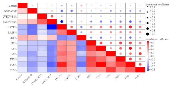
表 3 LVEF相关性分析
Table 3. LVEF correlation analysis
Index R P Age -0.237 0.264 LGE(%) -0.620 0.001 LVEDV/BSA -0.402 0.051 LVESV/BSA -0.836 < 0.001 RS(%) 0.674 < 0.001 CS(%) 0.722 < 0.001 LS(%) 0.785 < 0.001 TRS(%) 0.748 < 0.001 TCS(%) 0.900 < 0.001 TLS(%) 0.736 < 0.001 LAEF(%) 0.754 < 0.001 NT-proBNP -0.702 < 0.001 Gensini -0.429 0.036
下载: 导出CSV
-
[1] 《中国心血管健康与疾病报告》编写组. 《中国心血管健康与疾病报告2021》概述[J]. 中国心血管病研究, 2022(7): 577-96. doi: 10.3969/j.issn.1672-5301.2022.07.001 [2] Leiner T, Bogaert J, Friedrich MG, et al. SCMR Position Paper (2020) on clinical indications for cardiovascular magnetic resonance[J]. J Cardiovasc Magn Reson, 2020, 22(1): 76. doi: 10.1186/s12968-020-00682-4 [3] Avard E, Shiri I, Hajianfar G, et al. Non-contrast Cine Cardiac Magnetic Resonance image radiomics features and machine learning algorithms for myocardial infarction detection[J]. Comput Biol Med, 2022, 141: 105145. doi: 10.1016/j.compbiomed.2021.105145 [4] Reddy A, Singh V, Karthikeyan B, et al. Biventricular strain imaging with cardiac MRI in genotyped and histology validated amyloid cardiomyopathy[J]. Cardiogenetics, 2021, 11(3): 98-110. doi: 10.3390/cardiogenetics11030011 [5] Zhang SR, Li RX, Jiao YD, et al. The prognostic value of microvascular obstruction in patients with acute ST-segment elevation myocardial infarction[J]. Chin J Cardiovasc Res, 2021, 19(3): 193-97. [6] Galand V, Ghoshhajra B, Szymonifka J, et al. Left ventricular wall thickness assessed by cardiac computed tomography and cardiac resynchronization therapy outcomes[J]. Europace, 2020, 22(3): 401-11. doi: 10.1093/europace/euz322 [7] Yu SQ, Zhou JY, Yang K, et al. Correlation of myocardial strain and late gadolinium enhancement by cardiac magnetic resonance after a first anterior ST-segment elevation myocardial infarction[J]. Front Cardiovasc Med, 2021, 8: 705487. doi: 10.3389/fcvm.2021.705487 [8] 中华医学会心血管病学分会, 中华心血管病杂志编辑委员会. 非ST段抬高型急性冠状动脉综合征诊断和治疗指南[J]. 中华心血管中华心血管病杂志, 2017, 45(5): 359-76. https://www.cnki.com.cn/Article/CJFDTOTAL-ZJYE201713001.htm [9] 中华医学会心血管病学分会, 中华心血管病杂志编辑委员会. 急性ST段抬高型心肌梗死诊断和治疗指南[J]. 中华心血管病杂志, 2019, 47(10): 766-83. https://www.cnki.com.cn/Article/CJFDTOTAL-YXQY201902036.htm [10] Smith SC Jr, Dove JT, Jacobs AK, et al. ACC/AHA guidelines for percutaneous coronary intervention (revision of the 1993 PTCA guidelines)-executive summary[J]. Circulation, 2001, 103(24): 3019-41. doi: 10.1161/01.CIR.103.24.3019 [11] Gensini GG. A more meaningful scoring system for determining the severity of coronary heart disease[J]. Am J Cardiol, 1983, 51(3): 606. doi: 10.1016/S0002-9149(83)80105-2 [12] Liu T, Howarth AG, Chen Y, et al. Intramyocardial hemorrhage and the "wave front" of reperfusion injury compromising myocardial salvage[J]. J Am Coll Cardiol, 2022, 79(1): 35-48. doi: 10.1016/j.jacc.2021.10.034 [13] Borlotti A, Jerosch-Herold M, Liu D, et al. Acute microvascular impairment post-reperfused STEMI is reversible and has additional clinical predictive value[J]. JACC Cardiovasc Imaging, 2019, 12(9): 1783-93. doi: 10.1016/j.jcmg.2018.10.028 [14] Kumar A, Dharmakumar R. Editorial for "detection of intramyocardial iron in patients Following ST-elevation myocardial infarction using diffusion tensor imaging"[J]. Magnetic Resonance Imaging, 2022, 56(4): 1182-3. doi: 10.1002/jmri.28099 [15] Giulia PA, Georgios G, Gabriella V, et al. Head-to-head comparison of multiple cardiovascular magnetic resonance techniques for the detection and quantification of intramyocardial haemorrhage in patients with ST-elevation myocardial infarction[J]. Eur Radiol, 2020, 31: 1-12. [16] Arnold JR, McCann GP. Cardiovascular magnetic resonance: applications and practical considerations for the general cardiologist[J]. Heart, 2020, 106(3): 174-81. doi: 10.1136/heartjnl-2019-314856 [17] Voigt JU, Cvijic M. 2-and 3-dimensional myocardial strain in cardiac health and disease[J]. JACC Cardiovasc Imaging, 2019, 12(9): 1849-63. doi: 10.1016/j.jcmg.2019.01.044 [18] Johnson C, Kuyt K, Oxborough D, et al. Practical tips and tricks in measuring strain, strain rate and twist for the left and right ventricles[J]. Echo Res Pract, 2019, 6(3): R87-98. doi: 10.1530/ERP-19-0020 [19] Domenech-Ximenos B, Sanz-de la Garza M, Sepulveda-Martinez Á, et al. Assessment of myocardial deformation with CMR: a comparison with ultrasound speckle tracking[J]. Eur Radiol, 2021, 31(10): 7242-50. doi: 10.1007/s00330-021-07857-2 [20] Imperiale A, Chapelle D, Moireau P. Sequential data assimilation for mechanical systems with complex image data: application to tagged-MRI in cardiac mechanics[J]. Adv Modeling Simul Eng Sci, 2021, 8(1): 1-47. doi: 10.1186/s40323-020-00187-w [21] Yang ZY, Wang H, Chang SS, et al. Left ventricular strain-curve morphology to distinguish between constrictive pericarditis and restrictive cardiomyopathy[J]. ESC Heart Fail, 2021, 8(6): 4863-72. doi: 10.1002/ehf2.13679 [22] Montgomery DE, Puthumana JJ, Fox JM, et al. Global longitudinal strain aids the detection of non-obstructive coronary artery disease in the resting echocardiogram[J]. Eur J Echocardiogr, 2012, 13(7): 579-87. [23] Wei L, Zhao CX, Dong JX, et al. Prognostic implications of left ventricular torsion measured by feature-tracking cardiac magnetic resonance in patients with ST-elevation myocardial infarction[J]. Eur Heart J Cardiovasc Imaging, 2023, 24(6): 785-95. doi: 10.1093/ehjci/jeac177 [24] Reindl M, Tiller C, Holzknecht M, et al. Prognostic implications of global longitudinal strain by feature-tracking cardiac magnetic resonance in ST-elevation myocardial infarction[J]. Circ Cardiovasc Imaging, 2019, 12: e009404. doi: 10.1161/CIRCIMAGING.119.009404 [25] Gavara J, Rodriguez-Palomares JF, Valente F, et al. Prognostic value of strain by tissue tracking cardiac magnetic resonance after ST-segment elevation myocardial infarction[J]. JACC Cardiovasc Imaging, 2018, 11(10): 1448-57. [26] Podlesnikar T, Pizarro G, Fernández-Jiménez R, et al. Left ventricular functional recovery of infarcted and remote myocardium after ST-segment elevation myocardial infarction (METOCARD-CNIC randomized clinical trial substudy)[J]. J Cardiovasc Magn Reson, 2020, 22(1): 44. [27] Dastidar AG, Harries I, Pontecorboli G, et al. Native T1 mapping to detect extent of acute and chronic myocardial infarction: comparison with late gadolinium enhancement technique[J]. Int J Cardiovasc Imaging, 2019, 35(3): 517-27. [28] Reindl M, Holzknecht M, Tiller C, et al. Impact of infarct location and size on clinical outcome after ST-elevation myocardial infarction treated by primary percutaneous coronary intervention[J]. Int J Cardiol, 2020, 301: 14-20. [29] Ito H, Ishida M, Makino W, et al. Cardiovascular magnetic resonance feature tracking for characterization of patients with heart failure with preserved ejection fraction: correlation of global longitudinal strain with invasive diastolic functional indices[J]. J Cardiovasc Magn Reson, 2020, 22(1): 42. [30] 李慧娣, 向定成, 张金霞, 等. 急性心肌梗死早期脑钠肽浓度的动态演变规律及其对心力衰竭的诊断价值[J]. 南方医科大学学报, 2018, 38(1): 112-6. https://www.cnki.com.cn/Article/CJFDTOTAL-DYJD201801019.htm -

 点击查看大图
计量
- 文章访问数: 179
- HTML全文浏览量: 66
- PDF下载量: 12
- 被引次数: 0