


Value of magnetic resonance imaging in patients with adult orbital xanthogranulomatous disease
-
摘要:
目的 分析成人眼眶黄色肉芽肿病的磁共振表现,提高对该病的认识。 方法 回顾性分析11例患者的MR图像,分析MR图像病变部位、双侧/单侧、形态、边缘、眶内结构、T1WI、T2WI信号特点、DWI特点、ADC及强化方式。 结果 11例眼眶黄色肉芽肿经手术病理证实,分别是成人起病的黄色肉芽肿(n=6)、渐进性坏死性黄色肉芽肿(n=4)和Erdheim-Chester病(n=1)。成人起病的黄色肉芽肿6例,3例累及单侧眼睑,1例累及右侧鼻泪管,1例累及双侧眼睑和泪腺,1例累及右侧眼睑、翼腭窝和左侧颞肌;渐进性坏死性黄色肉芽肿4例,2例累及左侧眼睑,2例累及双侧眼睑、鼻部、额颞部;Erdheim-chester病1例,双侧眶内多发病变并累及垂体、心包、肾脏。4例边界清晰,7例范围弥漫,边界不清。10例T1WI低、T2WI及压脂高信号,1例T1WI低、T2WI及压脂低信号。弥散加权成像低信号(n=7);平均表观扩散系数值为1.18×10⁻³ mm2/s(n=7)。6例行动态增强检查,时间-信号曲线(TIC)均表现为持续上升型(Ⅰ型)。 结论 眼眶黄色肉芽肿病多有眼睑肿胀,无破溃,累及眼睑及皮下组织、眶前部、眶后部和泪腺,可累及眼外肌肌腱,未见累及视神经,累及眶内病变未见骨壁破坏。MRI可以通过显示化学位移伪影来辅助此病的诊断。 Abstract:Objective To analyze the MRI features of adult orbital xanthogranuloma and improve recognition of the disease. Methods The MRI images of 11 patients were retrospectively analyzed. We analyzed the location, unilateral or bilateral involvement, shape, margins, orbital structures, T1WI and T2WI signal characteristics, diffusion-weighted imaging characteristics, apparent diffusion coefficient and enhancement patterns. Results Eleven cases of orbital xanthogranuloma were confirmed by surgical pathology, including 6 cases of adult-onset xanthogranuloma, 4 cases of necrobiotic xanthogranuloma, and 1 case of Erdheim-Chester disease. Of the 6 cases of adult-onset xanthogranuloma, 3 involved unilateral eyelids, 1 involved the right nasolacrimal duct, 1 involved bilateral eyelids and lacrimal glands and 1 involved right eyelid, pterygopalatine fossa and left temporal muscle. Of the 4 cases of necrobiotic xanthogranuloma, 2 involved the left eyelid, and 2 involved bilateral eyelids, nasal and temporal regions. The case of Erdheim-Chester disease involved multiple bilateral orbital lesions and also affected the pituitary gland, pericardium, and kidneys. The lesions of 4 cases had clear boundaries, while those of 7 cases had diffuse boundaries. Ten cases showed low signal intensity on T1WI, high signal intensity on T2WI and fatsuppressed imaging, and 1 case showed low signal intensity on T1WI, T2WI and fat-suppressed imaging. Diffusion-weighted imaging showed low signal intensity (n=7). The value of apparent diffusion coefficient was 1.18×10⁻³ mm2/s (n=7). Dynamic contrast- enhanced MRI showed continuous rising patterns (type Ⅰ) in all 6 cases. Conclusion Orbital xanthogranuloma typically presents as eyelid swelling without ulceration, involving eyelids and subcutaneous tissue, the anterior and posterior orbit, and lacrimal glands, and may involve extraocular muscle tendons but not optic nerves. No bone destruction was observed in the involved orbit. MRI can assist in the diagnosis of this disease by displaying chemical shift artifacts. -

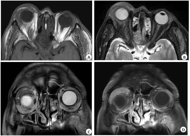
图 1 患者男,59岁,右侧眼睑、翼腭窝和左侧颞肌的AOX的MRI表现
Figure 1. MRI findings of a 59- year- old AOX male patient, which involved right eyelid, pterygopalatine fossa, and left temporal muscle
A: T1WI axial image showed the lesions in the right eyelid, pterygopalatine fossa, and left temporal muscle appeared as irregular low signals; B: T2WI axial image showed the lesions appeared as irregular high signals; C: T2WI coronal image shows the lesion showed a curved low signal at the edge of the adjacent lateral and superior rectus muscles(→); D: T1WI coronal image showed that the enhancement was significantly and uniformly enhanced.

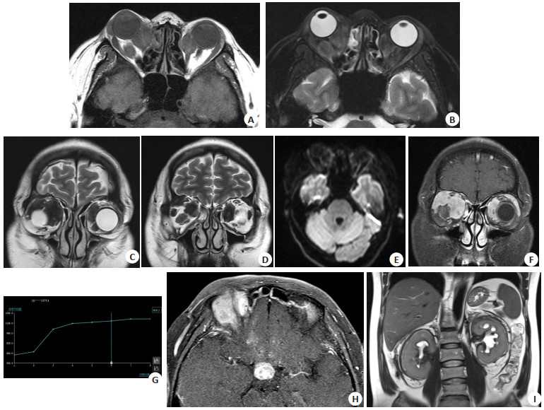
图 2 患者男,43岁,双侧眶内多发病变并累及垂体、肾脏的ECD MRI表现
Figure 2. MRI findings of a 43-year-old ECD male patient, which involved multiple bilateral orbital lesions and also affected the pituitary gland and kidneys
A: T1WI axial image showed multiple lesions with low signal intensity in both orbits, with clear boundaries; B: T2WI axial image showed comparing with the gray matter of the brain, the lesion appeared as a low signal; C, D: T2WI coronal image showed the lesion appeared as a low signal; E: The DWI lesion appeared as a low signal, ADC was 1.01×10⁻³mm2/s; F: T1WI coronal image showed the lesion with obvious and uniform enhancement, and encircled the bilateral rectus internus muscle and the right superior rectus muscle; G: TIC showed continuous rising patterns (type Ⅰ); H: T1WI axial image showed a lesion with a diameter of approximately 15mm in the sellar region; I: T2WI coronal image showed dilation and hydronephrosis of both renal pelvises, and infiltration shadows around both kidneys.
表 1 11例AOXGD的临床资料和MRI表现
Table 1. Clinical data and MR findings of 11 cases of AOXGD
Subtype Age(years) Chief complaint Lesion location MRI findings AOX(n=6) 33-66 Five cases of eyelid swelling ranging from 3 months to 3 years, and one case of right eye tearing for more than 3 years. Three cases involved unilateral eyelids, one case involved the right nasolacrimal duct, one case involved both eyelids and lacrimal gland, and one case involved the right eyelid, pterygopalatine fossa, and left temporal muscle. T1WI showed low signal, T2WI showed high signal, with unclear boundaries in four cases and clear boundaries in two cases. The lesions had irregular shapes, diffuse and unrestricted distribution, and homogeneous enhancement. NXG(n=4) 41-58 Eyelid swelling for more than 3 months to 7 years. Two cases involving the left eyelid, and two cases involving both eyelids, nose, forehead and temporal region. T1WI showed low signal, T2WI showed high signal. Three cases had unclear boundaries, and one case had clear boundaries. The shape was irregular, with diffuse and unrestricted enhancement. ECD(n=1) 43 Bilateral eyelid swelling with protrusion of the eyeballs for more than 2 months. Bilateral lesions in the anterior and posterior orbit and medial and lateral rectus muscles. T1WI showed low signal intensity, T2WI images showed low signal intensity, and T2WI fatsuppressed images show low signal intensity. The border was clear, the shape was irregular, the diffusion was unrestricted, and with homogeneous enhancement. AOX: Adult-onset xanthogranuloma; NXG: Necrobiotic xanthogranuloma; ECD: Erdheim-Chester disease.  下载: 导出CSV
-
[1] Kerstetter J, Wang J. Adult orbital xanthogranulomatous disease[J]. Dermatol Clin, 2015, 33(3): 457-63. doi: 10.1016/j.det.2015.03.010 [2] 王玉川, 李静, 林锦镛. 成人眼眶黄色肉芽肿病伴泪腺反应性淋巴组织增生的临床病理学特征[J]. 中华眼科杂志, 2022, 58(9): 682-7. [3] 王婷婷, 林婷婷, 刘勋, 等. 眼眶成人型黄色肉芽肿的临床分析[J]. 中华眼科杂志, 2019, 55(5): 381-6. https://www.cnki.com.cn/Article/CJFDTOTAL-GJYK202201035.htm [4] Detiger SE, Hötte GJ, Verdijk RM, et al. Adult orbital xanthogranuloma: long-term follow-up of treated cases[J]. Eye, 2022. doi: 10.1038/s41433-022-02357-z. [5] Maeng MM, Godfrey KJ, Jalaj S, et al. Adult xanthogranulomatous disease of the orbit: case report of spontaneous regression and review of treatment modalities[J]. Orbit, 2020, 39(1): 31-7. doi: 10.1080/01676830.2019.1590421 [6] Sahu K, Sethy M, Sirka C, et al. Adult-onset asthma with periocular xanthogranuloma (AAPOX), a variant of periorbital xanthogranulomatous disease: an uncommon entity[J]. Indian Dermatol Online J, 2020, 11(5): 792. doi: 10.4103/idoj.IDOJ_541_19 [7] Sim JK, Lee JY, Jang SY, et al. MRI findings of adult-onset orbital xanthogranulomatous disease[J]. Clin Neuroradiol, 2018, 28(4): 601-4. doi: 10.1007/s00062-018-0673-5 [8] Adam Z, Veselý K, Motyčková I, et al. Eyelids with yellow granulomas and cough-periocular xanthogranuloma associated with adult-onset asthma: a case study and an overview of clinical forms of juvenile xanthogranuloma and its therapy[J]. Vnitr Lek, 2012, 58 (5): 365-77. [9] 张嘉莹, 李瑾. 成人眼眶黄色肉芽肿病的临床研究进展[J]. 国际眼科杂志, 2017, 17(12)2274-2277 https://www.cnki.com.cn/Article/CJFDTOTAL-GJYK201712025.htm [10] Jakobiec F, Mills MD, Hidayat A, et al. Periocular xanthogranulomas associated with severe adult-onset asthma[J]. Trans Am Ophthalmol Soc, 1993, 91(2): 99-125. [11] Diamond EL, Dagna L, Hyman DM, et al. Consensus guidelines for the diagnosis and clinical management of Erdheim-Chester disease[J]. Blood, 2014, 124(4): 483-92. doi: 10.1182/blood-2014-03-561381 [12] Haroun F, Millado K, Tabbara I. Erdheim-Chester disease: comprehensive review of molecular profiling and therapeutic advances[J]. Anticancer Res, 2017, 37(6): 2777-83. [13] Haroche J, Arnaud L, Cohen-Aubart F, et al. Erdheim-Chester disease[J]. Curr Rheumatol Rep, 2014, 16(4): 412. doi: 10.1007/s11926-014-0412-0 [14] Mazor RD, Manevich-Mazor M, Shoenfeld Y. Erdheim-Chester Disease: a comprehensive review of the literature[J]. Orphanet J Rare Dis, 2013, 8: 137. doi: 10.1186/1750-1172-8-137 [15] Cives M, Simone V, Rizzo FM, et al. Erdheim-Chester disease: a systematic review[J]. Crit Rev Oncol, 2015, 95(1): 1-11. doi: 10.1016/j.critrevonc.2015.02.004 [16] 李锋, 刘克, 孙国龙. 常规MRI联合磁共振表观扩散系数在眼眶肿瘤鉴别诊断中的应用价值[J]. 磁共振成像, 2020, 11(12): 1163-6. https://www.cnki.com.cn/Article/CJFDTOTAL-CGZC202012019.htm [17] Lecler A, Duron L, Zmuda M, et al. Intravoxel incoherent motion (IVIM) 3 T MRI for orbital lesion characterization[J]. Eur Radiol, 2021, 31(1): 14-23. doi: 10.1007/s00330-020-07103-1 [18] 李靖, 林毅, 王紫仪, 等. 泪腺IgG4相关性病变与易混淆泪腺炎性假瘤及淋巴瘤的磁共振扩散加权成像对比分析[J]. 临床放射学杂志, 2019, 38(12): 2275-9. https://www.cnki.com.cn/Article/CJFDTOTAL-LCFS201912014.htm [19] Shams PN, Rasmussen SL, Dolman PJ. Adult-onset asthma associated with simultaneous conjunctival, eyelid, and orbital xanthogranulomatosis responsive to systemic immunosuppression [J]. Ophthalmic Plast Reconstr Surg, 2015, 31(6): e162-3. doi: 10.1097/IOP.0000000000000191 [20] Minami-Hori M, Takahashi I, Honma M, et al. Adult orbital xanthogranulomatous disease: adult-onset xanthogranuloma of periorbital location[J]. Clin Exp Dermatol, 2011, 36(6): 628-31. doi: 10.1111/j.1365-2230.2011.04041.x [21] Guo J, Wang J. Adult orbital xanthogranulomatous disease: review of the literature[J]. Arch Pathol Lab Med, 2009, 133(12): 1994-7. doi: 10.5858/133.12.1994 [22] Cavallazzi R, Hirani A, Vasu TS, et al. Clinical manifestations and treatment of adultonset asthma and periocular xanthogranuloma[J]. Can Respir J, 2009, 16(5): 15962. [23] Ferreira TA, Saraiva P, Genders SW, et al. CT and MR imaging of orbital inflammation[J]. Neuroradiology, 2018, 60(12): 1253-66. [24] 聂嘉敏, 何茜, 邵举薇, 等. 扩散加权成像结合动态对比增强扫描对鉴别诊断眼眶淋巴瘤和炎性假瘤的价值[J]. 实用放射学杂志, 2019, 35(11): 1739-42, 1839. [25] Tiegs-Heiden CA, Eckel LJ, Hunt CH, et al. Immunoglobulin G4-related disease of the orbit: imaging features in 27 patients[J]. Am J Neuroradiol, 2014, 35(7): 1393-7. [26] Umehara H, Okazaki K, Kawano M, et al. How to diagnose IgG4- related disease[J]. Ann Rheum Dis, 2017, 76(11): e46. [27] Mulay K, Aggarwal E, Jariwala M, et al. Orbital immunoglobulinG4-related disease: case series and literature review[J]. Clin Experiment Ophthalmol, 2014, 42(7): 682-7. [28] Haroche J, Abla O. Uncommon histiocytic disorders: Rosai-Dorfman, juvenile xanthogranuloma, and Erdheim-Chester disease[J]. Hematology, 2015, 2015(1): 571-8. -

 点击查看大图
计量
- 文章访问数: 115
- HTML全文浏览量: 39
- PDF下载量: 9
- 被引次数: 0