Clinical value of coronary CT angiography plaque quantitative parameters on predicting myocardial ischemic events in patients with coronary heart disease
-
摘要:
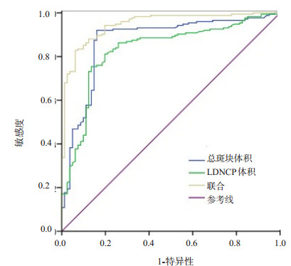
目的 分析冠脉CT血管成像(CTA)斑块定量参数预测冠心病患者心肌缺血事件的临床价值。 方法 纳入2020年1月~2022年6月256例初诊冠心病患者作为研究对象,均行冠脉CTA检查,检测斑块定量参数,根据血流储备分数检测结果将患者分为心肌缺血组和非心肌缺血组,比较两组冠脉CTA斑块定量参数,采用多元线性回归分析斑块定量参数与心肌缺血性损伤的关系,采用ROC曲线评估斑块定量参数对心肌缺血性损伤的预测价值。 结果 心肌缺血组总斑块体积、非钙化斑块体积、低密度非钙化斑块(LDNCP)体积、斑块长度、直径狭窄度均大于非心肌缺血组(P < 0.05),钙化斑块(CP)体积及血流储备分数小于非心肌缺血组(P < 0.05);多元线性回归分析显示,总斑块体积、LDNCP体积为冠心病患者心肌缺血的独立影响因素(P < 0.05);ROC曲线显示,总斑块体积、LDNCP体积联合预测心肌缺血性损伤的敏感度、特异性、曲线下面积分别为94.30%、77.80%、0.948。 结论 冠脉CTA斑块定量参数变化与冠心病患者心肌缺血性损伤有关,其中总斑块体积、LDNCP体积可作为心肌缺血事件的预测指标。 Abstract:Objective To analyze the clinical value of coronary CT angiography (CTA) plaque quantitative parameters on predicting myocardial ischemia events in patients with coronary heart disease. Methods A total of 256 patients with newly diagnosed coronary heart disease during the period from January 2020 and June 2022 were included as the research subjects. All patients underwent coronary CTA examination. The plaque quantitative parameters were detected. According to fractional flow reverse, the patients were divided into myocardial ischemia group and non-myocardial ischemia group. The coronary CTA plaque quantitative parameters were compared between the two groups. Multivariate linear regression analysis was used to analyze the relationship between plaque quantitative parameters and myocardial ischemic injury. ROC curve was drawn to evaluate the predictive value of plaque quantitative parameters on myocardial ischemic injury. Results The total plaque volume, non-calcified plaque volume, volume of low-density non-calcified plaque (LDNCP), plaque length and diameter stenosis in myocardial ischemia group were larger or higher than those in non-myocardial ischemia group (P < 0.05) while the calcified plaque volume and fractional flow reverse were smaller than that in non-myocardial ischemia group (P < 0.05). Multivariate linear regression analysis showed that total plaque volume and LDNCP volume were the independent influencing factors of myocardial ischemia in patients with coronary heart disease (P < 0.05). ROC curve showed that the sensitivity, specificity and area under curve of total plaque volume and LDNCP volume in combination on predicting myocardial ischemic injury were 94.30%, 77.80% and 0.948, respectively. Conclusion The changes in coronary CTA plaque quantitative parameters are related to myocardial ischemic injury in patients with coronary heart disease. Total plaque volume and LDNCP volume can be used as predictors of myocardial ischemic events. -
Key words:
- coronary CT angiography /
- coronary heart disease /
- myocardial ischemia /
- predictive value
-
表 1 两组一般资料比较
Table 1. Comparison of general data between the two groups [n(%)]
资料 心肌缺血组(n=175) 非心肌缺血组(n=81) t/χ2 P 性别 0.160 0.690 男 121(69.14) 58(71.60) 女 54(30.86) 23(28.40) 年龄(岁, Mean±SD) 59.73±10.12 58.65±7.41 0.859 0.391 BMI(kg/m2, Mean±SD) 24.07±3.62 24.02±2.14 0.115 0.908 高血压 93(53.14) 39(48.15) 0.553 9.457 糖尿病 82(46.86) 34(41.98) 0.533 0.466 高血脂 54(30.86) 23(28.40) 0.160 0.690 吸烟 59(33.71) 27(33.33) 0.004 0.952 饮酒 92(52.57) 37(45.68) 1.052 0.305 心血管家族史 33(18.86) 16(19.75) 0.029 0.865 表 2 两组冠脉CTA斑块定量参数及FFR比较
Table 2. Comparison of coronary CTA plaque quantitative parameters and FFR between the two groups (Mean±SD)
指标 心肌缺血组(n=175) 非心肌缺血组(n=81) t P 斑块体积(mm3) 总斑块体积 99.89±11.93 72.47±6.11 19.520 < 0.001 CP体积 7.25±1.72 9.09±1.26 8.614 < 0.001 NCP体积 92.64±11.69 63.38±5.89 21.295 < 0.001 LDNCP体积 14.62±3.83 8.54±1.79 13.605 < 0.001 斑块长度(mm) 4.97±1.64 3.42±1.13 7.698 < 0.001 直径狭窄度(%) 19.06±4.85 13.27±3.88 9.434 < 0.001 FFR 0.70±0.11 0.83±0.04 10.316 < 0.001 CP: 钙化斑块; NCP: 非钙化斑块; LDNCP: 低密度非钙化斑块; FFR: 血流储备分数. 表 3 斑块定量参数与冠心病患者心肌缺血性损伤的关系分析
Table 3. Analysis of the relationship between quantitative plaque parameters and myocardial ischemic injury in patients with coronary heart disease
指标 β SE Waldχ2 P OR 95% CI 总斑块体积 0.9712 0.219 10.570 0.001 2.038 1.327~3.131 CP体积 0.645 0.442 2.129 0.145 1.906 0.801~4.533 NCP体积 0.682 0.374 3.325 0.069 1.978 0.950~4.117 LDNCP体积 0.614 0.167 13.518 < 0.001 1.848 1.332~2.563 斑块长度 0.498 0.372 1.792 0.181 1.645 0.794~3.411 直径狭窄度 0.507 0.398 1.623 0.203 1.660 0.761~3.622 表 4 斑块定量参数对冠心病患者心肌缺血性损的预测效能
Table 4. Predictive efficacy of quantitative plaque parameters on myocardial ischemic damage in patients with coronary heart disease
指标 最佳截断值 曲线下面积 敏感度(%) 特异性(%) 总斑块体积 82.300 0.872 92.00 84.00 LDNCP体积 10.845 0.832 81.70 79.00 联合 - 0.948 94.30 77.80 -
[1] 官晓晖, 李传, 黄涛. 冠状动脉CTA图像模拟无创血流储备分数对功能性心肌缺血的价值[J]. 医学影像学杂志, 2021, 31(12): 2035-8. doi: 10.3969/j.issn.1006-9011.2021.12.yxyxxzz202112015 [2] 赵娜, 高扬, 徐波, 等. 基于冠状动脉CT血管成像的狭窄率与斑块特征联合分析对提高CT诊断心肌缺血效能的价值[J]. 中华放射学杂志, 2021, 55(1): 40-7. doi: 10.3760/cma.j.cn112149-2020330-00480 [3] 刘鑫, 杨林林, 王一婧, 等. 应用全模型迭代重建技术的低剂量冠脉CTA在疑似冠心病患者中诊断价值的初步研究[J]. 中国临床医学影像杂志, 2020, 31(4): 252-7. https://www.cnki.com.cn/Article/CJFDTOTAL-LYYX202004009.htm [4] 张岭岭, 石俊岭, 陈雪果, 等. SPECT心肌灌注显像和冠脉CTA及其融合图像对冠心病的诊断价值比较[J]. 中国CT和MRI杂志, 2020, 18(4): 64-7. doi: 10.3969/j.issn.1672-5131.2020.04.020 [5] 高艳, 顾慧, 杨世锋, 等. 基于冠状动脉CT血管成像的斑块定量分析及其与心肌缺血损伤的相关性研究[J]. 中华放射学杂志, 2020, 54(2): 129-35. doi: 10.3760/cma.j.issn.1005-1201.2020.02.008 [6] 司东雷, 李静, 李文洪, 等. CT冠脉成像斑块定量分析结合机器学习算法预测冠心病心肌缺血的价值[J]. 临床和实验医学杂志, 2021, 20(19): 2116-20. doi: 10.3969/j.issn.1671-4695.2021.19.028 [7] 孙明菲, 刘婷, 袁雪, 等. 冠脉斑块CT血管造影的定性特征对预测心肌缺血的诊断价值[J]. 中国临床医学影像杂志, 2021, 32(3): 190-4. https://www.cnki.com.cn/Article/CJFDTOTAL-LYYX202103011.htm [8] 中华医学会心血管病学分会. 心血管疾病防治指南和共识[M]. 北京: 人民卫生出版社, 2010: 265-7. [9] Taylor CA, Fonte TA, Min JK. Computational fluid dynamics applied to cardiac computed tomography for noninvasive quantification of fractional flow reserve: scientific basis[J]. J Am Coll Cardiol, 2013, 61(22): 2233-41. doi: 10.1016/j.jacc.2012.11.083 [10] Ru L, Lan PX, Xu CC, et al. The value of coronary CTA in the diagnosis of coronary artery disease[J]. Am J Transl Res, 2021, 13(5): 5287-93. [11] 魏芳. ECHO、DCG及MSCT冠脉成像在无症状心肌缺血临床诊断中的应用[J]. 中国CT和MRI杂志, 2022, 20(5): 116-8. https://www.cnki.com.cn/Article/CJFDTOTAL-CTMR202205039.htm [12] 李智群. 动态心电图联合CTA对冠心病心肌缺血的诊断价值[J]. 中国CT和MRI杂志, 2021, 19(5): 11-3, 32. doi: 10.3969/j.issn.1672-5131.2021.05.004 [13] Kamperidis V, Graaf MA, Uusitalo V, et al. Atherosclerotic plaque characteristics on quantitative coronary computed tomography angiography associated with ischemia on positron emission tomography in diabetic patients[J]. Int J Cardiovasc Imaging, 2022, 38(7): 1639-50. doi: 10.1007/s10554-022-02611-1 [14] 何燕, 钟捷, 杨小娟. 动态心电图联合CT首过灌注成像对冠心病心肌缺血患者的诊断价值[J]. 中国CT和MRI杂志, 2021, 19(2): 77-9. doi: 10.3969/j.issn.1672-5131.2021.02.024 [15] Gao YG, Shi YB, Xia P, et al. Diagnostic efficacy of CCTA and CT-FFR based on risk factors for myocardial ischemia[J]. J Cardiothorac Surg, 2022, 17(1): 39. doi: 10.1186/s13019-022-01787-w [16] 任雪会, 崔胜宏, 马秀梅, 等. 多层螺旋CT血管造影在评价冠状动脉粥样硬化性心脏病心肌缺血程度价值分析[J]. 中国CT和MRI杂志, 2021, 19(4): 30-2. doi: 10.3969/j.issn.1672-5131.2021.04.010 [17] Hoshino M, Yang S, Sugiyama T, et al. Peri-coronary inflammation is associated with findings on coronary computed tomography angiography and fractional flow reserve[J]. J Cardiovasc Comput Tomogr, 2020, 14(6): 483-9. doi: 10.1016/j.jcct.2020.02.002 [18] 梁洁, 李葆青, 王月卿. 冠状动脉CTA定量评估稳定型心绞痛患者斑块进展及其在心血管事件中的预测价值[J]. 影像科学与光化学, 2020, 38(1): 94-100. https://www.cnki.com.cn/Article/CJFDTOTAL-GKGH202001014.htm [19] Kofoed KF, Engstrøm T, Sigvardsen PE, et al. Prognostic value of coronary CT angiography in patients with non-ST-segment elevation acute coronary syndromes[J]. J Am Coll Cardiol, 2021, 77(8): 1044-52. doi: 10.1016/j.jacc.2020.12.037 [20] 何赟, 丁凯, 许建兴. CTA对冠脉狭窄的定量分析及诊断冠脉病变的应用价值[J]. 医学临床研究, 2021, 38(1): 154-6. [21] Villines TC. Plaque quantification on coronary CTA: the March towards standardization[J]. J Cardiovasc Comput Tomogr, 2020, 14(5): 462-3. doi: 10.1016/j.jcct.2020.08.004 [22] 孙欣杰, 徐怡, 朱晓梅, 等. 基于冠状动脉CTA的FFRCT与斑块特征对冠心病患者主要不良心脏事件的预测价值[J]. 中国医学计算机成像杂志, 2021, 27(4): 296-301. https://www.cnki.com.cn/Article/CJFDTOTAL-YJTY202104005.htm [23] 宋军锋. 冠状动脉造影结合斑块钙化积分对冠心病患者冠状动脉狭窄的评估价值[J]. 中国药物与临床, 2020, 20(20): 3455-7. https://www.cnki.com.cn/Article/CJFDTOTAL-YWLC202020047.htm [24] Barbieri F, Bleckwenn S, Stoessl L, et al. Bicuspid aortic valve is associated with less coronary artery calcium and coronary artery disease burden by computed tomography[J]. Eur Heart J, 2021, 42(1): 192-5. [25] Stanescu AG, Benedek I, Opincariu D, et al. Assessment of lesion-associated myocardial ischemia based on fusion coronary CT imaging-the FUSE-HEART study: a protocol for non-randomized clinical trial[J]. Medicine (Baltimore), 2021, 100(14): e25378. -


 下载:
下载:


