


Evaluation of the efficaay of rectal cancer after chemotherapy by texture analysis based on magnetic resonance T2 weighted images
-
摘要:
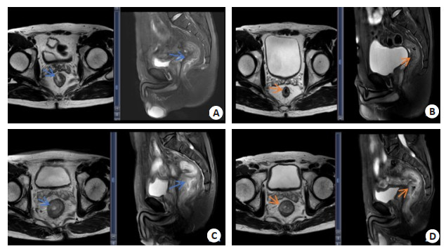
目的 通过磁共振T2加权图像的纹理分析评估直肠癌化疗后疗效的价值。 方法 选取我院2019年1月~2021年4月经临床与病理明确诊断的直肠癌患者117例,分为完全缓解组(n=38)和非完全缓解组(n=79);受试者化疗前和治疗结束后均采用3.0T MRI扫描;采用Omni Kineitics软件提取受试者纹理特征,计算纹理特征的峰度、方差、熵和能量参数;绘制ROC曲线分析磁共振T2加权图像的纹理,评估直肠癌化疗后病理完全缓解的价值。 结果 治疗后完全缓解组受试者方差、熵明显低于未完全缓解组(1534±312 vs 2312±586、5.43±0.41 vs 6.42±0.29)峰度、能量明显高于未完全缓解组(4.85±0.66 vs 3.72±0.49、0.016±0.002 vs 0.013±0.02),差异有统计学意义(P < 0.05)。 结论 采用磁共振T2加权图像的纹理分析可评估化疗后的疗效,并可预测直肠癌化疗后病理完全缓解情况。 Abstract:Objective To evaluate the efficacy of rectal cancer after chemotherapy by texture analysis of magnetic resonance T2-weighted images. Methods A total of 117 patients with rectal cancer who were clinically and pathologically diagnosed in our hospital from January 2019 to April 2021. The patients were divided into complete remission group (n=38) and incomplete remission group (n=79), they were scanned with 3.0T MRI before and after chemotherapy. Omni Kineitics software was used to extract texture features of subjects, and the kurtosis, variance, entropy and energy parameters of texture features were calculated. ROC curves were drawn to analyze the texture of magnetic resonance T2-weighted images, and the rectal value of pathological complete response after cancer chemotherapy. Results After treatment, the variance and entropy of the subjects in the complete remission group were significantly lower than those in the incomplete remission group (1534±312 vs 2312±586, 5.43±0.41 vs 6.42±0.29), the kurtosis and energy were significantly higher than those in the incomplete remission group (4.85±0.66 vs 3.72±0.49, 0.016±0.002 vs 0.013±0.02), and the difference was statistically significant (P < 0.05). Conclusion Texture analysis using magnetic resonance T2-weighted images can evaluate the efficacy of chemotherapy and predict the pathological complete response of rectal cancer after chemotherapy. -
[1] Aker M, Ganeshan B, Afaq A, et al. Magnetic resonance texture analysis in identifying complete pathological response to neoadjuvant treatment in locally advanced rectal cancer[J]. Dis Colon Rectum, 2019, 62(2): 163-70. doi: 10.1097/DCR.0000000000001224 [2] Nakamoto T, Takahashi W, Haga A, et al. Prediction of malignant glioma grades using contrast‑enhanced T1‑weighted and T2-weighted magnetic resonance images based on a radiomic analysis[J]. Sci Rep, 2019, 9(1): 19411. doi: 10.1038/s41598-019-55922-0 [3] 高树全, 薛军, 张迎春, 等. 新辅助化疗对中晚期低位直肠癌患者的疗效研究[J]. 中国中西医结合消化杂志, 2020, 28(3): 215-8, 226. https://www.cnki.com.cn/Article/CJFDTOTAL-ZXPW202003013.htm [4] Huynh S, Lheure C, Franck N, et al. Induced sarcoid-like reactions in patients with metastatic melanoma treated with dabrafenib and trametinib: a monocentric retrospective study[J]. Melanoma Res, 2020, 30(3): 317-20. doi: 10.1097/CMR.0000000000000649 [5] Detappe A, Thomas E, Tibbitt MW, et al. Ultrasmall silica‑based bismuth gadolinium nanoparticles for dual magnetic resonance-computed tomography image guided radiation therapy[J]. Nano Lett, 2017, 17(3): 1733-40. doi: 10.1021/acs.nanolett.6b05055 [6] Hong J, Wu JY, Huang O, et al. Early response and pathological complete remission in Breast Cancer with different molecular subtypes: a retrospective single center analysis[J]. J Cancer, 2020, 11(23): 6916-24. doi: 10.7150/jca.46805 [7] Brancati GE, Tripodi B, Novi M, et al. P. 310 Response to electroconvulsive therapy of different clinical features in 670 bipolar patients with depression or mixed state[J]. Eur Neuropsychopharmacol, 2020, 40: S179. [8] 林涵, 李庆姝, 张涛. 磁共振肿瘤退缩分级在经新辅助治疗后的局部进展期直肠癌中的应用[J]. 现代医药卫生, 2020, 36(21): 3410-3. doi: 10.3969/j.issn.1009-5519.2020.21.013 [9] Nomori H, Shiraishi A, O'Uchi T, et al. Positron emission tomography in T3/T4 non‑small cell lung cancer after induction chemoradiotherapy[J]. Ann Thorac Surg, 2020, 109(1): 255-61. doi: 10.1016/j.athoracsur.2019.06.089 [10] Cooley JR, Hebert JJ, de Zoete A, et al. Assessing lumbar paraspinal muscle cross-sectional area and fat composition with T1 versus T2‑weighted magnetic resonance imaging: reliability and concurrent validity[J]. PLoS One, 2021, 16(2): e0244633. doi: 10.1371/journal.pone.0244633 [11] Chida K, Shimada Y, Fujimoto K, et al. Identification of the distal end of carotid plaque using 3‑dimensional fast spin echo T1-weighted magnetic resonance plaque imaging[J]. J Stroke Cerebrovasc Dis, 2020, 29(4): 104680. doi: 10.1016/j.jstrokecerebrovasdis.2020.104680 [12] 朱海桃, 魏正强, 黄旺, 等. CT检查测量直肠上静脉和肠系膜下静脉直径变化评估局部进展期直肠癌新辅助治疗效果的应用价值[J]. 中华消化外科杂志, 2019, 18(8): 797-802. doi: 10.3760/cma.j.issn.1673-9752.2019.08.016 [13] Lee H, Kim HO, Huh YM. T2-weighted and ultra‑short TE molecular magnetic resonance imaging for gastric cancer diagnosis using polymer-based magnetic nanoparticles[J]. J Magn, 2020, 25(4): 567-76. doi: 10.4283/JMAG.2020.25.4.567 [14] 柴广金, 许曼, 李围围, 等. 新辅助放化疗对局部进展期直肠癌术后并发症及LARS发生影响因素分析[J]. 河北医学, 2020, 26(8): 1380-5. doi: 10.3969/j.issn.1006-6233.2020.08.034 [15] Huang Q, Liu Y, Zheng LL, et al. Biocompatible iron(Ⅱ)‑doped carbon dots as T1-weighted magnetic resonance contrast agents and fluorescence imaging probes[J]. Microchim Acta, 2019, 186(8): 492. doi: 10.1007/s00604-019-3593-4 [16] Filho OM, Viale G, Stein S, et al. Impact of HER2 heterogeneity on treatment response of early‑stage HER2‑positive breast cancer: phase Ⅱ neoadjuvant clinical trial of T‑DM1 combined with pertuzumab[J]. Cancer Discov, 2021, 11(10): 2474-87. doi: 10.1158/2159-8290.CD-20-1557 [17] Li FL, Yang YQ, Wei YN, et al. Deep learning‑based predictive biomarker of pathological complete response to neoadjuvant chemotherapy from histological images in breast cancer[J]. J Transl Med, 2021, 19(1): 348. doi: 10.1186/s12967-021-03020-z [18] Huo MJ, Ye JZ, Dong Z, et al. Quantification of brown adipose tissue in vivo using synthetic magnetic resonance imaging: an experimental study with mice model[J]. Quant Imaging Med Surg, 2022, 12(1): 526-38. doi: 10.21037/qims-20-1344 [19] Rama E, Mohapatra SR, Melcher C, et al. Monitoring the remodeling of biohybrid tissue‑engineered vascular grafts by multimodal molecular imaging[J]. Adv Sci (Weinh), 2022, 9(10): e2105783. doi: 10.1002/advs.202105783 [20] Lee S, Choi SH, Cho HR, et al. Multiparametric magnetic resonance imaging features of a canine glioblastoma model[J]. PLoS One, 2021, 16(7): e0254448. doi: 10.1371/journal.pone.0254448 -

 下载:
下载:
 点击查看大图
图(1)
计量
- 文章访问数: 141
- HTML全文浏览量: 88
- PDF下载量: 6
- 被引次数: 0