


Comparation of humoral immune function and CT findings between Mycoplasma pneumoniae combined with Streptococcus pneumoniae pneumonia and Mycoplasma pneumoniae alone in children
-
摘要:
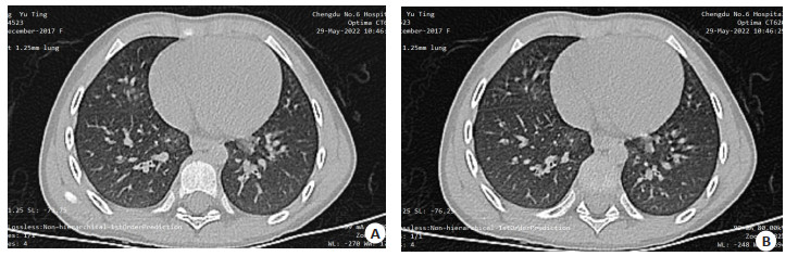
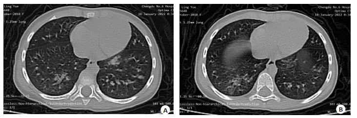
目的 探讨小儿肺炎支原体(MP)合并肺炎链球菌(SP)性肺炎与单纯MP体液免疫功能及胸部CT表现的差异。 方法 选取本院2020年3月~2022年3月收治的肺炎患儿70例,其中MP感染46例,MP合并SP感染24例,通过检测患儿机体体液免疫指标和观察CT表现,分析两种不同感染类型患儿体液免疫和CT表现间的差异。 结果 在病灶分布上两组差异无统计学意义(P>0.05);与SP组比较,MP合并SP组支气管血管束增厚、支气管壁增厚、网状影、磨玻璃影和扇形薄片影等影像征象发生率较低,实变影发生率较高,差异有统计学意义(P<0.05),MP合并SP组胸腔积液厚度较厚、淋巴结最大横径较大(P<0.05);两组患儿IgA、IgG、IgM水平的差异无统计学意义(P>0.05)。 结论 与单纯SP感染相比,MP合并SP感染引起的小儿肺炎体液免疫功能紊乱差异不明显,但在CT影像征象上差异较大,有助于鉴别诊断。 Abstract:Objective To investigate the differences of humoral immune function and chest CT findings between Mycoplasma pneumoniae (MP) complicated with Streptococcus pneumoniae (SP) pneumonia and MP alone in children. Methods Seventy children with pneumonia admitted to our hospital from March 2020 to March 2022 were selected, including 46 cases of MP infection and 24 cases of MP combined with SP infection. The humoral immunity indexes and CT manifestations of the children were detected, and the differences between humoral immunity and CT manifestations of the two different infection types were analyzed. Results There was no significant difference in lesion distribution between the two groups (P > 0.05). Compared with the SP group, the incidence of bronchial vascular bundle thickening, bronchial wall thickening, reticular shadow, ground glass shadow and fan-shaped slice shadow in MP combined with SP group was significantly lower, and the incidence of consolidation shadow was significantly higher(P < 0.05). In MP combined with SP group, the thickness of pleural effusion was significantly thicker and the maximum transverse diameter of lymph nodes was significantly larger (P < 0.05). There were no significant differences in IgA, IgG and IgM levels between the two groups (P > 0.05). Conclusion Compared with SP infection alone, the humoral immune dysfunction caused by MP combined with SP infection in children with pneumonia is not obvious. but there is a big difference in CT image signs, which is helpful for differential diagnosis. -
Key words:
- pneumonia /
- Mycoplasma pneumoniae /
- Streptococcus pneumoniae /
- humoral immunity /
- computed tomography
-
表 1 两组患儿CT征象比较
Table 1. Comparison of CT signs between the two groups [n(%)]
CT征象 MP(n=46) MP合并SP(n=24) χ2 P 支气管充气相 36(78.26) 14(58.33) 3.069 0.080 支气管血管束增厚 31(67.39) 9(37.50) 5.754 0.016 支气管壁增厚 33(71.74) 8(33.33) 9.587 0.002 网状影 34(73.91) 7(29.17) 13.014 <0.001 磨玻璃影 24(52.17) 5(20.83) 6.384 0.012 扇形薄片影 32(69.57) 8(33.33) 8.454 0.004 实变影 25(54.35) 20(83.33) 5.771 0.016 MP: 肺炎支原体; SP: 肺炎链球菌.  下载: 导出CSV
表 2 两组患儿胸腔积液厚度与淋巴结最大横径比较
Table 2. Comparison of pleural effusion thickness and maximum transverse diameter of lymph nodes between the two groups (mm, Mean±SD)
组别 胸腔积液厚度 淋巴结最大横径 MP(n=46) 3.22±1.03 7.63±2.16 MP合并SP(n=24) 11.84±3.52 10.86±3.13 t 15.476 5.070 P <0.001 <0.001
下载: 导出CSV
表 3 两组患儿体液免疫指标比较
Table 3. Comparison of humoral immune indexes between the two groups (g/L, Mean±SD)
组别 IgA IgG IgM MP(n=46) 1.53±0.44 9.47±2.04 1.76±0.51 MP合并SP(n=24) 1.62±0.48 9.86±3.13 1.93±0.42 t 0.787 0.629 1.402 P 0.434 0.532 0.165
下载: 导出CSV
-
[1] Smith DK, Kuckel DP, Recidoro AM. Community-acquired pneumonia in children: rapid evidence review[J]. Am Fam Physician, 2021, 104(6): 618-25. [2] Hayashi D, Akashi Y, Suzuki H, et al. Implementation of point-of-care molecular diagnostics for Mycoplasma pneumoniae ensures the correct antimicrobial prescription for pediatric pneumonia patients[J]. Tohoku J Exp Med, 2018, 246(4): 225-31. doi: 10.1620/tjem.246.225 [3] 王瑶, 黄健, 骆诗露, 等. 肺炎链球菌感染的相关因素及防治策略[J]. 医学综述, 2021, 27(18): 3557-62. doi: 10.3969/j.issn.1006-2084.2021.18.005 [4] 吴传飞, 唐红平, 吴仕筠. 小儿肺炎支原体急性感染快速诊断的研究现状与进展[J]. 医学临床研究, 2019, 36(3): 512-4. doi: 10.3969/j.issn.1671-7171.2019.03.033 [5] Lu YY, Luo R, Fu Z. Pathogen distribution and bacterial resistance in children with severe community-acquired pneumonia[J]. Zhongguo Dang Dai Er Ke Za Zhi, 2017, 19(9): 983-8. [6] Khan N, Whitla L, Kenosi M, et al. Follow up chest radiography in paediatric pneumonia: is it avoidable?[J]. Ir Med J, 2018, 111(3): 721. [7] 王上林. 儿童肺炎链球菌合并支原体感染的体液免疫功能状态分析[J]. 中国社区医师, 2021, 37(35): 75-6. https://d.wanfangdata.com.cn/periodical/zgsqys202135036 [8] 兰彬, 曾廷蓉, 李刚. 胸部CT鉴别小儿MP肺炎与MP并SP肺炎的价值探析[J]. 中国CT和MRI杂志, 2019, 17(7): 76-8, 85. https://www.cnki.com.cn/Article/CJFDTOTAL-CTMR201907023.htm [9] 王卫平, 孙锟, 常立文. 儿科学[M]. 9版. 北京: 人民卫生出版社, 2018. [10] 余静, 张慧. 小儿肺炎支原体感染的免疫学研究进展[J]. 中国儿童保健杂志, 2018, 26(11): 1214-6. https://www.cnki.com.cn/Article/CJFDTOTAL-ERTO201811015.htm [11] Miyashita N. Atypical pneumonia: pathophysiology, diagnosis, and treatment[J]. Respir Investig, 2022, 60(1): 56-67. doi: 10.1016/j.resinv.2021.09.009 [12] Ti-An, Tsai, Rational stepwise approach for Mycoplasma pneumoniae pneumonia in children[J]. J Microbiol Immunol Infect, 2021, 54(4): 557-65. [13] 蓝天航, 潘江峰. 胸部电子计算机断层扫描鉴别小儿肺炎不同感染类型的价值[J]. 中国妇幼保健, 2021, 36(20): 4858-61. https://www.cnki.com.cn/Article/CJFDTOTAL-ZFYB202120069.htm [14] Andronikou S, Goussard P, Sorantin E. Computed tomography in children with community-acquired pneumonia[J]. Pediatr Radiol, 2017, 47(11): 1431-40. [15] Esser M, Tsiflikas I, Kraus MS, et al. Effectiveness of chest CT in children: CT findings in relation to the clinical question[J]. Rofo, 2022, 194(3): 281-90. [16] Fu XL, Yang NF, Ji JW. Application of CT images based on the optimal atlas segmentation algorithm in the clinical diagnosis of Mycoplasma Pneumoniae Pneumonia in Children[J]. Pak J Med Sci, 2021, 37(6): 1647-51. [17] 王玮, 仲颖岚. CT鉴别小儿肺炎支原体肺炎及肺炎支原体肺炎合并肺炎链球菌性肺炎的价值观察[J]. 中国CT和MRI杂志, 2020, 18(12): 74-6. https://www.cnki.com.cn/Article/CJFDTOTAL-CTMR202012024.htm [18] 肖波涛, 高斌, 邹晓倩, 等. 小儿肺炎支原体肺炎MSCT影像学表现及对合并SP的鉴别诊断价值探讨[J]. 中国CT和MRI杂志, 2020, 18(10): 37-9, 43. https://www.cnki.com.cn/Article/CJFDTOTAL-CTMR202010012.htm [19] 宋双生, 钱丹. 胸部CT对小儿链球菌感染肺炎和支原体感染肺炎的鉴别诊断价值研究[J]. 中国CT和MRI杂志, 2019, 17(6): 52-5. https://www.cnki.com.cn/Article/CJFDTOTAL-CTMR201906016.htm [20] Krafft C, Christy C. Mycoplasma pneumonia in children and adolescents[J]. Pediatr Rev, 2020, 41(1): 12-9. [21] Loughran AJ, Orihuela CJ, Tuomanen EI. Streptococcus pneumoniae: invasion and inflammation[J]. Microbiol Spectr, 2019, 7(2): 10.1128/microbiolspec. GPP3-0004-2018. doi: 10.1128/microbiolspec.GPP3-0004-2018 [22] 郁飞, 孙杭, 张宪伟. 儿童肺炎链球菌合并支原体感染的体液免疫功能状态分析[J]. 浙江临床医学, 2017, 19(3): 419-21. http://med.wanfangdata.com.cn/Paper/Detail?id=PeriodicalPaper_zjlcyx201703009 -

 点击查看大图
图(2) / 表(3)
计量
- 文章访问数: 95
- HTML全文浏览量: 100
- PDF下载量: 9
- 被引次数: 0