


Application of ultrasound and dynamic contrast- enhanced magnetic resonance imaging combined with serum prostate specific antigen detection in the differential diagnosis of benign and malignant prostate tumors
-
摘要:
目的 观察经直肠超声(TRUS)、磁共振动态增强扫描(DCE-MRI)联合血清前列腺特异性抗原(PSA)检测在前列腺肿瘤良恶性鉴别诊断中的应用价值。 方法 选取2019年1月~2022年10月我院收治的前列腺肿瘤患者102例,对所有入选患者行TRUS及DCE-MRI检查,以穿刺活检诊断结果为金标准,观察良恶性前列腺肿瘤患者的血清PSA水平,并比较TRUS、DCE-MRI、PSA以及三者联合诊断对前列腺肿瘤良恶性鉴别的价值。 结果 102例前列腺肿瘤患者中,经穿刺活检发现38例为恶性肿瘤,64例为良性肿瘤。采用TRUS鉴别前列腺肿瘤良恶性的敏感度、特异性及准确性分别为63.16%、76.56%及71.57%,DCE-MRI检查为73.68%、78.13%及76.47%,PSA检测为89.47%、70.31%及76.47%,且经穿刺活检诊断为恶性前列腺肿瘤的患者,PSA水平及DCE-MRI检查参数值均高于良性肿瘤患者(P<0.05)。三者联合检测的敏感度及准确性分别为97.37%、88.24%,高于单一检查方法(P<0.05)。 结论 恶性前列腺肿瘤患者与良性患者相比,其血清PSA水平更高,采用TRUS、DCE-MRI及PSA三者联合检测的方法能显著提高前列腺肿瘤良恶性鉴别的诊断效能,值得推广。 Abstract:Objective To observe the application value of transrectal ultrasound (TRUS) and dynamic contrast-enhanced magnetic resonance imaging (DCE-MRI) combined with serum prostate specific antigen (PSA) detection in the differential diagnosis of benign and malignant prostate tumors. Methods A total of 102 patients with prostate tumors admitted to the hospital were enrolled from January 2019 to October 2022. All the patients underwent TRUS and DCE- MRI examination. Taking the diagnostic results of needle biopsy as the golden standard, level of serum PSA in patients with benign and malignant prostate tumors was observed. The value of TRUS, DCE- MRI, PSA and combined detection in the differential diagnosis of benign and malignant prostate tumors was compared. Results Among the 102 patients with prostate tumors, needle biopsy showed that there were 38 cases with malignant tumors and 64 cases with benign tumors. The sensitivity, specificity and accuracy of TRUS, DCE-MRI and PSA in the differential diagnosis of benign and malignant prostate tumors were 63.16%, 76.56%, 71.57%; 73.68%, 78.13%, 76.47% and 89.47%, 70.31%, 76.47%, respectively. Needle biopsy showed that PSA level and DCE- MRI parameters in patients with malignant tumors were significantly higher than those with benign tumors (P < 0.05). The sensitivity and accuracy of combined detection were 97.37% and 88.24%, significantly higher than those of single single (P < 0.05). Conclusion Compared with patients with benign prostate tumors, level of serum PSA is higher in patients with malignant tumors. The combined detection of TRUS, DCE-MRI and PSA can significantly improve differential diagnosis efficiency of benign and malignant prostate tumors. -

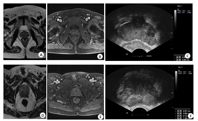
图 1 典型患者的TRUS及DCE-MRI图像
A~C: 病例1, 男, 72岁, 经穿刺活检诊断为PCa; A~B: DCE-MRI检查显示T1WI呈等信号, T2WI呈低信号, 边界欠清晰, DWI呈高信号, ADC呈低信号, 动态增强扫描DIC曲线呈流出型; C: TRUS检查显示左侧外周带见低回声, 形态不规则, 边界不清晰, 回声尚均匀, 向前列腺包膜外膨出; D~F: 病例2, 男, 78岁, 经穿刺活检诊断为PCa; D~E: DCE-MRI检查显示右侧外周带可见不规则等长T1T2信号, DWI序列为稍高信号, ADC图为低信号, ADC值约0.5×10⁻³ mm2/s, 增强后可见轻度强化; F: TRUS检查显示右侧外周带可见不规则稍高回声, 形态不规则, 边界不清晰, 回声不均匀.
Figure 1. TRUS and DCE-MRI images of typical patients.
表 1 TRUS、DCE-MRI及血清PSA检测结果
Table 1. TRUS, DCE-MRI and serum PSA test results (n)
确诊结果 TRUS DCE-MRI 血清PSA 恶性 良性 合计 恶性 良性 合计 恶性 良性 合计 恶性 24 14 38 28 10 38 34 4 38 良性 15 49 64 14 50 64 19 45 64 合计 39 63 102 42 60 102 53 49 102 TRUS: 经直肠超声; DCE-MRI: 磁共振动态增强扫描; PSA: 前列腺特异性抗原.  下载: 导出CSV
表 2 良恶性患者PSA水平及DCE-MRI检查参数比较
Table 2. Comparison of PSA level and DCE-MRI parameters of benign and malignant patients (Mean±SD)
确诊结果 PSA(ng/mL) Ktrans Kep Ve 良性(n=64) 5.85±2.06 0.19±0.03 1.41±0.32 0.19±0.04 恶性(n=38) 35.46±8.73 0.62±0.17 2.38±0.57 0.23±0.08 t 26.022 19.787 11.020 3.362 P <0.001 <0.001 <0.001 <0.001 Ktrans: 容量转移常数; Kep: 速率常数; Ve: 血管外细胞间隙容积.
下载: 导出CSV
表 3 TRUS、DCE-MRI联合血清PSA检测鉴别前列腺肿瘤良恶性结果
Table 3. Results of TRUS, DCE-MRI combined with serum PSA detection in differentiating benign and malignant prostate tumors (n)
TRUS+DCE-MRI+PSA 确诊结果 合计 恶性 良性 恶性 37 11 48 良性 1 53 54 合计 38 64 102
下载: 导出CSV
表 4 三种检查方法的敏感性、特异性及准确性结果
Table 4. Results of sensitivity, specificity and accuracy of three test methods (%)
检查方法 敏感度 特异性 准确性 TRUS 63.16 76.56 71.57 DCE-MRI 73.68 78.13 76.47 PSA 89.47 70.31 77.45 三者联合 97.37 82.81 88.24 χ2 45.063 4.509 8.755 P <0.001 0.211 0.033
下载: 导出CSV
-
[1] 李友芳, 杨小杰, 张栋, 等. 良性前列腺增生与前列腺肿瘤MSCT征象及鉴别诊断分析[J]. 中国CT和MRI杂志, 2021, 19(2): 112-3, 143. doi: 10.3969/j.issn.1672-5131.2021.02.036 [2] 江志国, 杜尉, 陈家存, 等. 前列腺穿刺术后行前列腺癌根治术的间隔时间对患者围手术期及预后的影响[J]. 国际外科学杂志, 2020, 47 (5): 321-5. doi: 10.3760/cma.j.cn115396-20190603-00085 [3] 连碧珺, 李晶, 陈欢, 等. 多西他赛联合内分泌疗法治疗转移性激素敏感性前列腺癌的疗效研究[J]. 中华泌尿外科杂志, 2020, 41(1): 26-31. doi: 10.3760/cma.j.issn.1000-6702.2020.01.005 [4] 张挺维, 韦煜, 潘剑, 等. TP53突变在中国前列腺癌患者中的临床特征及预后价值研究[J]. 中华外科杂志, 2021, 59(11): 897-901. doi: 10.3760/cma.j.cn112139-20210715-00312 [5] 冯亮, 师琳, 蒋益民. 经直肠超声联合前列腺特异抗原密度诊断前列腺癌的临床应用价值研究[J]. 影像科学与光化学, 2020, 38 (2): 323-7. https://www.cnki.com.cn/Article/CJFDTOTAL-GKGH202002027.htm [6] 李静, 黄宝生, 魏铭, 等. 动态对比增强磁共振成像结合表观扩散系数在前列腺癌诊断中的应用[J]. 实用放射学杂志, 2020, 36(12): 1976-9. doi: 10.3969/j.issn.1002-1671.2020.12.023 [7] 王宝华, 沙宇婷, 何凤蝶, 等. 前列腺特异性抗原对中国人群前列腺癌早期检测价值的Meta分析[J]. 中国癌症杂志, 2020, 30(11): 879-8. doi: 10.19401/j.cnki.1007-3639.2020.11.005 [8] 李华君, 罗文元, 温志安, 等. 苯肌动蛋白结合蛋白在前列腺癌中的表达及意义探索[J]. 现代泌尿生殖肿瘤杂志, 2020, 12(1): 38-42. doi: 10.3870/j.issn.1674-4624.2020.01.011 [9] 吴万文, 吕蔡, 刘振湘. 16G和18G穿刺针在超声引导下经直肠前列腺活检对前列腺癌的诊断效果及并发症比较[J]. 中华男科学杂志, 2020, 26(1): 31-5. https://www.cnki.com.cn/Article/CJFDTOTAL-NKXB202001007.htm [10] 朱云开, 陈亚青, 钟芙蓉, 等. 经直肠超声/多参数磁共振融合靶向穿刺在临床显著前列腺癌诊断中的价值[J]. 中华超声影像学杂志, 2021, 30(2): 145-50. [11] 符仕宝, 莫智波, 肖劲逐, 等. 海南地区Gleason评分≥7分的前列腺癌患者血清PSA和睾酮水平与预后相关性探讨[J]. 现代肿瘤医学, 2022, 30(11): 1998-2002. doi: 10.3969/j.issn.1672-4992.2022.11.016 [12] 梁晓秋, 曹凌玲, 陈溢旭. 经直肠超声造影引导前列腺穿刺活检诊断前列腺癌[J]. 中国介入影像与治疗学, 2020, 17(2): 93-7. https://www.cnki.com.cn/Article/CJFDTOTAL-JRYX202002012.htm [13] 陆健美, 葛建钢, 王静, 等. 联合应用3.0T磁共振T2WI、DWI及DCE-MRI成像诊断前列腺癌[J]. 实用肿瘤杂志, 2020, 35(4): 355-9. https://www.cnki.com.cn/Article/CJFDTOTAL-SYZZ202004017.htm [14] 吴艳艳, 陆俐, 陈雨薇, 等. 经直肠超声结合前列腺特异性抗原密度检测对前列腺癌的诊断意义[J]. 实用癌症杂志, 2020, 35(12): 2064-6. doi: 10.3969/j.issn.1001-5930.2020.12.038 [15] 龙玉屏, 黄珊珊, 赵中千, 等. 经直肠常规超声、实时组织弹性成像及超声造影联合应用对侵袭性前列腺癌的诊断价值[J]. 临床超声医学杂志, 2022, 24(4): 291-4. https://www.cnki.com.cn/Article/CJFDTOTAL-LCCY202204011.htm [16] 闫微微, 封淏, 李念芬, 等. 前列腺周缘区外侧偏前部穿刺在经直肠超声阴性前列腺癌患者诊断中的应用价值[J]. 国际泌尿系统杂志, 2021(5): 789-93. [17] Paulsen FO, Kang D, Becker F, et al. Targeting cyclin-dependent kinase 7-association between CDK7 and pMED1 expression in prostate cancer tissue[J]. Carcinogenesis, 2022, 43(8): 779-86. [18] 王环震, 席玉. MRI、TRUS联合血清PSA诊断前列腺癌的价值观察[J]. 中国CT和MRI杂志, 2022, 20(11): 132-4. https://www.cnki.com.cn/Article/CJFDTOTAL-CTMR202211048.htm [19] 陈爽, 陈子健, 钟建锋, 等. 动态对比增强磁共振联合PSA诊断前列腺癌价值探讨[J]. 中国CT和MRI杂志, 2022, 20(1): 142-5. https://www.cnki.com.cn/Article/CJFDTOTAL-CTMR202201044.htm [20] 刘婉君, 方毅, 刘辉, 等. 经直肠超声造影和直肠超声剪切波弹性成像联合血清前列腺特异性抗原诊断早期前列腺癌的价值[J]. 中华实用诊断与治疗杂志, 2022, 36(4): 407-10. https://www.cnki.com.cn/Article/CJFDTOTAL-HNZD202206016.htm [21] 刘毅豪, 黄智峰, 吴松, 等. 血清PSA、PSAD和HMGB1水平检测对老年前列腺癌的诊断价值[J]. 海南医学, 2021, 32(12): 1527-30. https://www.cnki.com.cn/Article/CJFDTOTAL-HAIN202112008.htm [22] 李红兵, 邹晓旭, 廖清松, 等. 血清PSA、F-PSA、HE4在前列腺癌中的表达及意义[J]. 实用癌症杂志, 2020, 35(5): 724-7. https://www.cnki.com.cn/Article/CJFDTOTAL-SYAZ202005008.htm [23] 卢晓冬, 杨明, 杨维, 等. DWI联合DCE-MRI对前列腺肿瘤术前鉴别诊断的效能分析[J]. 中国男科学杂志, 2022, 36(2): 69-73. https://www.cnki.com.cn/Article/CJFDTOTAL-NXXX202202010.htm [24] 李磊, 桂赞龙, 程华根, 等. DCE-MRI和ADC值对前列腺良恶性病变的鉴别及ADC值与血清TPSA的关系[J]. 实用放射学杂志, 2022, 38(4): 612-5. [25] 单连强, 瞿色华, 石士奎, 等. 磁共振表观扩散系数对前列腺癌的筛查价值[J]. 分子影像学杂志, 2019, 42(2): 151-4. doi: 10.12122/j.issn.1674-4500.2019.02.02 [26] 王涛, 马丹丹, 杨明, 等. 经直肠超声引导下徒手经会阴前列腺穿刺在前列腺病变中的应用[J]. 分子影像学杂志, 2020, 43(2): 221-4. doi: 10.12122/j.issn.1674-4500.2020.02.09 [27] 贺修宝, 李灿, 胡小梅. 经直肠超声造影、血清前列腺特异性抗原检测在前列腺癌穿刺活检诊断中的应用[J]. 中国性科学, 2022, 31(2): 9-12. https://www.cnki.com.cn/Article/CJFDTOTAL-XKXZ202202003.htm -

 点击查看大图
计量
- 文章访问数: 145
- HTML全文浏览量: 47
- PDF下载量: 6
- 被引次数: 0