


Application of ultrasound measurement of uterine artery blood flow parameters combined with detection of potential marker serum Serpin family member 1 in the diagnosis of uterine leiomyoma
-
摘要:
目的 分析超声测定子宫动脉血流参数结合潜在标志物血清Serpin家族成员1(SERPINA1)检测在子宫肌瘤诊断中的应用价值。 方法 选择2018年2月~2022年5月我院收治的67例子宫肌瘤患者作为观察组,103例子宫腺肌病患者作为对照组;对患者行超声检查记录患者子宫动脉阻力指数(RI)、子宫动脉搏动指数(PI)、子宫动脉收缩期血流速度、子宫动脉舒张期血流速度;检测受试者血清中SERPINA1蛋白水平;采用Logistic回归模型分析存显著性差异各指标联合应用诊断评估模型并绘制ROC曲线分析各指标单独及联合应用对预测子宫肌瘤的价值。 结果 观察组患者RI低于对照组(0.53±0.13 vs 0.69±0.08),PI高于对照组(1.56±0.21 vs 1.25±0.19),差异有统计学意义(P < 0.05);观察组患者血中SERPINA1水平高于对照组(138.59±20.28 vs 46.71±4.39),差异有统计学意义(P < 0.05);采用子宫动脉血流参数联合SERPINA1对子宫肌瘤患者进行诊断预测模型为Log(P)=0.658×子宫动脉阻力指数+0.617×子宫动脉搏动指数+0.642×SERPINA1+0.809;采用RI、PI、SERPINA1单独应用预测子宫肌瘤的AUC均大于0.85;采用三指标联合应用预测子宫肌瘤的AUC均高于各指标单独应用,差异有统计学意义P < 0.05)。 结论 采用超声子宫动脉血流参数结合SERPINA1检测可有效诊断和评估子宫肌瘤,具有较高对的临床应用价值。 -
关键词:
- 子宫动脉血流参数 /
- Serpin家庭成员1 /
- 子宫肌瘤 /
- 诊断
Abstract:Objective To analyze the application value of ultrasonic measurement of uterine artery blood flow parameters combined with the detection of serum Serpin family member 1(SERPINA1) as a potential marker in the diagnosis of uterine fibroids. Methods A total of 67 patients with uterine fibroids who were treated in our hospital from February 2018 to May 2022 were selected as the observation group, and 103 patients with adenomyosis were selected as the control group. The ultrasound examination was performed to record the uterine artery resistance index (RI), uterine artery pulsatility index (PI), uterine artery systolic blood flow velocity, and uterine artery diastolic blood flow velocity. We detected the serum SERPINA1 protein level of the subjects. We used the logistic regression model to analyze significant differences in the combined application of each index for diagnosis. The model was evaluated and ROC curve was drawn to analyze the value of each index alone and in combination in predicting uterine fibroids. Results The RI of the observation group was significantly lower than that of the control group (0.53±0.13 vs 0.69±0.08), and the PI was significantly higher than that of the control group (1.56±0.21 vs 1.25±0.19) (P < 0.05). The level of SERPINA1 in the blood of the observation group was significantly higher than that of the control group (138.59±20.28 vs 46.71±4.39), and the difference was statistically significant (P < 0.05). The uterine artery blood flow parameters combined with SERPINA1 were used to diagnose uterine fibroids. The prediction model is Log(P)=0.658×uterine artery resistance index+0.617×uterine artery pulsatility index+0.642×SERPINA1+0.809; the AUCs of RI, PI, and SERPINA1 used alone to predict uterine fibroids were all greater than 0.85. The combination of three indicators was used to predict uterine fibroids. The AUC of the application to predict uterine fibroids was significantly higher than that of each index alone(P < 0.05). Conclusion Ultrasound uterine artery blood flow parameters combined with SERPINA1 detection can effectively diagnose and evaluate uterine fibroids, and have high clinical application value. -
Key words:
- uterine artery blood flow parameters /
- Serpin family member 1 /
- fibroid /
- diagnosis
-

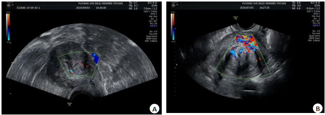
图 1 典型案例图
A: 宫腔内见一低回声, 大小约18 mm×18 mm, 向肌层扩展度大于50%; B: 子宫后壁见大小约65 mm×50 mm低回声, 内侧缘凸向宫腔, 突出部小于肿块大小的50%.
Figure 1. Typical case diagram.
表 1 受试者子宫动脉血流参数调查结果
Table 1. Investigation results of uterine artery blood flow parameters of subjects (Mean±SD)
组别 子宫动脉阻力指数 子宫动脉搏动指数 子宫动脉收缩期血流速度(mm/s) 子宫动脉舒张期血流速度(mm/s) 对照组(n=103) 0.69±0.08 1.25±0.19 45.63±0.94 5.69±0.98 观察组(n=67) 0.53±0.13 1.56±0.21 45.84±1.12 5.73±1.02 t 9.024 7.816 1.236 0.205 P < 0.001 < 0.001 0.218 0.838  下载: 导出CSV
表 2 各指标联合应用预测子宫肌瘤模型
Table 2. Prediction model of hysteromyoma by combined application of all indicators
指标 b SE χ2 P OR 95% CI 下限 上限 子宫动脉阻力指数 0.658 0.210 9.818 0.002 1.931 1.279 2.914 子宫动脉搏动指数 0.617 0.198 9.710 0.002 1.853 1.257 2.732 SERPINA1 0.642 0.207 9.619 0.002 1.900 1.267 2.851 常数项 0.809 0.225 12.928 < 0.001 2.246 1.445 3.490 SERPINA1: Serpin家族成员1.
下载: 导出CSV
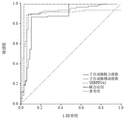
表 3 各指标及联合应用预测子宫肌瘤价值
Table 3. The value of each index and its combined application in predicting hysteromyoma
指标 AUC SE P 95% CI 下限 上限 子宫动脉阻力指数 0.875 0.030 < 0.001 0.816 0.935 子宫动脉搏动指数 0.899 0.029 < 0.001 0.843 0.955 SERPINA1 0.889 0.029 < 0.001 0.832 0.947 联合应用 0.964 0.021 < 0.001 0.924 1.000
下载: 导出CSV
-
[1] Zaria I, Garba I, Dung C, et al. Doppler ultrasound evaluation of blood flow patterns of the uterine arteries in pre-and postmenopausal women with cervical cancer and controls in Zaria[J]. West Afr J Radiol, 2020, 27(1): 18. doi: 10.4103/wajr.wajr_13_19 [2] Sen E, Ozdemir O, Ozdemir S, et al. The relationship between serum ischemia-modified albumin levels and uterine artery Doppler parameters in patients with primary dysmenorrhea[J]. Rev Bras Ginecol Obstet, 2020, 42(10): 630-3. doi: 10.1055/s-0040-1715141 [3] Akdemir Y, Ayvaci H, Uludogan M. Effect of multiple thrombophilic gene mutations on uterine artery blood flow in nonpregnant recurrent pregnancy loss patients: are we searching enough?[J]. J Matern Fetal Neonatal Med, 2020, 33(14): 2466-72. doi: 10.1080/14767058.2019.1569618 [4] 王海霞, 宋倩, 刘景萍. 彩色多普勒超声检查对乳腺癌鉴别诊断及新辅助化疗疗效评估的临床价值[J]. 临床和实验医学杂志, 2021, 20(15): 1666-9. doi: 10.3969/j.issn.1671-4695.2021.15.027 [5] Mansour GM, Hussein SH, Abd El Hady RM, et al. Uterine artery flow velocity waveform (FVW) type and subednometrial vascularity in recurrent pregnancy loss[J]. J Matern Fetal Neonatal Med, 2020, 33(4): 527-32. doi: 10.1080/14767058.2018.1495190 [6] Babaei MR. Survey of changes in ovarian and uterine artery blood flow and indexes by transvaginal Doppler sonography in POI patients and effect of acupuncture on their fertility[J]. Pak J Med Health Sci, 2021, 15(6): 2011-5. [7] 戴爱丽, 周全. 经阴道彩色多普勒超声结合血清CA199鉴别诊断子宫肌瘤与子宫腺肌症[J]. 中国计划生育学杂志, 2022, 30(5): 1150-3, 1212. https://www.cnki.com.cn/Article/CJFDTOTAL-JHSY202205039.htm [8] 张丽玲. 经阴道彩色多普勒超声诊断子宫肌瘤、腺肌症及腺肌瘤的临床价值[J]. 中国现代药物应用, 2022, 16(7): 71-3. https://www.cnki.com.cn/Article/CJFDTOTAL-ZWYY202207024.htm [9] Vedmedovska N, Bokucava D, Kivite-Urtane A, et al. The correlation between abnormal uterine artery flow in the first trimester and genetic thrombophilic alteration: a prospective case-controlled pilot study[J]. Diagnostics (Basel), 2020, 10(9): 654. doi: 10.3390/diagnostics10090654 [10] Psilopatis I, Fleckenstein FN, Collettini F, et al. Short and long-term evaluation of disease-specific symptoms and quality of life following uterine artery embolization of fibroids[J]. Insights Imaging, 2022, 13(1): 106. doi: 10.1186/s13244-022-01244-1 [11] 赵宁, 胡晓康, 车德红, 等. 子宫腺肌瘤鉴别诊断中经腹部彩色多普勒超声的应用及有效性分析[J]. 中国妇幼保健, 2021, 36(23): 5581-3. https://www.cnki.com.cn/Article/CJFDTOTAL-ZFYB202123065.htm [12] Otonkoski S, Sainio T, Komar G, et al. Oxytocin selectively reduces blood flow in uterine fibroids without an effect on myometrial blood flow: a dynamic contrast enhanced MRI evaluation[J]. Int J Hyperthermia, 2020, 37(1): 1293-300. doi: 10.1080/02656736.2020.1846792 [13] 徐静. 经阴道超声检查联合宫腔镜检查诊断子宫内膜病变的临床价值分析[J]. 医学影像学杂志, 2022, 32(9): 1618-20. https://www.cnki.com.cn/Article/CJFDTOTAL-XYXZ202209041.htm [14] Xia YC, Lei C, Yang DH, et al. Identification of key modules and hub genes associated with lung function in idiopathic pulmonary fibrosis[J]. PeerJ, 2020, 8: e9848. doi: 10.7717/peerj.9848 [15] Niemietz C, et al. SERPINA1 modulates expression of amyloidogenic transthyretin[J]. Exp Cell Res, 2020, 395(2): 112217. doi: 10.1016/j.yexcr.2020.112217 [16] 李欢, 王世全, 李铎. 乳腺浸润性导管癌中丝氨酸蛋白酶抑制剂A1表达及对预后判断的临床价值研究[J]. 陕西医学杂志, 2021, 50(10): 1292-5. doi: 10.3969/j.issn.1000-7377.2021.10.030 [17] Zhao J, Xie X, Xia B, et al. Identification of an Autophagy-Related Gene SERPINA1 as a Superior Biomarker Associated with the Occurrence and Distant Metastasis in Osteosarcoma[J]. J Biol Regul Homeost Agents, 2022, 36(3): 535-45. [18] Tiensuu H, Haapalainen AM, Tissarinen P, et al. Human placental proteomics and exon variant studies link AAT/SERPINA1 with spontaneous preterm birth[J]. BMC Med, 2022, 20(1): 141. [19] 李艳, 闫丽明, 杨敏, 等. 联合检测血清GC、SERPINA1和EDN1在子宫肌瘤中的诊断价值[J]. 川北医学院学报, 2020, 35(6): 1043-6. https://www.cnki.com.cn/Article/CJFDTOTAL-NOTH202006025.htm -

 点击查看大图
计量
- 文章访问数: 103
- HTML全文浏览量: 81
- PDF下载量: 5
- 被引次数: 0