


Diagnostic significance of echocardiography in coronary heart disease without obvious ST-T changes in electrocardiogram: Based on coronary flow velocity and ventricular wall motion
-
摘要:
目的 研究超声心动图检测患者冠状动脉相关血流速度联合节段性室壁运动对经心电图检测ST-T段未见明显改变的冠状动脉粥样硬化性心脏病中的诊断意义。 方法 回顾性分析2020年1月~2021年6月在我院心内科住院的经冠脉造影证实为冠心病且血管狭窄大于50%但未完全闭塞的患者并进行心电图检查,将其中无明显ST-T段改变的患者40例作为冠心病组;同时选取冠状动脉造影证实血管狭窄小于50%的患者30例作为对照组,所有的患者均在入院期间进行超声心动图检查,观察冠状动脉血流及室壁运动情况。 结果 冠心病组中冠脉舒张期血流速度高于对照组,差异有统计学意义(P < 0.05)。超声心动图显示舒张期节段性血流峰值流速对诊断冠心病的准确性为92.3%,敏感度为82.4%,特异性为90%,临界值为94.5 cm/s;节段性室壁运动诊断冠心病的敏感度为55.9%,特异性为86.7%,二者联合诊断冠心病的敏感度为84.2%,特异性为92.5%。 结论 超声心动图对冠状动脉血流速度联合节段性室壁运动情况可以明显提高无明显ST-T改变的冠心病患者的诊断率,有利于减少临床上对冠心病患者的误诊率。 -
关键词:
- 超声心动图 /
- 节段性室壁运动 /
- 心电图无明显ST-T改变 /
- 冠心病 /
- 冠脉血流速度
Abstract:Objective To explore the diagnostic significance of echocardiographic of coronary artery related blood flow velocity combined with segmental wall motion in patients with atherosclerotic heart disease with no significant change in ST-T segment detected by electrocardiogram. Methods A retrospective study was conducted on patients admitted to the Department of Cardiology in our hospital from January 2020 to June 2021 who were confirmed by coronary angiogrophy as coronary heart disease with vascular stenosis greater than 50% but not completel occlusion. The patients were performed with electrocardiogram examination. Forty patients without significant ST-T segment changes were selected as coronaty artery disease group. Meanwhile, 30 patients with vascular stenosis less than 50% confirmed by coronary angiography were selected as control group. All patients underwent echocardiography during admission to observe coronary blood flow and ventricular wall motion. Results The diastolic blood flow velocity in the coronaty artery disease group was significantly higher than that in the control group (P < 0.05). Echocardiography showed that the accuracy of diastolic segmental peak flow velocity in the diagnosis of coronaty artery disease was 92.3%, the sensitivity was 82.4%, the specificity was 90%, the critical value was 94.5 cm/s, the sensitivity of segmental wall motion in the diagnosis of coronary heart disease was 55.9%, the specificity was 86.7%, and the sensitivity and specificpatity of combined diagnosis of coronary heart disease were 84.2% and 92.5%. Conclusion Echocardiographic coronary flow velocity combined with segmental ventricular wall motion can improve the diagnostic rate of patients with coronary heart disease without obvious ST-T change. It is beneficial to reduce the misdiagnosis rate of clinical misdiagnusis of coronary heart disease patients. -

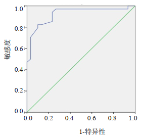
图 1 冠脉造影血管狭窄程度与超声心动图舒张期血流
A:冠状动脉造影示左前支近端狭窄90%; B:超声心动图示左冠脉舒张期流速121 cm/s; C:冠状动脉造影示右冠脉近段狭窄80%; D:超声心动图示右冠脉舒张期流速140 cm/s.
Figure 1. Coronary angiography stenosis degree and echocardiographic diastolic blood flow.

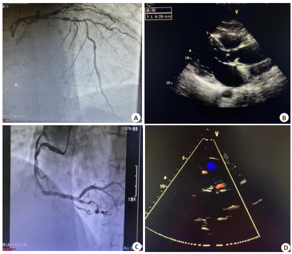
图 2 冠状动脉血流速度在冠心病诊断中的ROC曲线
Figure 2. ROC curve of coronary blood flow velocity in the coronary heart disease.
表 1 冠状动脉血流速度与室壁运动在冠心病诊断中的ROC分析
诊断 曲线下面积 阈值 敏感度(%) 特异性(%) 舒张期血流速度 0.92 94.5 cm/s 82.4 90.0 节段性室壁运动 55.9 86.7 二者联合 84.2 92.5  下载: 导出CSV
-
[1] 胡玲爱, 张洪生, 尉希清, 等. 老年冠心病患者危险因素及预后危险因素分析[J]. 心血管康复医学杂志, 2017, 26(5): 467-70. doi: 10.3969/j.issn.1008-0074.2017.05.02 [2] Bitarafan S, Yari M, Broumand MA, et al. Association of increased levels of lncRNA H19 in PBMCs with risk of coronary artery disease[J]. Cell J, 2019, 20(4): 564-8. [3] 孙静, 杜志祥, 卢英. 心电图aVR导联ST段及T波改变在病变冠状动脉血管定位的应用价值[J]. 江苏医药, 2019, 45(11): 1141-4. https://www.cnki.com.cn/Article/CJFDTOTAL-YIYA201911017.htm [4] 邱方, 程令刚, 林晖. 以超声心动图为标准分析静息心电图在心脏形态学诊断中的临床价值[J]. 中国医师杂志, 2021, 23(3): 428-31. doi: 10.3760/cma.j.cn431274-20200226-00190 [5] 王琼, 李娟. 彩色多普勒冠状动脉血流显像评价左冠状动脉前降支狭窄的应用研究[J]. 中国药物与临床, 2013, 13(2): 212-3. https://www.cnki.com.cn/Article/CJFDTOTAL-YWLC201302038.htm [6] 马克霞. 冠心病节段性室壁运动异常采用超声心动图诊断准确性分析[J]. 影像研究与医学应用, 2022, 6(10): 140-2. https://www.cnki.com.cn/Article/CJFDTOTAL-YXYY202210046.htm [7] 吴国强, 何真. 心脏彩超在冠心病节段性室壁运动异常诊断中的应用[J]. 当代医学, 2022, 28(18): 134-6. https://www.cnki.com.cn/Article/CJFDTOTAL-DDYI202218039.htm [8] 杨欣, 张新艳, 姜阳. 超声心动图诊断冠心病节段性室壁运动异常的临床价值分析[J]. 现代消化及介入诊疗, 2019, 24(S2): 1416. https://www.cnki.com.cn/Article/CJFDTOTAL-ZWYY201820015.htm [9] 陈斌, 邓又斌. 经胸超声冠状动脉显像技术检测冠状动脉粥样硬化及狭窄的应用[J]. 临床心血管病杂志, 2005, 21(3): 187-9. https://www.cnki.com.cn/Article/CJFDTOTAL-LCXB20050300O.htm [10] 李琳, 李影, 柴静, 等. 多巴酚丁胺超声心动图联合平板运动心电图对冠心病的筛查价值[J]. 中国运动医学杂志, 2020, 39(3): 173-7. https://www.cnki.com.cn/Article/CJFDTOTAL-YDYX202003001.htm [11] 周琦, 王其涛, 蔡芹芹, 等. 冠心病患者左室心尖形态及功能与斑块易损性显著相关[J]. 分子影像学杂志, 2021, 44(2): 332-5. doi: 10.12122/j.issn.1674-4500.2021.02.23 [12] 孙卫红, 李文华, 石红建. 冠心病患者心电图表现与冠状动脉造影的相关性研究[J]. 交通医学, 2016, 30(2): 162-3, 166. https://www.cnki.com.cn/Article/CJFDTOTAL-JTYX201602021.htm [13] 邹文彪. 心电图检查结果正常的冠心病患者冠脉病变特点分析[J]. 中国医疗器械信息, 2017, 23(20): 145-7. https://www.cnki.com.cn/Article/CJFDTOTAL-ZGQX201720072.htm [14] 沈燕. 常规心电图与平板运动试验在冠心病诊断中的应用[J]. 当代医学, 2015, 21(5): 73-4. https://www.cnki.com.cn/Article/CJFDTOTAL-DDYI201505049.htm [15] 刘全. 心电图ST-T段改变对早期诊断冠心病的临床应用价值评价[J]. 中国实用医药, 2019, 14(1): 48-9. https://www.cnki.com.cn/Article/CJFDTOTAL-ZSSA201901021.htm [16] 曹建林. 经胸多普勒超声心动图评价冠状动脉狭窄[D]. 太原: 山西医科大学, 2008. [17] 许黎阳. 经胸多普勒超声在冠状动脉病变诊断中的应用价值[J]. 山东医药, 2011, 51(6): 94-5. https://www.cnki.com.cn/Article/CJFDTOTAL-SDYY201106074.htm [18] 薛继平, 王健, 曹建林, 等. 经胸多普勒超声心动图对冠状动脉狭窄的研究[J]. 中西医结合心脑血管病杂志, 2010, 8(8): 914-6. https://www.cnki.com.cn/Article/CJFDTOTAL-ZYYY201008014.htm [19] 吴娟, 陈荣华, 萧少武. 二维超声心动图联合心电图检查在急性心肌梗死患者左前降支病变评估中的应用[J]. 中国临床研究, 2020, 33 (4): 493-6. https://www.cnki.com.cn/Article/CJFDTOTAL-ZGCK202004016.htm [20] 王欣怡, 彭琰君, 韩雪晶, 等. 血脂亚组与冠状动脉粥样硬化性心脏病患者冠状动脉狭窄程度相关性的研究[J]. 中华预防医学杂志, 2021, 55(12): 1435-41. https://www.cnki.com.cn/Article/CJFDTOTAL-SWCX202202022.htm -

 点击查看大图
图(2) / 表(1)
计量
- 文章访问数: 166
- HTML全文浏览量: 78
- PDF下载量: 4
- 被引次数: 0