


Evaluation of pericoronal adipose tissue changes in patients with coronary atherosclerosis by texture analysis
-
摘要:
目的 应用纹理分析技术初步评估冠状动脉粥样硬化患者冠周脂肪组织学的改变。 方法 选取解放军总医院海南医院220例行冠状动脉CT血管成像的患者, 其中正常组110例, 病变组(包含钙化、非钙化及混合斑块)110例, 分别对其在冠状动脉三支主干近端(右冠状动脉、左冠前降支、回旋支)多平面重建图像上选取显示靶斑块周围脂肪组织较清晰层面进行纹理特征分析, 选取角二阶矩、对比度、自相关、逆差距、熵5个纹理特征参数, 采用Mann-Whitney U检验各组之间的纹理特征差异。 结果 正常组与病变组的5个纹理特征参数的差异均有统计学意义(P角二阶矩 < 0.001, P对比度 < 0.001, P自相关 < 0.001, P逆差距 < 0.001, P熵 < 0.001), 两组间冠周脂肪组织纹理特征存在差异, 各纹理参数均具有较好的诊断价值。ROC曲线证实5个纹理特征的曲线下面积均大于0.8, 具有较高的诊断价值。 结论 正常组与病变组的冠状动脉周围脂肪组织的纹理特征不同, 纹理特征分析技术可基于冠状动脉CT血管成像检查, 初步评估冠状动脉粥样硬化患者的冠周脂肪组织学改变。 -
关键词:
- 冠状动脉粥样硬化 /
- 冠状动脉周围脂肪组织 /
- 纹理分析
Abstract:Objective To explore preliminary evaluation of pericoronal adipose tissue changes in patients with coronary atherosclerosis by texture analysis. Methods We selected 220 patients with coronary artery CT angiography from Hainan Hospital of PLA General Hospital, including 110 in the normal group and 110 in the coronary atherosclerosis group (including calcified, non-calcified and mixed plaques).The texture features of pericoronal adipose tissue around the target plaque were analyzed.We analyzed MPR images of the proximal three main coronary arteries (including right coronary artery, left anterior descending branch and circumflex branch), selected five texture feature parameters: angular second moment, contrast, correlation, inverse difference moment, and entropy.Mann-Whitney U was used to test differences in texture characteristics between groups. Results All the texture feature parameters showed significant differences between the two groups (PASM < 0.001, Pcontrast < 0.001, Pcorelation < 0.001, PIDM < 0.001, Pentropy < 0.001).There were differences in the texture features of pericoronary adipose tissue between the normal group and the coronary atherosclerosis group, and each texture parameter has good diagnostic value.ROC analysis confirmed that the area under the curve of the five texture features was greater than 0.8, which had good diagnostic value. Conclusion The texture features of pericoronary adipose tissue differ between the normal group and the coronary atherosclerosis group.Therefore, the texture feature analysis can be based on coronary CT angiography examination, preliminary evaluation of pericoronal adipose tissue changes in patients with coronary atherosclerosis. -

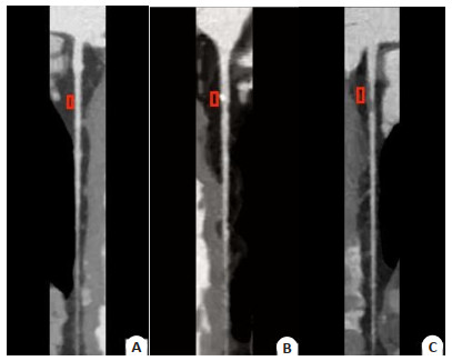
图 1 在冠状动脉MPR图像上选取显示靶斑块及正常管壁边缘较为均匀的最大截面区域作为感兴趣区
A: 正常; B: 钙化斑块; C: 非钙化斑块.
Figure 1. Selected the largest section area that displays the target plaque and the edge of normal pipe wall more evenly as the region of interest on the MPR image of coronary artery.

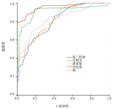
图 2 正常组及病变组5个纹理特征参数的ROC曲线
Figure 2. ROC curve of texture parameters for ASM, contrast, correlation, entropy and cut-off value.
表 1 正常组及病变组5个纹理特征参数比较
Table 1. Comparison of five texture characteristic parameters in the normal group and coronary atherosclerosis group (n=110)
分组 角二阶矩 对比度 自相关 逆差距 熵 正常组 0.042(0.040) 15.625(13.050) 0.028(0.030) 0.347(0.220) 3.433(0.690) 病变组 0.025(0.010) 107.536(59.940) 0.004(0.000) 0.192(0.140) 3.843(0.460) U 2321.000 11564.000 1090.500 2118.500 9955.500 P < 0.001 < 0.001 < 0.001 < 0.001 < 0.001 注: 以中位数(四分位数间距)表示.  下载: 导出CSV
表 2 正常组与病变组5个纹理特征参数(角二阶矩、对比度、自相关、逆差距、熵)、临界值及ROC曲线下面积
Table 2. Five texture characteristic parameters of the normal groupand coronary atherosclerosis group (including ASM, contrast, correlation, IDM, entropy), cut-off value and the area under the ROC curve
参数 临界值 AUC 敏感度 特异性 临界值以上 临界值以下 角二阶矩 0.026 0.808 0.855 0.600 正常 冠状动脉粥样硬化 对比度 72.702 0.956 0.791 0.991 冠状动脉粥样硬化 正常 自相关 0.006 0.910 0.936 0.909 正常 冠状动脉粥样硬化 逆差距 0.281 0.825 0.691 0.827 正常 冠状动脉粥样硬化 熵 3.455 0.823 0.945 0.518 冠状动脉粥样硬化 正常
下载: 导出CSV
-
[1] Goeller M, Achenbach S, Cadet S, et al. Pericoronary adipose tissue computed tomography attenuation and high-risk plaque characteristics in acute coronary syndrome compared with stable coronary artery disease[J]. JAMA Cardiol, 2018, 3(9): 858-63. doi: 10.1001/jamacardio.2018.1997 [2] Lin A, Dey D, Wong DTL, et al. Perivascular adipose tissue and coronary atherosclerosis: from biology to imaging phenotyping[J]. Curr Atheroscler Rep, 2019, 21(12): 47. doi: 10.1007/s11883-019-0817-3 [3] Sun Y, Li XG, Xu K, et al. Relationship between epicardial fat volume on cardiac CT and atherosclerosis severity in three-vessel coronary artery disease: a single-center cross-sectional study[J]. BMC Cardiovasc Disord, 2022, 22(1): 76. doi: 10.1186/s12872-022-02527-7 [4] Klüner LV, Oikonomou EK, Antoniades C. Assessing cardiovascular risk by using the fat attenuation index in coronary CT angiography[J]. Radiol Cardiothorac Imaging, 2021, 3(1): e200563. doi: 10.1148/ryct.2021200563 [5] 段慧, 韩丹, 江杰, 等. 人工智能冠状动脉周围脂肪参数测量的多中心研究[J]. 临床放射学杂志, 2022, 41(8): 1443-50. https://www.cnki.com.cn/Article/CJFDTOTAL-LCFS202208010.htm [6] Liu MQ, Zhang XW, Fan WP, et al. Functional changes of the lateral pterygoid muscle in patients with temporomandibular disorders: a pilot magnetic resonance images texture study[J]. Chin Med J (Engl), 2020, 133(5): 530-6. doi: 10.1097/CM9.0000000000000658 [7] 白建良, 牛金亮. SPECT和MRI诊断冠心病心肌缺血的临床价值分析[J]. 中国CT和MRI杂志, 2020, 18(12): 51-3, 73, 2. doi: 10.3969/j.issn.1672-5131.2020.12.017 [8] Lin A, Nerlekar N, Yuvaraj J, et al. Pericoronary adipose tissue computed tomography attenuation distinguishes different stages of coronary artery disease: a cross-sectional study[J]. Eur Heart J Cardiovasc Imaging, 2020, 22(3): 298-306. [9] Gać P, Macek P, Poręba M, et al. Thickness of epicardial and pericoronary adipose tissue measured using 128-slice MSCT as predictors for risk of significant coronary artery diseases[J]. Ir J Med Sci, 2021, 190(2): 555-66. doi: 10.1007/s11845-020-02339-8 [10] Okubo R, Nakanishi R, Toda M, et al. Pericoronary adipose tissue ratio is a stronger associated factor of plaque vulnerability than epicardial adipose tissue on coronary computed tomography angiography[J]. Heart Vessels, 2017, 32(7): 813-22. doi: 10.1007/s00380-017-0943-1 [11] Verhagen SN, et al. Perivascular adipose tissue as a cause of atherosclerosis[J]. Atherosclerosis, 2011, 214(1): 3-10. doi: 10.1016/j.atherosclerosis.2010.05.034 [12] Tanaka K, Sata M. Roles of perivascular adipose tissue in the pathogenesis of atherosclerosis[J]. Front Physiol, 2018, 9: 3. doi: 10.3389/fphys.2018.00003 [13] Fitzgibbons TP, Lee N, Tran KV, et al. Coronary disease is not associated with robust alterations in inflammatory gene expression in human epicardial fat[J]. JCI Insight, 2019, 4(20): e124859. doi: 10.1172/jci.insight.124859 [14] Cabrera-Rego JO, Iacobellis G, Castillo-Herrera JA, et al. Epicardial fat thickness correlates with carotid intima-media thickness, arterial stiffness, and cardiac geometry in children and adolescents[J]. Pediatr Cardiol, 2014, 35(3): 450-6. doi: 10.1007/s00246-013-0799-9 [15] Greenstein AS, Khavandi K, Withers SB, et al. Local inflammation and hypoxia abolish the protective anticontractile properties of perivascular fat in obese patients[J]. Circulation, 2009, 119(12): 1661-70. doi: 10.1161/CIRCULATIONAHA.108.821181 [16] Cheng VY, et al. Pericardial fat burden on ECG-gated noncontrast CT in asymptomatic patients who subsequently experience adverse cardiovascular events[J]. JACC Cardiovasc Imaging, 2010, 3(4): 352-60. doi: 10.1016/j.jcmg.2009.12.013 [17] Mahabadi AA, Massaro JM, Rosito GA, et al. Association of pericardial fat, intrathoracic fat, and visceral abdominal fat with cardiovascular disease burden: the Framingham Heart Study[J]. Eur Heart J, 2009, 30(7): 850-6. [18] 邹颖, 王平怀, 陈志晔. 纹理分析技术评估颞下颌关节紊乱病患者盘后附着组织的组织学改变初探[J]. 中华口腔医学杂志, 2020, 55(9): 629-33. doi: 10.3760/cma.j.cn112144-20200514-00272 [19] 王波涛, 刘刚, 樊文萍, 等. 纹理特征分析在肝囊肿及肝血管瘤磁共振成像鉴别诊断中的价值[J]. 中国医学科学院学报, 2017, 39(2): 169-76. doi: 10.3881/j.issn.1000-503X.2017.02.002 [20] Karahaliou AN, Boniatis IS, Skiadopoulos SG, et al. Breast cancer diagnosis: analyzing texture of tissue surrounding microcalcifications[J]. IEEE Trans Inf Technol Biomed, 2008, 12(6): 731-8. doi: 10.1109/TITB.2008.920634 [21] Zheng R, Gu L, Jiang W, et al. Value of pericoronary adipose tissue texture analysis in diagnosis of coronary artery disease[J]. Biomed J Sci Tech Res, 2021, 35(5): 103-7. -

 点击查看大图
计量
- 文章访问数: 150
- HTML全文浏览量: 91
- PDF下载量: 8
- 被引次数: 0