


Construction and evaluation of radiotherapy effect prediction model for lung cancer patients: Based on tumor volume evaluation
-
摘要:
目的 分析基于肿瘤体积的肺癌患者放射治疗效果预测模型的构建与评价。 方法 选择我院2019年1月~2021年12月收治的173例肺癌患者作为研究对象。受试者接受放疗方案进行干预治疗;治疗前及放疗结束后1月行影像学检查读取病灶体积,计算患者治疗前后最大直径减少率与体积减少率,扫描并分析患者病灶区Ve、Ktrans、Kep;治疗后对患者的临床疗效进行评估。利用Pearson检验对最大直径的减少率以及体积的减少率间的相关性给予分析,采用Logistic回归模型分析Ve、Ktrans、Kep联合应用预测患者基于肿瘤体积的疗效评估模型,利用ROC曲线对各指标的单独和联合诊断在疗效方面的价值给予预测。 结果 完全缓解+部分缓解组患者最大直径减少率和体积减少率均低于疾病稳定+疾病进展组(P < 0.05);肿瘤最大直径减少率与体积减少率与肺癌患者治疗临床疗效呈正相关关系(P < 0.05);完全缓解+部分缓解组Ve、Ktrans、Kep均高于疾病稳定+疾病进展组(P < 0.05);采用MRI指标联合肿瘤体积和最大径预测患者临床疗效的多元回归预测模型为Log(P)=0.685×最大直径减少率+0.651×体检减少率+0.604×Ve+0.612×Ktrans+0.644×Kep+0.849;采用MRI指标联合肿瘤体积和最大径预测患者临床疗效模型的价值明显高于各指标单独应用(P < 0.05)。 结论 通过评估肺癌患者肿瘤体积及MRI扫描检查可有效分析肺癌患者的预后质量,具有较高的应用价值。 Abstract:Objective To analyze the construction and evaluation of radiotherapy effect prediction model for lung cancer patients based on tumor volume. Methods A total of 173 patients with lung cancer treated in our hospital from January 2019 to December 2021 were selected as the research objects. The subjects received radiotherapy regimen for intervention treatment. Imaging examination was performed before treatment and 1 month after radiotherapy to read the lesion volume, calculate the maximum diameter reduction rate and volume reduction rate before and after treatment. We scaned and analyzed the lesion areas Ve, Ktrans and Kep. After treatment, the clinical efficacy of the patients was evaluated. Pearson correlation test was used to analyze the correlation between maximum diameter reduction rate and volume reduction rate. Logistic regression model was used to analyze the efficacy evaluation model of Ve, Ktrans and Kep combined application to predict patients' tumor volume. ROC curve was drawn to analyze the value of each index alone and combined application to predict patients' efficacy. Results The maximum diameter reduction rate and volume reduction rate in CR+PR group were significantly lower than those in SD+PD group (P < 0.05). There was a significant positive correlation between the maximum diameter reduction rate and volume reduction rate and the clinical efficacy of lung cancer patients (P < 0.05). Ve, Ktrans and Kep in CR+PR group were significantly higher than those in SD+PD group (P < 0.05). The multiple regression prediction model of MRI combined with tumor volume and maximum diameter was log (P)=0.685× maximum diameter reduction rate+0.651×physical examination reduction rate+0.604×Ve+ 0.612×Ktrans+0.644×Kep+0.849; The value of using MRI index combined with tumor volume and maximum diameter to predict the clinical efficacy of patients was significantly higher than that of using each index alone (P < 0.05). Conclusion The evaluation of tumor volume and MRI scan in patients with lung cancer can effectively analyze the prognosis quality of patients with lung cancer. -
Key words:
- lung cancer /
- radiotherapy /
- prediction model /
- tumor volume
-

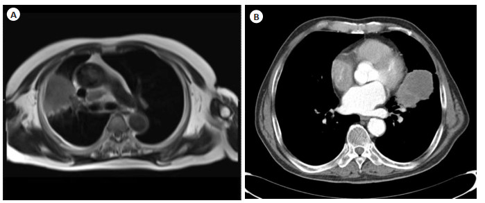
图 1 典型案例图
A: 右上肺癌的MRI影像图(男性, 60岁, 右上肺癌, T1WI呈等信号, 信号欠均匀); B: 左上肺癌胸部CT影像图(男性, 72岁, 增强扫描后左上肺病灶明显增强).
Figure 1. Typical case diagrams.
表 1 不同疗效患者肿瘤最大直径减少率与体积减少率调查结果
Table 1. The survey results of tumor maximum diameter reduction rate and tumor volume reduction rate in patients with different curative effects(Mean±SD)
疗效 最大直径减少率(%) 体积减少率(%) CR+PR(n=101) 58.39±5.03 65.49±7.94 SD+PD(n=72) 28.93±4.39 32.83±6.95 t 30.580 21.870 P < 0.001 < 0.001 PD: 疾病进展; SD: 疾病稳定; PR: 部分缓解; CR: 完全缓解.  下载: 导出CSV
表 2 不同疗效患者MRI指标调查结果
Table 2. Survey results of MRI indicators in patients with different curative effects
疗效 Ve(%) Ktrans(min) Kep(min) CR+PR(n=101) 0.63±0.05 0.39±0.03 0.60±0.03 SD+PD(n=72) 0.51±0.04 0.31±0.03 0.51±0.04 t 14.251 13.353 16.130 P < 0.001 < 0.001 < 0.001
下载: 导出CSV
表 3 MRI指标联合肿瘤体积和最大径预测病人临床疗效模型
Table 3. MRI indicators combined with tumor volume and maximum diameter to predict the clinical efficacy model of patients
指标 b SE χ2 P OR 95%CI 下限 上限 最大直径减少率 0.685 0.231 8.793 0.003 1.984 1.261 3.120 体积减少率 0.651 0.203 10.284 0.001 1.917 1.288 2.854 Ve 0.604 0.191 10.000 0.002 1.829 1.258 2.660 Ktrans 0.612 0.214 8.179 0.004 1.844 1.212 2.805 Kep 0.644 0.198 10.579 0.001 1.904 1.292 2.807 常数项 0.849 0.211 16.190 < 0.001 2.337 1.546 3.534
下载: 导出CSV
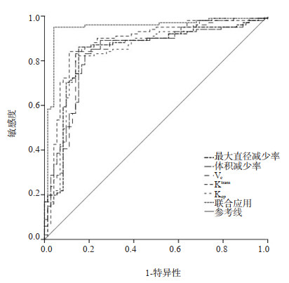
表 4 各指标单独及联合应用预测患者临床疗效价值
Table 4. The individual and combined application of each index to predict the value of clinical curative effect of patients
指标 区域 标准误差 渐近显著性 95% CI 下限 上限 最大直径减少率 0.844 0.033 < 0.001 0.779 0.908 体积减少率 0.834 0.033 < 0.001 0.770 0.898 Ve 0.841 0.033 < 0.001 0.777 0.905 Ktrans 0.882 0.029 < 0.001 0.825 0.938 Kep 0.848 0.032 < 0.001 0.786 0.910 联合应用 0.948 0.020 < 0.001 0.910 0.987
下载: 导出CSV
-
[1] Bourbonne V, Lucia F, Dissaux G, et al. Pulmonary and esophageal toxicity in lung cancer treated by (chemo)-radiotherapy: a radiomics-based prediction model[J]. Int J Radiat Oncol, 2020, 108(3): S31. [2] Tambe NS, Pires IM, Moore C, et al. Validation of in-house knowledge-based planning model for advance-stage lung cancer patients treated using VMAT radiotherapy[J]. Br J Radiol, 2020, 93(1106): 20190535. doi: 10.1259/bjr.20190535 [3] Wang SY, Abujarad F, Chen TG, et al. "Radiotherapy for older women (ROW)": a risk calculator for women with early-stage breast cancer[J]. J Geriatr Oncol, 2020, 11(5): 850-9. doi: 10.1016/j.jgo.2019.12.010 [4] 王辉. 图像引导放射治疗技术在腹部肿瘤放疗治疗中的临床应用[J]. 中国医药指南, 2020, 18(4): 110-1. https://www.cnki.com.cn/Article/CJFDTOTAL-YYXK202004086.htm [5] 廖辰, 郑贝贝, 崔鹤清, 等. 营养支持对卡瑞利珠单抗联合化疗一线治疗晚期非小细胞肺癌疗效的影响[J]. 临床肺科杂志, 2022, 27(12): 1808-10, 1824. doi: 10.3969/j.issn.1009-6663.2022.12.006 [6] Lee SF, Luk H, Wong A, et al. Prediction model for short-term mortality after palliative radiotherapy for patients having advanced cancer: a cohort study from routine electronic medical data[J]. Sci Rep, 2020, 10(1): 5779. doi: 10.1038/s41598-020-62826-x [7] 杨华菊. MRI在肺癌放疗的应用进展[J]. 实用放射学杂志, 2020, 36(3): 487-90. doi: 10.3969/j.issn.1002-1671.2020.03.037 [8] Feng M, Yan L, Du X, et al. 873P Early efficacy prediction of nasopharyngeal carcinoma based on 3D-ADC acquired during radiotherapy: a phase Ⅱ prospective study[J]. Ann Oncol, 2021, 32: S792. [9] 张丽丽, 姜仁伟. 精确放射治疗技术在老年肿瘤患者临床治疗中的运用分析[J]. 影像研究与医学应用, 2020, 4(2): 27-8. https://www.cnki.com.cn/Article/CJFDTOTAL-YXYY202002014.htm [10] Yang H, Zhang X, Zhang Y, et al. A deep learning framework for automatic segmentation and prediction model establishment from T2-weighted MRI in cervical cancer patients receiving radiotherapy[J]. Int J Radiat Oncol, 2020, 108(3): e783. [11] Gkantaifi A, Papadopoulos C, Spyropoulou D, et al. Evaluation of the irradiated volume of the heart and cardiac substructures after left breast radiotherapy[J]. Anticancer Res, 2020, 40(5): 3003-9. doi: 10.21873/anticanres.14281 [12] 苏贞. 磁共振全身弥散加权成像联合常规CT/MRI在肺癌转移灶诊断中的应用[J]. 黑龙江医学, 2021, 45(9): 998-1000. doi: 10.3969/j.issn.1004-5775.2021.09.040 [13] 张晓冬. 精确放射治疗技术在老年肿瘤患者临床治疗中的价值探讨[J]. 中西医结合心血管病电子杂志, 2020, 8(23): 185. https://www.cnki.com.cn/Article/CJFDTOTAL-ZXJH202023169.htm [14] Someya M, Fukushima Y, Hasegawa T, et al. Radiotherapy for HPV-related cancers: prediction of therapeutic effects based on the mechanism of tumor immunity and the application of immunoradiotherapy[J]. Jpn J Radiol, 2022, 40(5): 458-65. doi: 10.1007/s11604-021-01231-4 [15] 付靓, 李凯, 李则锋, 等. 动态对比增强MRI定量参数评估孤立实性结节肺癌病理分级及临床分期的价值[J]. 实用放射学杂志, 2021, 37(11): 1785-9. doi: 10.3969/j.issn.1002-1671.2021.11.010 [16] 王旎力. 分析肿瘤化疗患者血管通路选择中的静疗小组应用价值[J]. 临床医药文献电子杂志, 2020, 7(40): 110. https://www.cnki.com.cn/Article/CJFDTOTAL-LCWX202040096.htm [17] Peng C, Hao YJ, Ren ZN, et al. Prognostic factors of chondroblastic osteosarcoma and nomogram development for prediction: a population-based, STROBE-compliant study[J]. Medicine (Baltimore), 2021, 100(23): e26021. doi: 10.1097/MD.0000000000026021 [18] Li XJ, Ye ZM, Lin S, et al. Predictive factors for survival following stereotactic body radiotherapy for hepatocellular carcinoma with portal vein tumour thrombosis and construction of a nomogram[J]. BMC Cancer, 2021, 21(1): 701. doi: 10.1186/s12885-021-08469-1 [19] 刘阳阳. CyCAR标准与RECIST标准在一线应用贝伐珠单抗治疗晚期非鳞非小细胞肺癌疗效评价中的比较[J]. 山西医药杂志, 2021, 50(20): 2870-3. doi: 10.3969/j.issn.0253-9926.2021.20.004 [20] Abdah-Bortnyak R, Keidar Z, Billan S, et al. PET radiomics analysis for prediction of metabolic complete response of primary tumor after chemo-radiotherapy for squamous cell carcinoma of cervix uteri[J]. Int J Radiat Oncol, 2020, 108(3): e314. [21] 易芹芹, 周宙, 罗燕, 等. 基于术前MRI影像组学及临床特征的早期宫颈癌中危因素预测模型构建[J]. 磁共振成像, 2022, 13(4): 124-7, 136. https://www.cnki.com.cn/Article/CJFDTOTAL-CGZC202204024.htm -

 点击查看大图
计量
- 文章访问数: 163
- HTML全文浏览量: 93
- PDF下载量: 21
- 被引次数: 0