


Effect of complete right bundle branch block on right ventricular volume and function assessed by three-dimensional speckle-tracking echocardiography
-
摘要:
目的 研究三维散斑跟踪超声心动图评估在高血压右束支传导阻滞(CRBBB)对右心室大小及功能的影响。 方法 选取2018年1月~2021年1月收治的106例CRBBB患者纳入观察组,另选取50例经心电图、超声心动图以其他实验室检查确定无心脏器质性病变者作为对照组。所有患者均具备完整三维散斑跟踪超声心动图检查资料,根据患者是否具有右心室机械不同步,将其分为低标准差组(n=51)与高标准差组(n=55),比较两组右心室大小及功能,分析三维散斑跟踪技术在CRBBB中的应用价值。 结果 观察组患者心脏个节段处,入口外侧、入口偏下、入口间隔、流出道间隔的达峰时间面积应变水平均高于对照组(P < 0.05),两组流出道游离壁、心尖游离壁、心尖间隔达峰时间面积应变水平差异无统计学意义(P>0.05)。CRBBB患者中,低标准差组患者QRS间隔、左心室射血分数、右心室舒张末期容积指数、三尖瓣反流峰值梯度及右心室收缩期末期容积指数水平均低于高标准差组(P < 0.05),左心室舒张末期容积、左心室舒张末期容积、右心室面积分数变化、右心室射血分数及三尖瓣环收缩期位移水平均高于高标准差组(P < 0.05)。 结论 3D斑点追踪超声心动图能有效反映CRBBB患者心脏各节段运动延迟状况以及右心室容积、功能状况,在指导临床CRBBB心脏同步性治疗以及病情评估中具有良好的应用价值。 -
关键词:
- 三维散斑跟踪超声心动图 /
- 高血压右束传导阻滞 /
- 右心室大小 /
- 右心室功能
Abstract:Objective To investigate the effect of complete right bundle branch block (CRBBB) on right ventricular volume and function assessed by three-dimensional speckle-tracking echocardiography. Methods A total of 106 patients with CRBBB in our hospital from January 2018 to January 2021 were enrolled, and another 50 patients without cardiac organic lesions confirmed by electrocardiogram, echocardiography and other laboratory examinations were selected as the control group. According to the absence or presence of right ventricular mechanical dyssynchrony presented in the images of three-dimensional speckletracking echocardiography, patients were classified into synchrony group and dyssynchrony group. The right ventricular volume and function of two groups were compared, then the value of three-dimensional speckle tracking echocardiography in CRBBB was analyzed. Results The time-to-peak area change ratio of RA inlet lateral, inlet inferior, inlet septum and outflow free wall was significantly higher in observation group than in control, apical free wall and apical septum showed no significant difference between groups (P>0.05). Among CRBBB patients, the QRS interval, left ventricular ejection fraction, right ventricular end- diastolic volume index, tricuspid regurgitation pressure gradient, and right ventricular end- systolic volume index of low standard deviation group were significantly lower than those of high standard deviation group (P < 0.05), while the left ventricular end-diastolic volume, left ventricular end- systolic volume, right ventricular fractional area change, right ventricular ejection fraction, and tricuspid annular plane systolic excursion were significantly higher than those of high standard deviation group (P < 0.05). Conclusion Application of three-dimensional speckle-tracking echocardiography can effectively reflect the contraction delay of each cardiac segment, right ventricular volume and function in patients with CRBBB, which is of great clinical value in guiding the clinical cardiac synchronization treatment and disease evaluation of CRBBB. -

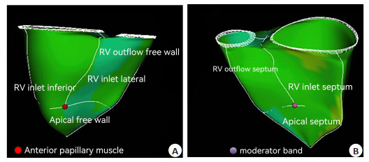
图 1 RV 3D斑点追踪超声心动图模型
注: 白色实线表示7个节段边界, 包括外侧入口、下入口、入口间隔、流出道间隔、流出道游离壁、心尖游离壁、心尖间隔.A: RV游离壁; B: RV间隔壁侧. 前乳头肌与游离壁(A, 红圈)和隔膜上调节束(B, 紫圈)连接定义为入口、流出道和顶点之间边界点.
Figure 1. Right ventricular measurement by three-dimensional speckle tracking echocardiography.
表 1 3D斑点追踪超声心动图评估RV段QRS模式和达峰时间面积应变
Table 1. QRS pattern and time-to-peak change ratio of RV under the 3D speckle tracking echocardiography assessment (Mean±SD)
组别 入口外侧 入口偏下 入口间隔 流出道间隔 流出道游离壁 心尖游离壁 心尖间隔 对照组(n=50) 394.15±66.25 376.66±53.26 351.11±49.52 410.21±58.85 431.15±103.21 377.66±63.58 361.11±58.55 观察组(n=106) 461.15±73.48 423.15±61.12 374.11±50.14 493.15±73.64 428.69±98.15 381.49±71.15 363.74±61.14 t 5.480 4.614 2.684 6.978 0.144 0.324 0.254 P < 0.001 < 0.001 0.008 < 0.001 0.886 0.746 0.800  下载: 导出CSV
表 2 CRBBB患者RV不同步者RV体积及功能比较
Table 2. Comparison of RV volume and function in CRBBB patients with RV dyssynchrony (Mean±SD)
指标 低标准差组(n=51) 高标准差组(n=55) t P QRS间隔 141.69±16.15 156.47±17.47 4.513 < 0.001 左心室舒张末期容积(mL) 90.41±13.33 80.30±12.54 4.024 < 0.001 左心室舒张末期容积(mL) 42.35±6.74 33.14±7.73 6.516 < 0.001 左心室射血分数(%) 60.14±15.41 67.19±16.16 2.295 0.024 左心房容积指数(mL/m2) 27.74±5.14 28.03±6.21 0.261 0.795 E/A 1.49±0.33 1.41±0.36 1.190 0.237 E/e' 9.41±3.15 9.71±2.73 0.525 0.601 右心房压(mmHg) 4.61±1.13 5.63±1.07 4.773 < 0.001 三尖瓣反流峰值梯度(mmHg) 38.49±6.62 53.69±7.74 10.825 < 0.001 RVTDIS,(cm/s) 10.36±2.15 10.43±2.21 0.165 0.869 RV面积分数变化(°%) 39.41±5.66 25.47±6.63 11.598 < 0.001 RV舒张末期容积指数(mL/m2) 86.41±15.34 123.41±17.48 11.545 < 0.001 RV收缩期末期容积指数(mL/m2) 57.41±13.25 95.74±20.56 11.311 < 0.001 RV射血分数(%) 33.69±4.15 23.69±5.14 10.968 < 0.001 三尖瓣环收缩期位移(mm) 16.66±3.87 14.59±4.13 2.657 0.010
下载: 导出CSV
-
[1] Alventosa-Zaidin M, Guix Font L, Benitez Camps M, et al. Right bundle branch block: Prevalence, incidence, and cardiovascular morbidity and mortality in the general population[J]. Eur J Gen Pract, 2019, 25(3): 109-15. doi: 10.1080/13814788.2019.1639667 [2] Nakagawa K, Nagase S, Morita H, et al. Impact of premature activation of the right ventricle with programmed stimulation in Brugada syndrome[J]. J Cardiovasc Electrophysiol, 2018, 29(1): 71-8. doi: 10.1111/jce.13336 [3] Wada T, Nagase S, Morita H, et al. Incidence and clinical significance of brugada syndrome masked by complete right bundle-branch block[J]. Circ J, 2015, 79(12): 2568-75. doi: 10.1253/circj.CJ-15-0618 [4] 李国治, 尹立雪, 沈洁, 等. 速度向量成像技术评价完全性右束支传导阻滞患者左心室收缩期不同步[J]. 中华医学超声杂志: 电子版, 2015, 12(9): 689-95. doi: 10.3877/cma.j.issn.1672-6448.2015.09.005 [5] 魏渠成, 叶沈锋, 王亚萍, 等. Brugada综合征合并间歇性右束支传导阻滞经奎尼丁治疗成功一例[J]. 中华心血管病杂志, 2020, 48(2): 154-6. [6] 顾敏, 华伟. 心脏再同步治疗方法新进展[J]. 中华心律失常学杂志, 2019, 23(4): 352-4. doi: 10.3760/cma.j.issn.1007-6638.2019.04.017 [7] 朱慧, 谢剑, 肖青叶, 等. 应用二维斑点追踪显像技术比较左束支传导阻滞与右束支传导阻滞患者的心室收缩同步性[J]. 中国社区医师, 2017, 33(35): 126-7. doi: 10.3969/j.issn.1007-614x.2017.35.72 [8] Yeh SSF, Chen CYJ, Wu IC, et al. Prognostic value and prevalence of complete right bundle branch block in an elderly population: a community-based 10-year prospective study[J]. Aging, 2020, 12 (19): 19073-82. doi: 10.18632/aging.103702 [9] Nakazawa N, Ishizu T, Seo Y, et al. The impact of right bundle branch block on right ventricular size and function assessed by three-dimensional speckle-tracking echocardiography[J]. Heart Vessels, 2020, 35(4): 576-85. doi: 10.1007/s00380-019-01523-w [10] Rahm AK, Helmschrott M, Darche FF, et al. Newly acquired complete right bundle branch block early after heart transplantation is associated with lower survival[J]. ESC Heart Fail, 2021, 8(5): 3737-47. doi: 10.1002/ehf2.13494 [11] 丁雪晏, 吕秀章, 李一丹, 等. 二维斑点追踪技术评价心脏淀粉样变性患者左心室收缩功能及同步性改变[J]. 中国超声医学杂志, 2020, 36(7): 625-8. doi: 10.3969/j.issn.1002-0101.2020.07.017 [12] 张拓伟, 李成祥. 实时三维超声心动图及斑点追踪技术评价原发性肺动脉高压患者右心功能的研究[J]. 海南医学, 2016, 27(17): 2811-4. doi: 10.3969/j.issn.1003-6350.2016.17.020 [13] 汪咏莳, 巩雪, 宿燕岗, 等. 基于三维斑点追踪的左心室扭转功能预测心脏再同步化治疗疗效的实验研究[J]. 中华超声影像学杂志, 2014, 23(1): 57-61. https://www.cnki.com.cn/Article/CJFDTOTAL-ZGYK201110024.htm [14] Hidayet Ş, Yağmur J, Bayramoğlu A, et al. Fragmented QRS complexes are associated with subclinical left ventricular dysfunction in patients with Behcet's disease: four-dimensional speckle tracking echocardiography[J]. J Clin Ultrasound, 2021, 49 (3): 227-33. doi: 10.1002/jcu.22899 [15] Kang Y, Sun MM, Cui J, et al. Three-dimensional speckle tracking echocardiography for the assessment of left ventricular function and mechanical dyssynchrony[J]. Acta Cardiol, 2012, 67(4): 423-30. doi: 10.1080/AC.67.4.2170683 [16] 王佳玉, 张萍, 李学斌, 等. 超声心动图机械同步性指标与心脏再同步化治疗临床疗效的相关性研究[J]. 中国全科医学, 2019, 22(23): 2805-10. doi: 10.12114/j.issn.1007-9572.2019.00.245 [17] 易仁凤, 周燕翔, 宋宏宁, 等. 二维斑点追踪成像评价心脏移植术后左室收缩同步性的价值探讨[J]. 临床超声医学杂志, 2020, 22 (2): 108-12. https://www.cnki.com.cn/Article/CJFDTOTAL-LCCY202002012.htm [18] 周红, 李勇, 王吴刚, 等. 三维斑点追踪成像评价心房颤动患者左心房功能和同步性[J]. 中国超声医学杂志, 2020, 36(9): 802-5. https://www.cnki.com.cn/Article/CJFDTOTAL-ZGCY202009010.htm [19] Li YM, Zhang L, Gao Y, et al. Comprehensive assessment of right ventricular function by three-dimensional speckle-tracking echocardiography: comparisons with cardiac magnetic resonance imaging[J]. J Am Soc Echocardiogr, 2021, 34(5): 472-82. [20] Nemes A, Kormányos Á, Domsik P, et al. Normal reference values of three-dimensional speckle-tracking echocardiography-derived right atrial volumes and volume-based functional properties in healthy adults (Insights from the MAGYAR-Healthy Study)[J]. J Clin Ultrasound, 2020, 48(5): 263-8. [21] Badran HM, Faheem N, Soliman M, et al. Comparison of vector velocity imaging and three-dimensional speckle tracking echocardiography for assessment of left ventricular longitudinal strain in hypertrophic cardiomyopathy[J]. Gcsp, 2019, 2019(1): 6. -

 点击查看大图
计量
- 文章访问数: 139
- HTML全文浏览量: 129
- PDF下载量: 3
- 被引次数: 0