


Value of multi-slice spiral CT-based regression model in preoperative diagnosis of pericolonic tumor deposits in patients with colorectal cancer
-
摘要:
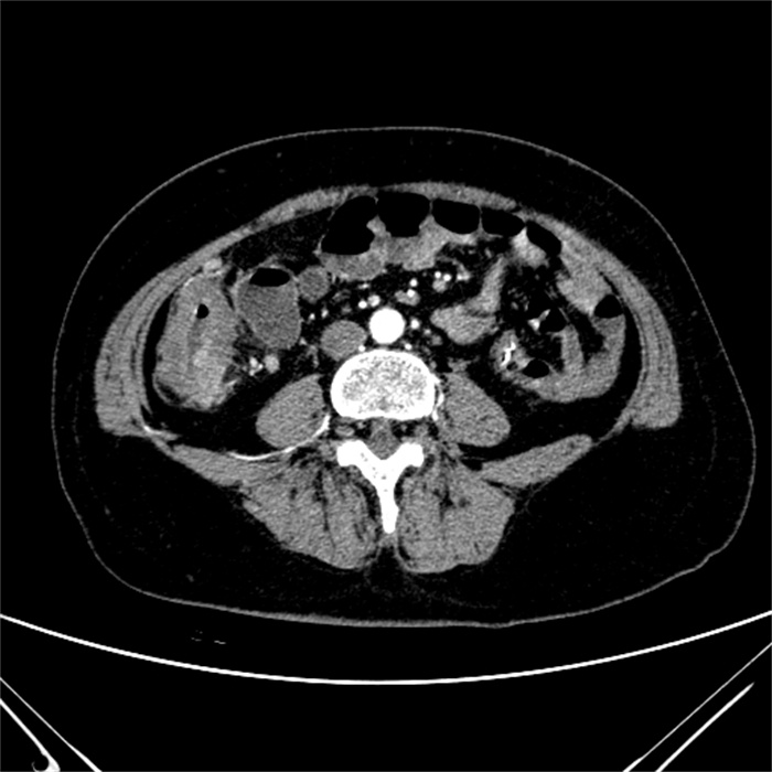
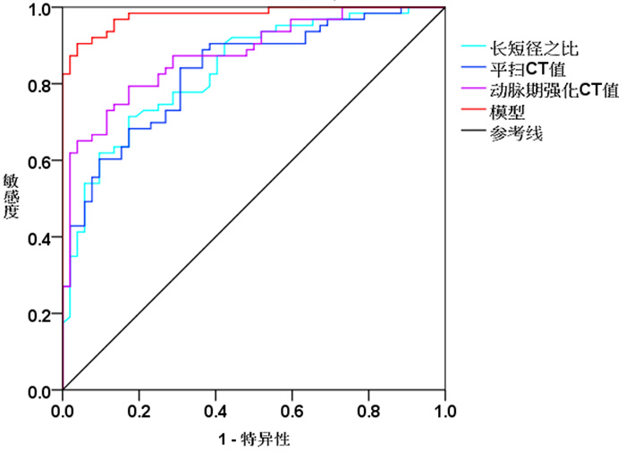
目的 探究结直肠癌患者癌旁肿瘤沉积(TD)多层螺旋CT(MSCT)表现及术前诊断价值。 方法 选择2018年12月~2021年12月医院收治的63例结直肠癌存在TD患者为TD组、52例结直肠癌出现淋巴结转移患者为淋巴结转移组,对两组患者相关资料进行回顾性分析。患者均于术前接受MSCT检查,分析TD与淋巴结转移患者MSCT表现及其在患者TD病情诊断中价值。 结果 TD患者病灶较大,形态多为不规则,与附近组织界线不清晰,平扫密度不均匀,增强扫可见均匀显著强化,强化程度与原发肿瘤相近,动脉增强扫显示为边界强化,少量病灶可以观察到液化坏死;淋巴结转移患者病灶较小,形态规则,多为圆形或者椭圆形,与附近组织比较边界相对清晰,密度相对均匀。TD组与淋巴结转移组患者病灶密度、边界、形状、长短径之比、平扫CT值、动脉期强化CT值差异有统计学意义(P < 0.05);多元Logistic回归分析显示,长短径之比、平扫CT值、动脉期强化CT值与TD有关(P < 0.05),以此构建Logistic回归模型公式为Logit (P)=65.212-6.001×长短径之比-0.315×平扫CT值-0.333×动脉期强化CT值。ROC曲线显示,长短径之比、平扫CT值、动脉期强化CT值用于TD诊断曲线下面积值分别为0.836、0.832以及0.878,回归模型曲线下面积值为0.979。 结论 MSCT检查可以为结直肠癌TD诊断提供有效影像学依据,基于MSCT回归模型有助于提高TD诊断价值。 Abstract:Objective To analyze the multi-slice spiral CT (MSCT) findings of pericolonic tumor deposits (TD) in patients with colorectal cancer, and its value in preoperative diagnosis. Methods The relevant data of 63 colorectal cancer patients with TD (TD group) and 52 colorectal cancer patients with lymph node metastasis (lymph node metastasis group) who were admitted to the hospital from December 2018 and December 2021 was retrospectively analyzed. All patients received MSCT examination before operation. MSCT findings of patients with TD and lymph node metastasis, and the value of MSCT in the diagnosis of TD were analyzed. Results The lesions in patients with TD were large, with irregular shapes, unclear boundaries with surrounding tissues, and uneven density on plain scan. Enhanced scan showed significant homogeneous enhancement which was similar to that of primary tumor. Arterial enhanced scan showed edge enhancement with liquefactive necrosis in a small amount of lesions. The lesions in patients with lymph node metastasis were small, with regular shape, mostly round or oval, relatively clear boundaries with surrounding tissues, and relatively uniform density. There were statistically significant differences in lesion density, boundary, shape, long-to-short diameter ratio, plain scan CT values, and arterial phase enhanced CT values between the TD group and the lymph node metastasis group (P < 0.05). Multivariate logistic regression analysis showed that long-to-short diameter ratio, plain scan CT value, and arterial phase enhanced CT value were related to TD (P < 0.05). The logistic regression model was constructed: Logit(P)=65.212-6.001×long-to-short diameter ratio-0.315×plain scan CT value-0.333×arterial phase enhanced CT value. The ROC curve showed that the area under the curve values of long-to-short diameter ratio, plain scan CT value, and arterial phase enhanced CT value to diagnose TD were 0.836, 0.832 and 0.878, respectively. The area under the curve of the regression model was 0.979. Conclusion MSCT examination provides an effective imaging basis for the diagnosis of TD in colorectal cancer. The MSCT-based regression model is helpful for the diagnosis of TD. -
Key words:
- colorectal cancer /
- pericolonic tumor deposits /
- multi-slice spiral CT /
- regression model /
- diagnosis
-

图 3 MSCT表现、CT参数对于TD诊断价值分析的ROC曲线
Figure 3. ROC curve analysis of the value of MSCT findings and CT parameters in the diagnosis of TD.
表 1 TD组与淋巴结转移组患者MSCT表现与CT测定参数比较
Table 1. Comparison of MSCT findings and CT parameters between TD group and lymph node metastasis group
指标 TD组(n=63) 淋巴结转移组(n=52) t/χ2 P 病灶密度[n(%)] 5.443 0.020 均匀 25(39.68) 32(61.54) 不均匀 38(60.32) 20(38.46) 边界[n(%)] 5.441 0.020 清晰 30(47.62) 36(69.23) 不清晰 33(52.38) 16(30.77) 形状[n(%)] 8.720 0.003 规则 18(28.57) 29(55.77) 不规则 45(71.43) 23(44.23) 液化坏死区[n(°%)] 2.255 0.133 有 28(44.44) 16(30.77) 无 35(55.56) 36(69.23) 平均径(cm, Mean±SD) 1.06±0.25 0.99±0.28 1.415 0.160 短径线(cm, Mean±SD) 1.16±0.21 1.12±0.24 0.953 0.343 长径线(cm, Mean±SD) 1.31±0.32 1.24±0.29 1.218 0.226 长短径之比(Mean±SD) 1.06±0.15 1.39±0.24 8.995 < 0.001 平扫CT值(Hu, Mean±SD) 26.94±6.23 18.15±5.19 8.111 < 0.001 静脉期强化CT值(Hu, Mean±SD) 48.26±10.58 44.81±9.62 1.813 0.073 动脉期强化CT值(Hu, Mean±SD) 29.55±6.76 21.64±5.29 6.876 < 0.001 TD: 肿瘤沉积.  下载: 导出CSV
表 2 MSCT表现、CT参数与TD之间关系分析
Table 2. The relationship between MSCT findings, CT parameters and TD
指标 β SE Wald χ2 OR 95% CI P 病灶密度 1.399 0.917 2.329 4.053 0.672~24.452 0.127 边界 1.327 0.912 2.114 3.768 0.63~22.532 0.146 形状 1.075 0.846 1.613 2.929 0.558~15.38 0.204 长短径之比 -6.001 1.965 9.322 0.002 0~0.117 0.002 平扫CT值 -0.315 0.093 11.332 0.73 0.608~0.877 0.001 动脉期强化CT值 -0.333 0.094 12.647 0.717 0.597~0.861 < 0.001 AUC: 曲线下面积.
下载: 导出CSV
表 3 MSCT表现、CT参数对于TD诊断价值分析
Table 3. Value of MSCT findings and CT parameters in the diagnosis of TD
指标 AUC 敏感度 特异性 95% CI P 长短径之比 0.836 71.38 82.67 0.764~0.908 < 0.001 平扫CT值 0.832 84.12 39.23 0.760~0.905 < 0.001 动脉期强化CT值 0.878 79.44 82.69 0.816~0.939 < 0.001 模型 0.979 90.48 96.23 0.958~1.00 < 0.001
下载: 导出CSV
-
[1] Mauri G, Sartore-Bianchi A, Russo AG, et al. Early-onset colorectal cancer in young individuals[J]. Mol Oncol, 2019, 13(2): 109-31. doi: 10.1002/1878-0261.12417 [2] 郭天安, 谢丽, 赵江, 等. 中国结直肠癌1988—2009年发病率和死亡率趋势分析[J]. 中华胃肠外科杂志, 2018(1): 33-40. doi: 10.3760/cma.j.issn.1671-0274.2018.01.007 [3] Piawah S, Venook AP. Targeted therapy for colorectal cancer metastases: a review of current methods of molecularly targeted therapy and the use of tumor biomarkers in the treatment of metastatic colorectal cancer[J]. Cancer, 2019, 125(23): 4139-47. doi: 10.1002/cncr.32163 [4] Xu M, Xu XN, Pan B, et al. LncRNA SATB2-AS1 inhibits tumor metastasis and affects the tumor immune cell microenvironment in colorectal cancer by regulating SATB2[J]. Mol Cancer, 2019, 18(1): 135. doi: 10.1186/s12943-019-1063-6 [5] Jakubowska K, Koda M, Kisielewski W, et al. Tumor- infiltrating lymphocytes in primary tumors of colorectal cancer and their metastases[J]. Exp Ther Med, 2019, 18(6): 4904-12. [6] Shen Y, Wang XH, Lu JY, et al. Reduction of liver metastasis stiffness improves response to bevacizumab in metastatic colorectal cancer[J]. Cancer Cell, 2020, 37(6): 800-17. e7. doi: 10.1016/j.ccell.2020.05.005 [7] Jin M, Frankel WL. Lymph node metastasis in colorectal cancer[J]. Surg Oncol Clin NAm, 2018, 27(2): 401-12. doi: 10.1016/j.soc.2017.11.011 [8] 罗超元, 岑东芝. 多层螺旋CT图像纹理分析在直肠癌淋巴结转移中的应用价值[J]. 中华胃肠外科杂志, 2019, 22(5): 488-90. [9] 曹丽娜, 张兆国, 张中旭, 等. 粘液性/非粘液性结直肠癌MSCT征象及鉴别诊断价值研究[J]. 中国CT和MRI杂志, 2022, 20(2): 155-7. doi: 10.3969/j.issn.1672-5131.2022.02.050 [10] Jilg CA, Drendel V, Rischke HC, et al. Detection rate of 18F-choline PET/CT and 68 Ga-PSMA-HBED-CC PET/CT for prostate cancer lymph node metastases with direct link from PET to histopathology: dependence on the size of tumor deposits in lymph nodes[J]. J Nucl Med, 2019, 60(7): 971-7. doi: 10.2967/jnumed.118.220541 [11] 王剑峰, 孙旭, 闫序波, 等. 白介素6、白介素7和Foxp3在结肠癌旁肿瘤沉积形成预测价值[J]. 中华胃肠外科杂志, 2018, 21(1): 87-9. doi: 10.3760/cma.j.issn.1671-0274.2018.01.017 [12] Tsai YC, Shih JH, Hwang HE, et al. Early prediction of 1-year tumor response of hepatocellular carcinoma with lipiodol deposition pattern through post-embolization cone-beam computed tomography during conventional transarterial chemoembolization [J]. Eur Radiol, 2021, 31(10): 7464-75. doi: 10.1007/s00330-021-07843-8 [13] 黄瑜亮, 黄斌, 严俊. 结直肠癌的早期诊断技术进展[J]. 分子影像学杂志, 2019, 42(1): 77-80. doi: 10.12122/j.issn.1674-4500.2019.01.18 [14] Zhang MF, Hu WW, Hu K, et al. Association of KRAS mutation with tumor deposit status and overall survival of colorectal cancer[J]. Cancer Causes Control, 2020, 31(7): 683-9. doi: 10.1007/s10552-020-01313-0 [15] 罗锦文, 王梦龙, 戴子扬, 等. 基于CT纹理分析在鉴别结直肠癌旁肿瘤沉积与转移淋巴结中的价值[J]. 临床放射学杂志, 2021, 40(3): 528-35. https://www.cnki.com.cn/Article/CJFDTOTAL-LCFS202103026.htm [16] 吴菊华, 郭磊. 术前多层螺旋CT检查对结直肠癌旁肿瘤沉积的诊断应用价值[J]. 临床与病理杂志, 2021, 41(8): 1787-92. https://www.cnki.com.cn/Article/CJFDTOTAL-WYSB202108011.htm [17] Athanasakis E, Xenaki S, Venianaki M, et al. Newly recognized extratumoral features of colorectal cancer challenge the current tumor-node-metastasis staging system[J]. Ann Gastroenterol, 2018, 31(5): 525-34. [18] 罗锦文, 李新春, 雷强, 等. 螺旋CT多参数测量对结直肠癌旁肿瘤沉积的诊断价值[J]. 放射学实践, 2019, 34(8): 901-5. https://www.cnki.com.cn/Article/CJFDTOTAL-FSXS201908013.htm [19] Sarioglu S, Kilicarslan E, Aydin B, et al. Tumor deposits in salivary gland tumors[J]. Pathol Int, 2018, 68(3): 183-9. doi: 10.1111/pin.12637 [20] Dai SD, Kanemitsu Y, Hamaguchi T, et al. Introducing the eighth edition of the tumor-node-metastasis classification as relevant to colorectal cancer, anal cancer and appendiceal cancer: a comparison study with the seventh edition of the tumor-node-metastasis and the Japanese Classification of Colorectal, Appendiceal, and Anal Carcinoma[J]. Jpn J Clin Oncol, 2019, 49(4): 321-8. doi: 10.1093/jjco/hyy198 [21] Mirkin KA, Kulaylat AS, Hollenbeak CS, et al. Prognostic significance of tumor deposits in stage Ⅲ colon cancer[J]. Ann Surg Oncol, 2018, 25(11): 3179-84. doi: 10.1245/s10434-018-6661-9 [22] 罗锦文, 李新春, 刘美玲, 等. 基于CT增强图像的影像组学模型对鉴别结直肠癌旁肿瘤沉积及转移淋巴结的诊断价值[J]. 放射学实践, 2020, 35(12): 1553-9. https://www.cnki.com.cn/Article/CJFDTOTAL-FSXS202012015.htm [23] 王晨, 孟闫凯, 崔莹莹, 等. CT影像学及临床病理学特征在预测结肠癌肿瘤沉积中的价值[J]. 临床放射学杂志, 2020, 39(8): 1537-42. https://www.cnki.com.cn/Article/CJFDTOTAL-LCFS202008019.htm -

 点击查看大图
计量
- 文章访问数: 187
- HTML全文浏览量: 104
- PDF下载量: 1
- 被引次数: 0