


Diagnostic efficacy of multi-slice spiral CT combined with serum MP-IgM and MP-Ab detection in children with mycoplasma pneumonia
-
摘要:
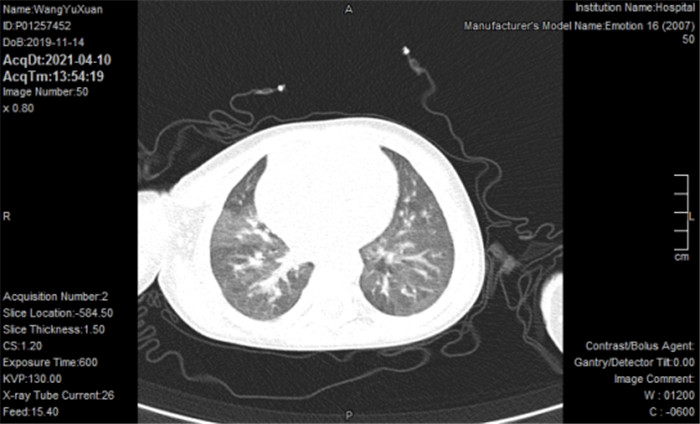
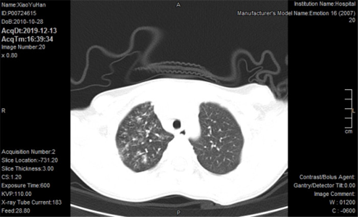
目的 分析多层螺旋CT(MSCT)联合血清肺炎支原体免疫球蛋白M(MP-IgM)、肺炎支原体抗体(MP-Ab)检测对小儿支原体感染肺炎的诊断效能。 方法 选取2019年1月~2021年1月本院收治的86例疑似支原体感染肺炎小儿为研究对象,采用金标准将患者分为感染组(n=38)和非感染组(n=48)。采用MSCT对所有患者进行扫描检查,并检测血清MP-IgM和MP-Ab水平,采用ROC曲线分析MSCT联合血清MP-IgM、MP-Ab对小儿支原体感染肺炎的诊断效能。 结果 MSCT图显示,小儿支原体感染肺炎表现为支气管充气相、支气管壁增厚、磨玻璃影、网状影和支气管血管束增厚,此外还可见明显的呈扇形分布的薄片影。在38例感染患儿中,发生在单侧左肺的有15例(39.47%),发生在单侧右肺的有17例(44.74%),发生在双肺的有6例(15.79%);MSCT与金标准具有较好的一致性(Kappa=0.72);感染组MP-IgM阳性和MP-Ab阳性率均高于非感染组(P < 0.05);根据临床诊断为准制作ROC曲线,其中3项联合的诊断价值最高,其与MSCT、MP-IgM、MP-Ab的差异均有统计学意义(P < 0.05)。 结论 MSCT联合MP-IgM、MP-Ab对支原体感染肺炎小儿具有较高的诊断价值。 -
关键词:
- 多层螺旋CT /
- 肺炎支原体免疫球蛋白M /
- 肺炎支原体抗体 /
- 支原体感染肺炎 /
- 诊断效能
Abstract:Objective To analyze the diagnostic efficacy of multi-slice spiral CT (MSCT) combined with serum mycoplasma pneumoniae immunoglobulin M (MP- IgM) and mycoplasma pneumoniae antibody (MP-Ab) in children with mycoplasma pneumonia. Methods A total of 86 children with suspected mycoplasma pneumonia admitted to our hospital from January 2019 to January 2021 were selected as the study subjects, and divided into the infection group (n=38) and non-infection group (n=48) according to the gold standard. MSCT was used to scan all children, and serum MP-IgM and MP-Ab levels were measured. ROC was used to analyze the diagnostic efficacy of MSCT combined with serum MP-igm and MP-Ab for mycoplasma pneumonia in children. Results MSCT images showed that mycoplasma infection pneumonia in children showed bronchial inflation phase, bronchial wall thickening, ground- glass opacities, reticular opacities and bronchovascular bundle thickening. In addition, there were obvious fan-shaped thin slices. Among 38 cases, 15 cases (39.47%) occurred in unilateral left lung, 17 cases (44.74%) in unilateral right lung, and 6 cases (15.79%) in bilateral lung. MSCT had good consistency with gold standard (Kappa=0.72). The positive rates of MP-IgM and MP-Ab in infected group were significantly higher than those in non-infected group (P < 0.05). ROC curve was made based on clinical diagnosis, among which three combinations had the highest diagnostic value, and there were significant differences compared with MSCT, MP-IgM and MP-Ab (P < 0.05). Conclusion MSCT combined with MP-IgM and MP-Ab has high diagnostic value in children with mycoplasma pneumonia. -

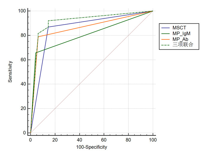
图 3 MSCT联合血清MP-IgM、MP-Ab对小儿支原体感染肺炎的诊断价值
Figure 3. Diagnostic value of MSCT combined with serum MP-IgM and MP-Ab in children with mycoplasma pneumonia.
表 1 38例患儿病变范围及部位分析
Table 1. Lesion range and location analysis of 38 children
病变范围 病变部位 病变数[n (%)] 构成比(%) 单侧左肺 上叶 3(7.89) 39.47 下叶 12(31.58) 单侧右肺 上叶 9(23.68) 44.74 下叶 8(21.05) 双肺 中叶及下叶 1(2.63) 15.79 5(13.16)  下载: 导出CSV
表 2 MSCT对小儿支原体感染肺炎的诊断情况
Table 2. MSCT diagnosis of mycoplasma pneumonia in children (n)
MSCT 金标准 合计 阳性 阴性 阳性 33 7 40 阴性 5 41 46 合计 38 48 86
下载: 导出CSV
表 3 两组血清MP-IgM、MP-Ab比较
Table 3. Comparison of serum MP-igm and MP-AB between the two groups [n(%)]
组别 MP-IgM阳性 MP-Ab阳性 感染组(n=38) 25(65.79) 30(78.95) 非感染组(n=48) 2(4.17) 3(6.25) χ2 37.393 47.399 P < 0.001 < 0.001 MP-IgM: 肺炎支原体免疫球蛋白M; MP-Ab: 肺炎支原体抗体.
下载: 导出CSV
表 4 MSCT联合血清MP-IgM、MP-Ab对小儿支原体感染肺炎的诊断价值
Table 4. Diagnostic value of MSCT combined with serum MP-IgM and MP-Ab in children with mycoplasma pneumonia
指标 AUC 敏感度(%) 特异性(%) 95% CI P* MSCT 0.861 86.84 85.42 0.770~0.926 0.015 MP-IgM 0.808 65.79 95.83 0.709~0.885 0.003 MP-Ab 0.863 78.98 93.75 0.772~0.928 0.036 3项联合 0.916 92.11 85.42 0.837~0.965 - *与3项联合比较, AUC: 曲线下面积.
下载: 导出CSV
-
[1] Mi YM, Qi Q, Zhang L, et al. Assessment of serum sialic acid correlated with C3 in children with Mycoplasma pneumoniae pneumonia[J]. J Clin LabAnal, 2020, 34(3): e23078. [2] Kumar S, Garg I, Sethi G, et al. Detection of immunoglobulin M and immunoglobulin G antibodies to Mycoplasma pneumoniae in children with community-acquired lower respiratory tract infections [J]. Indian J Pathol Microbiol, 2018, 61(2): 214. doi: 10.4103/IJPM.IJPM_21_17 [3] 刘婕, 茆占湖, 肖锡昌, 等. 多层螺旋CT动态增强成像在肺部孤立性结节诊断中的应用价值[J]. 临床和实验医学杂志, 2020, 19(17): 1903-5. doi: 10.3969/j.issn.1671-4695.2020.17.036 [4] Zhang Y, Yang X, Qian J, et al. Simultaneous detection of Mycoplasma pneumoniae IgG and IgM using dual-label time resolved fluoroimmunoassay[J]. Anal Biochem, 2018, 548: 1-6. doi: 10.1016/j.ab.2018.02.015 [5] Wu Y, Duan GK, Cheng MG, et al. Comparison of multiple detection methods of Mycoplasma pneumoniae antibody for the early diagnosis of pediatric Mycoplasma pneumonia[J]. Eur J Inflamm, 2019, 17: 1-14. https://www.sciencedirect.com/science/article/pii/S0732889309003290 [6] 祝亚军. MP-Ab、hs-CRP联合检测对小儿支原体肺炎感染的诊断价值分析[J]. 国外医学: 医学地理分册, 2019, 40(4): 354-6. doi: 10.3969/j.issn.1001-8883.2019.04.005 [7] 中华医学会重症医学分会. 呼吸机相关性肺炎诊断、预防和治疗指南(2013[) J]. 中华内科杂志, 2013, 52(6): 524-43. doi: 10.3760/cma.j.issn.0578-1426.2013.06.024 [8] Zheng BY, Zhao J, Cao L. The clinical characteristics and risk factors for necrotizing pneumonia caused by Mycoplasma pneumoniae in children[J]. BMC Infect Dis, 2020, 20: 391. doi: 10.1186/s12879-020-05110-7 [9] 曹建伟, 郑铠军, 冯淑芳, 等. 小儿肺炎支原体肺炎临床特征及病情程度的相关因素[J]. 分子影像学杂志, 2018, 41(3): 388-91. doi: 10.3969/j.issn.1674-4500.2018.03.25 [10] 陈志敏. 难治性肺炎支原体肺炎的诊断与药物治疗进展[J]. 实用儿科临床杂志, 2012, 27(4): 235-7. https://www.cnki.com.cn/Article/CJFDTOTAL-YXZS201222031.htm [11] Yoon SH, Min IK, Ahn JG. Immunochromatography for the diagnosis of Mycoplasma pneumoniae infection: a systematic review and meta-analysis[J]. PLoS One, 2020, 15(3): e0230338. doi: 10.1371/journal.pone.0230338 [12] Tashiro M, Fushimi K, Kawano K, et al. Comparison of efficacy of antimicrobial agents among hospitalized patients with Mycoplasma pneumoniae pneumonia in Japan during large epidemics of macrolide-resistant M. pneumoniae infections: a nationwide observational study[J]. Clin Infect Dis, 2017, 65(11): 1837-42. doi: 10.1093/cid/cix695 [13] Figallo CE, Bayes L, Lanata M, et al. A diagnostic dilemma: PCR or serology to detect Mycoplasma pneumoniae pneumonia in children [J]. Pediatrics, 2016, 137(Supplement 3): 348A. doi: 10.1542/peds.137.Supplement_3.348A [14] Sabri YY, Kamel KM, Hafez MAF, et al. Evaluation of the role of MSCT airway mapping in guiding trans-bronchial lung biopsy in cases of inaccessible lung lesions[J]. Egypt J Radiol Nucl Med, 2017, 48(4): 947-52. doi: 10.1016/j.ejrnm.2017.06.004 [15] 杨虓, 宋彬. 局灶性机化性肺炎的多层螺旋CT征象特征及与周围型肺癌的鉴别诊断分析[J]. 川北医学院学报, 2019, 34(6): 733-6. doi: 10.3969/j.issn.1005-3697.2019.06.20 [16] Kuwahara M, Samukawa M, Ikeda T, et al. Characterization of the neurological diseases associated with Mycoplasma pneumoniae infection and anti-glycolipid antibodies[J]. J Neurol, 2017, 264(3): 467-75. doi: 10.1007/s00415-016-8371-1 [17] 方茜茜, 何晶, 石慧芳, 等. 普米克令舒雾化联合左氧氟沙星对支原体肺炎的疗效及Mp-Ab的影响[J]. 南昌大学学报: 医学版, 2018, 58 (2): 37-41. https://www.cnki.com.cn/Article/CJFDTOTAL-JXYB201802010.htm [18] 袁娟, 白慧萍, 徐鹏飞, 等. MSCT检查对儿童肺炎支原体肺炎与肺炎支原体合并肺炎链球菌感染肺炎的鉴别诊断价值分析[J]. 中国CT和MRI杂志, 2021, 19(8): 72-3, 76. https://www.cnki.com.cn/Article/CJFDTOTAL-CTMR202108023.htm [19] 李若淳, 曾明燕, 苏艳丽, 等. MP-IgM抗体检测与微生物快速培养检测对小儿肺炎支原体感染的诊断价值[J]. 临床医学研究与实践, 2020, 5(13): 120-1. https://www.cnki.com.cn/Article/CJFDTOTAL-YLYS202013046.htm [20] 邢雅明. 肺炎支原体抗体联合超敏C反应蛋白检测在小儿肺炎支原体肺炎感染诊断中的临床价值[J]. 国际儿科学杂志, 2016, 43(12): 963-5. doi: 10.3760/cma.j.issn.1673-4408.2016.12.017 [21] 李智琼, 孙承谋, 朱玲娜, 等. MP-IgM、YKL-40、APOC1和IL-6在儿童支原体肺炎治疗及预后中的意义[J]. 标记免疫分析与临床, 2019, 26(4): 576-9, 619. https://www.cnki.com.cn/Article/CJFDTOTAL-BJMY201904009.htm -

 点击查看大图
计量
- 文章访问数: 494
- HTML全文浏览量: 190
- PDF下载量: 3
- 被引次数: 0