


Characteristics and diagnostic value of ultrasound and MSCT for thyroid cancer
-
摘要:
目的 分析甲状腺癌超声和多层螺旋CT(MSCT)扫描的影像学特征并分析其诊断价值。 方法 选取我院2018年1月1日~2021年10月8日收治的349例拟诊为甲状腺癌的患者为研究对象,根据超声检查结果,将患者分为超声恶性组203例和超声良性组146例; 根据MSCT检查结果,分为MSCT恶性组198例和MSCT良性组151例。分析其超声和MSCT影像学特征,并绘制受试者工作特征曲线分析超声和MSCT对甲状腺癌诊断的特异性和敏感度。 结果 超声恶性组与良性组患者在实性结节、低回声、钙化、形态不规则以及边界不清晰的差异有统计学意义(P < 0.05); MSCT恶性组与良性组患者在单发病灶、边界不清晰、形态不规则、钙化、囊变、不均匀强化以及淋巴结肿大的差异有统计学意义(P < 0.05); MSCT扫描诊断甲状腺癌的曲线下面积为0.789,敏感度为75.18%,特异性为82.69%,超声扫描诊断甲状腺癌的曲线下面积为0.862,敏感度为82.98%,特异性为89.42%。 结论 超声诊断甲状腺癌的影像学特征表现为实性结节、低回声、钙化、形态不规则以及边界不清晰等,MSCT诊断甲状腺癌的影像学特征表现为单发病灶、边界不清晰、形态不规则、钙化、囊变、不均匀强化以及淋巴结肿大等; 超声诊断甲状腺癌的特异性和敏感度高于MSCT诊断。 Abstract:Objective To explore the characteristics and diagnostic value of ultrasound and multi-slice spiral CT (MSCT) for thyroid cancer. Methods A total of 349 cases of patients suspected of thyroid cancer who were admitted to the hospital between January 1, 2018 and October 8, 2021 were selected. According to the results of ultrasonography, 203 cases were divided into malignant group and 146 cases were divided into benign group. According to the results of MSCT, 198 cases were divided into malignant group and 151 cases were divided into benign group. Their ultrasound and MSCT characteristics were analyzed. The ROC curve was plotted to analyze the specificity and sensitivity of ultrasound and MSCT in the diagnosis of thyroid cancer. Results There were significant differences between the malignant group and benign group which were examined by ultrasonography in solid nodules, hypoecho, calcification, irregular morphology and fuzzy boundaries (P < 0.05). There were significant differences between the malignant group and benign group which were examined by MSCT in single lesions, fuzzy boundaries, irregular morphology, calcification, cystic degeneration, heterogeneous enhancement, and enlarged lymph nodes (P < 0.05). The area under the curve value, sensitivity and specificity of MSCT to diagnose thyroid cancer were 0.789, 75.18% and 82.69%, which of ultrasound were 0.862, 82.98% and 89.42%. Conclusion The imaging features of thyroid carcinoma diagnosed by ultrasound are solid nodules, hypoechoic, calcification, irregular morphology and fuzzy boundaries. The imaging features of MSCT in the diagnosis of thyroid cancer are single focus, fuzzy boundaries, irregular morphology, calcification, cystic change, uneven enhancement and enlarged lymph nodes. Ultrasound is more specific and sensitive than MSCT in the diagnosis of thyroid cancer. -
Key words:
- thyroid cancer /
- ultrasound /
- multi-slice spiral CT /
- imaging characteristic /
- diagnosis
-

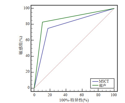
图 1 超声和MSCT诊断价值比较
Figure 1. Comparison of the diagnostic value between ultrasound and MSCT.

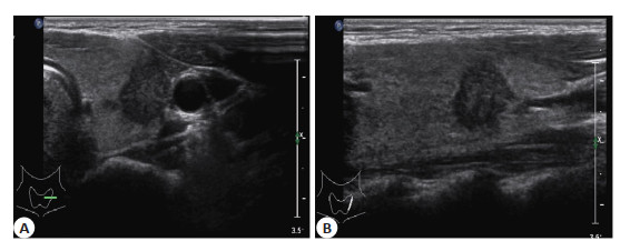
图 2 甲状腺癌超声特征
A: 甲状腺左侧叶横断面示: 中下份低回声结节, 边界欠清, 形态不规则, 纵横比 > 1, 边缘呈角, 内可见点状强回声; B: 甲状腺左侧叶纵断面示: 中下份低回声结节, 边界欠清, 形态不规则, 边缘呈角, 内可见点状强回声.
Figure 2. Ultrasonographic features of thyroid carcinoma.

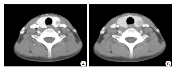
图 3 甲状腺癌MSCT特征
A: 甲状腺左叶低密度结节动脉期不均匀明显强化, 强化低于正常甲状腺; B: 甲状腺左叶低密度结节静脉期强化稍减退, 伴“咬饼征”.
Figure 3. MSCT features of thyroid carcinoma.
表 1 甲状腺癌超声影像学特征
Table 1. Ultrasound imaging features of thyroid cancer [n(%)]
影像学特征 恶性组(n=203) 良性组(n=146) χ2 P 实性结节 165(81.28) 43(28.48) 99.631 < 0.001 低回声 124(61.08) 33(21.85) 53.993 < 0.001 钙化 137(67.49) 34(22.52) 70.129 < 0.001 形态不规则 69(33.99) 9(5.96) 39.602 < 0.001 边界不清晰 142(69.95) 31(20.53) 84.640 < 0.001 血流丰富 69(33.99) 37(24.50) 3.715 0.054  下载: 导出CSV
表 2 甲状腺癌MSCT影像学特征
Table 2. MSCT imaging features of thyroid cancer [n(%)]
影像学特征 恶性组(n=198) 良性组(n=151) χ2 P 单发病灶 157(79.29) 63(41.72) 51.899 < 0.001 边界不清晰 161(81.31) 34(22.52) 120.120 < 0.001 形态不规则 105(53.03) 12(7.95) 78.131 < 0.001 钙化 102(51.52) 10(6.62) 79.223 < 0.001 囊变 97(48.99) 46(30.46) 12.157 < 0.001 不均匀强化 146(73.74) 70(46.36) 27.228 < 0.001 淋巴结肿大 105(53.03) 5(3.31) 98.112 < 0.001
下载: 导出CSV
表 3 超声和MSCT诊断价值比较
Table 3. Comparison of the diagnostic value between ultrasound and MSCT
参数 AUC(95% CI) Z P 约登指数 敏感度(%) 特异性(%) MSCT 0.789(0.743~0.831) 12.862 < 0.001 0.579 75.18 82.69 超声 0.862(0.821~0.896) 18.911 < 0.001 0.724 82.98 89.42
下载: 导出CSV
-
[1] Grani G, Sponziello M, Pecce V, et al. Contemporary thyroid nodule evaluation and management[J]. J Clin Endocrinol Metab, 2020, 105 (9): 2869-83. doi: 10.1210/clinem/dgaa322 [2] Wong R, Farrell SG, Grossmann M. Thyroid nodules: diagnosis and management[J]. Med J Aust, 2018, 209(2): 92-8. doi: 10.5694/mja17.01204 [3] Xu B, Ghossein R. Poorly differentiated thyroid carcinoma[J]. Semin Diagn Pathol, 2020, 37(5): 243-7. doi: 10.1053/j.semdp.2020.03.003 [4] Thomas CM, Asa SL, Ezzat S, et al. Diagnosis and pathologic characteristics of medullary thyroid carcinoma-review of current guidelines[J]. Curr Oncol, 2019, 26(5): 338-44. doi: 10.3747/co.26.5539 [5] 阚志文, 黄子杰, 崔亚云, 等. 电化学方法检测血浆游离DNA甲基化水平与甲状腺癌诊断以及相关临床特征的关系[J]. 实用医学杂志, 2021, 37(6): 792-6. doi: 10.3969/j.issn.1006-5725.2021.06.021 [6] 张艳, 马冰, 赵佳航, 等. 经淋巴管超声造影诊断甲状腺癌颈部淋巴结转移的价值[J]. 中国医学科学院学报, 2021, 43(3): 338-42. https://www.cnki.com.cn/Article/CJFDTOTAL-ZYKX202103005.htm [7] 张有为, 陈季松, 胡兵, 等. MSCT微血管半定量技术对甲状腺结节的诊断价值[J]. 放射学实践, 2017, 32(8): 831-4. https://www.cnki.com.cn/Article/CJFDTOTAL-FSXS201708012.htm [8] Yang DJ, Zhou YP, Peng ZW, et al. Effects of MSCT enhanced scan image diagnosis on clinical outcome of patients after radical gastrectomy and its influence on misdiagnosis rate[J]. J BUON, 2021, 26(4): 1479-84. [9] 李水平, 林敏, 赖莉萍, 等. 甲状腺髓样癌超声影像学特征分析及诊断价值评价[J]. 中国实验诊断学, 2017, 21(10): 1764-5. doi: 10.3969/j.issn.1007-4287.2017.10.028 [10] Zhao JZ, Zheng XQ, Gao M, et al. Ultrasound features of medullary thyroid cancer as predictors of biological behavior[J]. Cancer Imaging, 2021, 21(1): 33. doi: 10.1186/s40644-021-00402-w [11] 石木兰. 肿瘤影像学[M]. 北京: 科学出版社, 2003. [12] Filetti S, Durante C, Hartl D, et al. Thyroid cancer: ESMO Clinical Practice Guidelines for diagnosis, treatment and follow-up[J]. Ann Oncol, 2019, 30(12): 1856-83. doi: 10.1093/annonc/mdz400 [13] 苑娜红, 魏萍, 刘栓良, 等. 声速匹配技术对甲状腺癌的诊断价值[J]. 影像科学与光化学, 2021, 39(2): 219-23. https://www.cnki.com.cn/Article/CJFDTOTAL-GKGH202102010.htm [14] 黄声稀, 金伟奎, 司芩. 超声造影联合弹性成像诊断甲状腺癌的价值分析[J]. 临床肿瘤学杂志, 2017, 22(9): 823-6. doi: 10.3969/j.issn.1009-0460.2017.09.011 [15] Chasen NN, Wang JR, Gan Q, et al. Imaging of cervical lymph nodes in thyroid cancer: ultrasound and computed tomography[J]. Neuroimaging Clin N Am, 2021, 31(3): 313-26. doi: 10.1016/j.nic.2021.04.002 [16] 黄加鹏, 杨彤, 刘虎, 等. 以囊性成分为主的甲状腺癌影像学特征分析[J]. 中华内分泌外科杂志, 2019, 13(1): 13-6. [17] Shalaby M, Hadedeya D, Lee GS, et al. Impact of surgeon-performed ultrasound on treatment of thyroid cancer patients[J]. Am Surg, 2020, 86(9): 1148-52. doi: 10.1177/0003134820945229 [18] Lamartina L, Bidault S, Hadoux J, et al. Can preoperative ultrasound predict extrathyroidal extension of differentiated thyroid cancer?[J]. Eur J Endocrinol, 2021, 185(1): 13-22. doi: 10.1530/EJE-21-0091 [19] Figge JJ, Gooding WE, Steward DL, et al. Do ultrasound patterns and clinical parameters inform the probability of thyroid cancer predicted by molecular testing in nodules with indeterminate cytology?[J]. Thyroid, 2021, 31(11): 1673-82. doi: 10.1089/thy.2021.0119 [20] 黄东琼, 许林, 邱冬梅, 等. 多层螺旋CT和核素扫描对结节性甲状腺肿诊断的临床价值对比[J]. 实用放射学杂志, 2021, 37(1): 34-7. doi: 10.3969/j.issn.1002-1671.2021.01.009 [21] 杨文杰, 随涛, 程好堂. 超声检查与螺旋CT在诊断甲状腺癌中的对比分析[J]. 医学影像学杂志, 2022, 32(4): 694-6. https://www.cnki.com.cn/Article/CJFDTOTAL-XYXZ202204035.htm [22] 李文艳, 刘燕飞, 黄慧君. B超与CT对结节性甲状腺肿合并甲状腺癌的诊断价值分析[J]. 中国实用医药, 2022, 17(7): 107-9. https://www.cnki.com.cn/Article/CJFDTOTAL-ZSSA202207037.htm [23] Zhang Y, Zhang X, Li J, et al. Contrast- enhanced ultrasound: a valuable modality for extracapsular extension assessment in papillary thyroid cancer[J]. Eur Radiol, 2021, 31(7): 4568-75. doi: 10.1007/s00330-020-07516-y [24] 李源, 陈士新, 黄斌. 多层螺旋CT诊断甲状腺癌中的辐射和对比剂剂量选择[J]. 影像科学与光化学, 2019, 37(6): 564-70. https://www.cnki.com.cn/Article/CJFDTOTAL-GKGH201906007.htm [25] 辛世卿. 高分辨率超声联合CT对甲状腺癌颈部淋巴结转移的临床诊断价值[J]. 中国CT和MRI杂志, 2021, 19(1): 56-8. doi: 10.3969/j.issn.1672-5131.2021.01.019 -

 点击查看大图
计量
- 文章访问数: 146
- HTML全文浏览量: 126
- PDF下载量: 5
- 被引次数: 0