


Value of MRI diffusion weighted imaging parameter ADC value in evaluating the differentiation degree and tumor microstructure of rectal cancer
-
摘要:
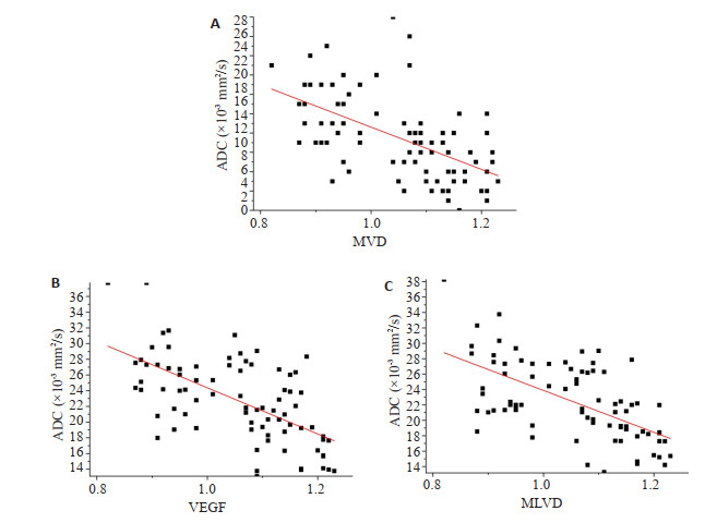
目的 探讨磁共振弥散加权成像参数表观扩散系数(ADC)值术前评估直肠癌分化程度及瘤体微观结构的价值。 方法 收集2018~2021年经病理证实的直肠癌患者82例,所有患者在接受3.0 T弥散加权成像检查后均测量瘤体ADC值,分析不同分化程度直肠癌ADC值的差异,通过ROC曲线确定ADC值区分直肠癌分化程度的效能,并研究ADC值与微血管密度、血管内皮生长因子和微淋巴管密度的相关性。 结果 高、中分化直肠癌ADC值[(1.16±0.05)×10-3 mm2/s、(1.04±0.08)×10-3 mm2/s]高于低分化直肠癌[(0.93±0.07)×10-3 mm2/s],差异有统计学意义(P < 0.05);ROC曲线显示,ADC值区分低分化与高、中分化直肠癌的最佳临界值为1.05×10⁻³ mm2/s,曲线下面积为0.902 (95% CI: 0.831~0.972);直肠癌瘤体ADC值与微血管密度、血管内皮生长因子阳性细胞百分比及微淋巴管密度均呈负相关关系(r=-0.56、-0.61、-0.59,P < 0.05)。 结论 直肠癌瘤体ADC值有助于术前评估肿瘤分化程度和微观结构特点,间接反映肿瘤血管新生和微淋巴管密度。 Abstract:Objective To investigate the diagnostic value of the apparent diffusion coefficient (ADC) of MRI diffusion weighted imaging parameters in preoperative assessment of the differentiation degree and tumor microstructure of rectal cancer. Methods A total of 82 patients with rectal cancer confirmed pathologically from 2018 to 2021 were collected. The ADC values of tumors were measured in all patients after 3.0 T diffusion weighted imaging, and the difference in ADC values of rectal cancers with different levels of differentiation was analyzed. ROC curve was used to determine the efficacy of ADC value in differentiating the differentiation degree of rectal cancer. The correlation between ADC value and microvessel density, vascular endothelial growth factor and microvessel density was analyzed. Results The ADC value of high [(1.16±0.05)×10-3 mm2/s] and moderately [(1.04±0.08)×10-3 mm2/s] differentiated rectal cancer was significantly higher than that of poorly differentiated rectal cancer [(0.93±0.07)×10-3 mm2/s, P < 0.05]. ROC curve showed that the optimal critical value of ADC value to distinguish poorly differentiated from well-differentiated and moderately differentiated rectal cancer was 1.05×10⁻³ mm2/s, and the AUC was 0.902 (95%CI: 0.831-0.972). The ADC value and microvessel density, the percentage of vascular endothelial growth factor positive cells and microlymphatic vessel density of rectal cancer tumors were all negatively correlated (r=- 0.56, -0.61, - 0.59, P < 0.05). Conclusion The ADC value of rectal cancer tumor is helpful to evaluate tumor differentiation and microstructure characteristics. It indirectly reflects tumor angiogenesis and microlymphatic vessel density. -

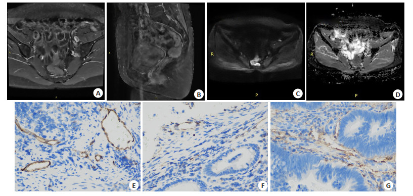
图 1 患者男,43岁,低分化直肠癌
A: 横轴位T2WI显示直肠壁肿块呈稍高信号; B: 矢状位T2WI显示肿块位于低位直肠壁; C~D: 横轴位DWI及ADC分别显示直肠壁肿块呈高信号和低信号; E~G: CD34、VEGF和MLVD抗体图像,呈明显阳性表达(SP, ×400).
Figure 1. A 43-year-old male patient with poorly differentiated rectal cancer.

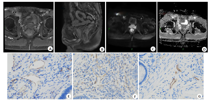
图 2 患者女,50岁,高分化直肠癌
A: 横轴位T2WI显示直肠壁肿块呈稍高信号;B: 矢状位T2WI显示肿块位于低位直肠壁; C~D: 横轴位DWI及ADC分别显示直肠壁肿块呈高信号和低信号; E~G: CD34、VEGF和MLVD抗体图像,呈明显阳性表达(SP, ×400).
Figure 2. A50-year-old female patient with well-differentiated rectal cancer.

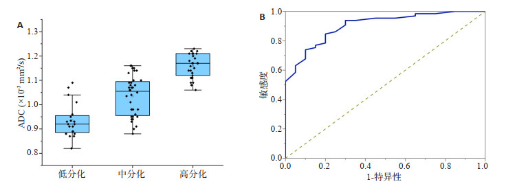
图 3 不同分化程度直肠癌瘤体ADC值比较结果(A)及ROC曲线评价ADC值区分低分化与高、中分化直肠癌的价值(B)
Figure 3. Comparison of ADC values of rectal cancer with different differentiation degrees (A) and The value of ROC curve to evaluate ADC value in differentiating poorly differentiated from well and moderately differentiated rectal cancer (B)
-
[1] 中华人民共和国国家卫生健康委员会医政医管局, 中华医学会肿瘤学分会. 中国结直肠癌诊疗规范(2020年版)[J]. 中国实用外科杂志, 2020, 40(6): 601-25. https://www.cnki.com.cn/Article/CJFDTOTAL-ZGWK202006001.htm [2] Zhu L, Pan ZL, Ma Q, et al. Diffusion kurtosis imaging study of rectal adenocarcinoma associated with histopathologic prognostic factors: preliminary findings[J]. Radiology, 2017, 284(1): 66-76. doi: 10.1148/radiol.2016160094 [3] 肖欢, 赵振华, 周莹, 等. 磁共振T2WI、DWI对直肠癌新辅助治疗疗效的评估价值[J]. 影像科学与光化学, 2022, 40(1): 151-5. https://www.cnki.com.cn/Article/CJFDTOTAL-GKGH202201030.htm [4] 韩超, 陈新晖. 磁共振弥散加权成像ADC值对结直肠癌放化疗疗效的评估价值[J]. 中国CT和MRI杂志, 2021, 19(8): 144-7. doi: 10.3969/j.issn.1672-5131.2021.08.047 [5] Peng Y, Tang H, Meng XY, et al. Histological grades of rectal cancer: whole-volume histogram analysis of apparent diffusion coefficient based on reduced field-of-view diffusion-weighted imaging[J]. Quant Imaging Med Surg, 2020, 10(1): 243-56. doi: 10.21037/qims.2019.11.17 [6] 王锡山. 从中美结直肠癌流行病学特征看结直肠癌早诊早治的重要性[J]. 中华结直肠疾病电子杂志, 2021, 10(1): 26-33. doi: 10.3877/cma.j.issn.2095-3224.2021.01.004 [7] 董健, 李垣婕, 谢宗源, 等. 进展期直肠癌新辅助放化疗疗效评估模型的建立及评价: 基于MRI和网状蛋白1C[J]. 分子影像学杂志, 2021, 44(3): 472-7. doi: 10.12122/j.issn.1674-4500.2021.03.11 [8] Ao WQ, Bao XD, Mao GQ, et al. Value of apparent diffusion coefficient for assessing preoperative T staging of low rectal cancer and whether this is correlated with ki-67 expression[J]. Can Assoc Radiol J, 2020, 71(1): 5-11. doi: 10.1177/0846537119885666 [9] Chen LG, Shen F, Li ZH, et al. Diffusion-weighted imaging of rectal cancer on repeatability and cancer characterization: an effect of b-value distribution study[J]. Cancer Imaging, 2018, 18(1): 43. doi: 10.1186/s40644-018-0177-1 [10] 刘玉霞, 谢瑞峰, 胡延涛. 多b值DWI联合MRI常规扫描对直肠癌检查图像质量的影响[J]. 实用癌症杂志, 2021, 36(7): 1111-5. doi: 10.3969/j.issn.1001-5930.2021.07.017 [11] 黄瑜亮, 黄斌, 严俊. 结直肠癌的早期诊断技术进展[J]. 分子影像学杂志, 2019, 42(1): 77-80. doi: 10.12122/j.issn.1674-4500.2019.01.18 [12] 陆志华, 蒋恒, 翁晓燕, 等. 优化选择高和超高b值直肠癌弥散加权成像[J]. 中国医学影像技术, 2020, 36(12): 1858-62. https://www.cnki.com.cn/Article/CJFDTOTAL-ZYXX202012023.htm [13] 孙海涛, 翟跃杰, 黄勇华. DWI及其ADC值对直肠癌病理分级分型的诊断价值研究[J]. 中国CT和MRI杂志, 2022, 20(3): 156-8, 179. doi: 10.3969/j.issn.1672-5131.2022.03.052 [14] 郭达, 杨陈, 周倩, 等. MR750 3.0T DWI ADC值与直肠癌患者临床病理分级及术后复发和吻合口炎性纤维化关系[J]. 放射学实践, 2020, 35(10): 1310-5. https://www.cnki.com.cn/Article/CJFDTOTAL-FSXS202010024.htm [15] Xiao CC, Zhou MH, Yang XH, et al. Novel nomogram with microvascular density in the surgical margins can accurately predict the risk for anastomotic leakage after anterior resection for rectal cancer[J]. J Surg Oncol, 2019, 120(8): 1412-9. doi: 10.1002/jso.25730 [16] 张茜, 刘影, 王昌新, 等. 直肠癌MRI与病理分期对照及其与MVD值的关系[J]. 安徽医科大学学报, 2014, 49(11): 1685-8. https://www.cnki.com.cn/Article/CJFDTOTAL-YIKE201411041.htm [17] 陈照宏, 高玉颖, 刘楠, 等. 直肠癌组织VEGF-C表达与磁共振成像功能学表征的相关性[J]. 现代肿瘤医学, 2019, 27(19): 3500-3. doi: 10.3969/j.issn.1672-4992.2019.19.033 [18] Meyer HJ, Höhn AK, Woidacki K, et al. Associations between IVIM histogram parameters and histopathology in rectal cancer[J]. Magn Reson Imaging, 2021, 77: 21-7. doi: 10.1016/j.mri.2020.12.008 [19] Meyer HJ, Wienke A, Surov A. Association between VEGF expression and diffusion weighted imaging in several tumors-A systematic review and meta-analysis[J]. Diagnostics (Basel), 2019, 9 (4): 126. doi: 10.3390/diagnostics9040126 [20] Barresi V, Reggiani Bonetti L, Vitarelli E, et al. Immunohistochemical assessment of lymphovascular invasion in stage I colorectal carcinoma: prognostic relevance and correlation with nodal micrometastases[J]. Am J Surg Pathol, 2012, 36(1): 66-72. doi: 10.1097/PAS.0b013e31822d3008 -

 下载:
下载:
 点击查看大图
计量
- 文章访问数: 258
- HTML全文浏览量: 104
- PDF下载量: 4
- 被引次数: 0