


Diagnostic value of salivary gland ultrasound score combined with radionuclide imaging in Sjogren's syndrome
-
摘要:
目的 分析唾液腺超声评分联合核素显像参数对干燥综合征(SS)的诊断价值。 方法 选取2018年1月~2021年12月本院收治的105例SS患者作为研究组,另选取同期105例有口干、眼干症状的非SS患者作为对照组。采用唾液腺超声检查和核素显像,观察两组患者超声图像、核素显像及唾液腺功能。采用ROC曲线分析超声评分联合核素显像对SS患者的诊断价值。 结果 超声声像图显示,SS患者可见腮腺呈弥漫性病变,腮腺或颌下腺有1个或多个低回声结节;核素显像显示,SS患者可见双侧腮腺和颌下腺摄取功能明显降低,唾液腺对99TcmO4-摄取减少,口腔99TcmO4-浓聚更少,口服维生素C后未见明显浓聚,双侧腮腺和颌下腺呈持续上升。研究组颌下腺评分、腮腺评分及唾液腺超声评分均高于对照组(P < 0.05),根据临床诊断为准制作ROC曲线,其中唾液腺超声总评分的诊断价值最高,其与颌下腺评分和腮腺评分的差异有统计学意义(P < 0.05)。研究组颌下腺、腮腺摄取指数和腺体排泄率水平均低于对照组(P < 0.05),根据临床诊断为准制作ROC曲线,其中唾液腺超声评分联合核素显像的诊断价值最高,其与单独诊断比较,差异有统计学意义(P < 0.05)。 结论 唾液腺超声评分联合核素显像对SS患者具有较高的诊断价值。 Abstract:Objective To explore the diagnostic value of salivary gland ultrasound scoring combined with nuclide imaging parameters in sjogren's syndrome (SS). Methods A total of 105 SS patients admitted to our hospital from January 2018 to December 2021 were selected as the study group, and 105 non-SS patients with dry mouth and dry eye were selected as the control group. Salivary gland ultrasonography and radionuclide imaging were used to observe the ultrasonic image, radionuclide imaging and salivary gland function in two groups. ROC curve was used to analyze the diagnostic value of ultrasonic scoring combined with radionuclide imaging in SS patients. Results Ultrasound imaging showed diffuse lesions in parotid gland and one or more hypoechoic nodules in parotid gland or submandibular gland. Radionuclide imaging showed that the uptake function of bilateral parotid gland and submandibular gland was significantly reduced in SS patients, the uptake of 99TcmO4- in salivary gland was reduced, and the concentration of 99TcmO4- in oral cavity was less. No obvious concentration was observed after oral vitamin C. The bilateral parotid gland and submandibular gland were continuously increased. The submandibular gland score, parotid gland score and salivary gland ultrasound score in the study group were significantly higher than those in the control group (P < 0.05). ROC curve was made based on clinical diagnosis, among which the total salivary gland ultrasound score had the highest diagnostic value and had significant differences with the submandibular gland score and parotid gland score (P < 0.05). The levels of UR and SR in the submandibular gland and parotid gland in the study group were significantly lower than those in the control group (P < 0.05). ROC curve was made based on clinical diagnosis, in which the ultrasonic score of salivary gland combined with radionuclide imaging had the highest diagnostic value, and there were significant differences compared with the diagnosis alone (P < 0.05). Conclusion Salivary gland ultrasound scoring combined with radionuclide imaging has high diagnostic value in SS patients. -
Key words:
- salivary gland ultrasonography /
- nuclide imaging /
- Sjogren's syndrome /
- diagnostic value
-

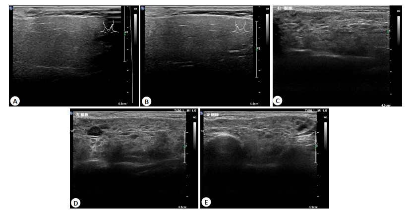
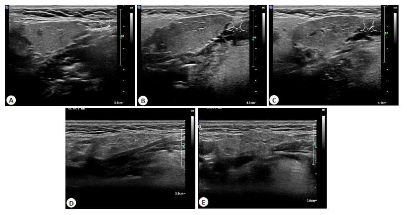
图 1 SS患者颌下腺超声评分
A: 正常颌下腺(0分); B: 颌下腺实质回声减低, 轻度不均匀, 可见少许高回声线(1分); C: 颌下腺实质明显回声不均匀, 弥漫分布的低回声结节, 直径<2 mm, 高回声线增多(2分); D: 颌下腺见结节状低回声区增大融合, 直径2~6 mm, 高回声线分布杂乱(3分); E: 颌下腺见低回声结节>6 mm, 或腺体萎缩, 多发高回声线(4分)
Figure 1. Submandibular gland ultrasound score of SS patients.

图 2 SS患者腮腺超声评分
A: 正常腮腺(0分); B: 腮腺实质回声减低, 轻度不均匀, 可见少许高回声线(1分); C: 腮腺实质明显回声不均匀, 弥漫分布的低回声结节, 直径<2 mm(2分); D: 腮腺见结节状低回声区增大融合, 直径2~6 mm, 高回声线分布杂乱(3分); E: 腮腺见低回声结节>6 mm, 或腺体萎缩, 多发高回声线(4分)
Figure 2. parotid ultrasound score of SS patients.

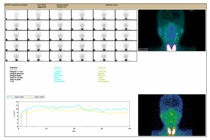
图 3 SS患者颌下腺核素显像
Figure 3. Radionuclide imaging of submandibular gland in patients with SS.

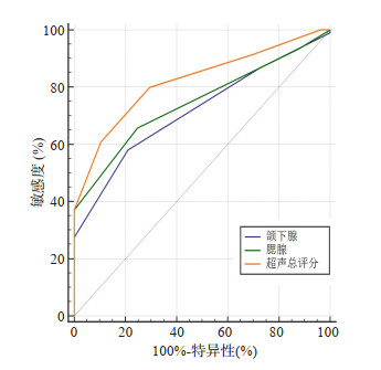
图 5 唾液腺超声评分诊断SS患者的ROC曲线
Figure 5. ROC curve of SS patients diagnosed by salivary gland ultrasound score.

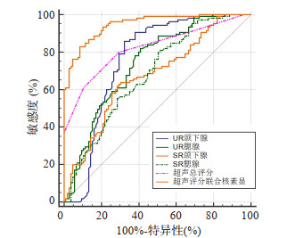
图 6 唾液腺超声评分联合核素显像诊断SS患者的ROC曲线
Figure 6. ROC curve of salivary gland ultrasound score combined with radionuclide imaging in the diagnosis of SS.
表 1 两组一般资料比较
Table 1. Comparison of two groups of general data (n=105)
项目 研究组 对照组 χ2/t P 性别(n) 0.494 0.482 男 45 40 女 60 65 年龄(岁, Mean±SD) 49.58±12.20 50.39±11.58 0.493 0.622 病程(年, Mean±SD) 4.25±1.26 4.00±1.35 1.387 0.167 口干症(n) 95 91 0.753 0.386 眼干症(n) 85 76 2.156 0.142  下载: 导出CSV
表 2 唾液腺超声评分比较
Table 2. Comparison of salivary gland ultrasound scores (score, n=105, Mean±SD)
组别 颌下腺 腮腺 超声总评分 研究组 2.71±1.04 2.22±1.33 4.93±1.64 对照组 1.93±0.70 1.12±0.60 3.06±1.02 t 6.376 4.263 10.270 P < 0.001 < 0.001 < 0.001
下载: 导出CSV
表 3 唾液腺超声评分对SS患者诊断价值
Table 3. Diagnostic value of salivary gland ultrasound score in patients with SS
指标 截点值 AUC 敏感度(%) 特异性(%) 95% CI P* 颌下腺 > 2分 0.719 58.10 79.05 0.652~0.778 < 0.001 腮腺 > 1分 0.747 65.71 75.24 0.682~0.804 0.002 总评分 > 3分 0.822 80.00 70.48 0.763~0.871 *与总评分比较; AUC: 曲线下面积.
下载: 导出CSV
表 4 对照组左右颌下腺、腮腺间UR和SR比较
Table 4. Comparison of UR and SR between left and right submandibular gland and parotid gland in the control group (n=105, Mean±SD)
部位 颌下腺 腮腺 UR SR UR SR 左侧 4.71±1.83 48.10±10.82 4.74±1.90 51.49±11.98 右侧 5.09±1.63 48.58±10.57 5.01±1.93 50.93±12.17 t 1.589 0.325 1.022 0.336 P 0.114 0.745 0.308 0.737 UR: 摄取指数; SR: 腺体排泄率.
下载: 导出CSV
表 5 研究组左右颌下腺、腮腺间UR和SR比较
Table 5. Comparison of UR and SR between left and right submandibular gland and parotid gland in the study group (n=105, Mean±SD)
部位 颌下腺 腮腺 UR SR UR SR 左侧 3.90±0.75 44.26±9.23 3.89±1.00 46.59±8.92 右侧 4.03±0.78 42.04±9.16 3.88±0.90 46.16±9.43 t 1.231 1.749 0.076 0.339 P 0.220 0.082 0.939 0.735
下载: 导出CSV
表 6 两组颌下腺、腮腺UR和SR比较
Table 6. Comparison of UR and SR in submandibular gland and parotid gland between the two groups (n=105, Mean±SD)
组别 颌下腺 腮腺 UR SR UR SR 研究组 3.96±0.57 43.13±7.96 3.89±0.79 46.38±7.10 对照组 4.90±1.28 48.34±8.60 4.87±1.33 51.21±9.45 t 6.874 4.556 6.492 4.187 P < 0.001 < 0.001 < 0.001 < 0.001
下载: 导出CSV
表 7 唾液腺超声评分联合核素显像对SS患者诊断价值
Table 7. Diagnostic value of salivary gland ultrasound score combined with radionuclide imaging in patients with SS
指标 截点值 AUC 敏感度(%) 特异性(%) 95% CI P* UR颌下腺 4.46 0.757 85.71 67.62 0.693~0.814 < 0.001 UR腮腺 4.50 0.736 81.90 59.05 0.671~0.795 < 0.001 SR颌下腺 44.15 0.671 61.90 71.43 0.603~0.735 < 0.001 SR腮腺 52.34 0.681 80.00 49.52 0.613~0.744 < 0.001 超声评分 > 3 0.822 80.00 70.48 0.763~0.871 < 0.001 五项联合 - 0.947 82.86 91.43 0.908~0.973 - *与五项联合比较.
下载: 导出CSV
-
[1] El Hasbani G, Chahine R, Uthman I, et al. Intravenous immunoglobulin, neonatal alloimmune thrombocytopenia, and Sjogren's syndrome: a case report[J]. Eur J Obstet Gynecol Reprod Biol, 2021, 258: 476-7. doi: 10.1016/j.ejogrb.2021.01.053 [2] 单悦, 王俊峰. 唾液腺超声检查诊断干燥综合征的研究进展[J]. 临床超声医学杂志, 2019, 21(6): 449-51. doi: 10.3969/j.issn.1008-6978.2019.06.017 [3] Jousse-Joulin S, Coiffier G. Current status of imaging of Sjogren's syndrome[J]. Best Pract Res Clin Rheumatol, 2020, 34(6): 101592. doi: 10.1016/j.berh.2020.101592 [4] Mariette X, Criswell LA. Primary Sjögren's syndrome[J]. N Engl J Med, 2018, 378(10): 931-9. doi: 10.1056/NEJMcp1702514 [5] la Paglia GMC, Sanchez-Pernaute O, Alunno A, et al. Ultrasound salivary gland involvement in Sjogren's syndrome vs. other connective tissue diseases: is it autoantibody and gland dependent? [J]. Clin Rheumatol, 2020, 39(4): 1207-15. doi: 10.1007/s10067-019-04780-2 [6] 周科, 贾志云. 核素动态显像对131Ⅰ治疗分化型甲状腺癌患者唾液腺损伤程度的评价[J]. 解放军预防医学杂志, 2019, 37(1): 83-5, 99. https://www.cnki.com.cn/Article/CJFDTOTAL-JYYX201901025.htm [7] Anjos DA, Etchebehere ECSC, Santos AO, et al. Normal values of 99mTc pertechnetate uptake and excretion fraction by major salivary glands[J]. Nucl Med Commun, 2006, 27(4): 395-403. doi: 10.1097/01.mnm.0000202864.52046.b1 [8] 田甜, 韦智晓, 李俊红, 等. 腮腺99TcmO4-摄取指数和残留比诊断干燥综合征的价值[J]. 中华核医学与分子影像杂志, 2016, 36(6): 521-4. doi: 10.3760/cma.j.issn.2095-2848.2016.06.009 [9] 安锐, 黄钢. 核医学[M]. 3版. 北京: 人民卫生出版社, 2015. [10] Ozek D, Kemer OE, Omma A. Association between systemic activity ındex and dry eye severity in patients with primary Sjögren syndrome[J]. Arq Bras Oftalmol, 2018, 82(1): 45-50. [11] Johri R, Peter J, Peravali V, et al. Diagnostic performance of dry eye tests, serology and labial salivary gland biopsy in primary Sjogren's syndrome in an Indian setting[J]. Clin Epidemiol Glob Heal, 2020, 8 (1): 301-4. doi: 10.1016/j.cegh.2019.03.007 [12] Martel A, Coiffier G, Bleuzen A, et al. What is the best salivary gland ultrasonography scoring methods for the diagnosis of primary or secondary Sjögren's syndromes?[J]. Joint Bone Spine, 2019, 86(2): 211-7. doi: 10.1016/j.jbspin.2018.06.014 [13] 孟娟. 定量分析核素唾液腺动态显像评估干燥综合征患者的睡眠质量及口腔干燥情况[J]. 世界睡眠医学杂志, 2018, 5(9): 1072-4. doi: 10.3969/j.issn.2095-7130.2018.09.022 [14] Costa S, Quintin-Roué I, Lesourd A, et al. Reliability of histopathological salivary gland biopsy assessment in Sjögren's syndrome: a multicentre cohort study[J]. Rheumatology: Oxford, 2014, 54(6): 1056-64. [15] van Nimwegen JF, Mossel E, Delli K, et al. Incorporation of salivary gland ultrasonography into the American college of rheumatology/ European league against rheumatism criteria for primary sjögren's syndrome[J]. Arthritis Care Res (Hoboken), 2020, 72(4): 583-90. doi: 10.1002/acr.24017 [16] 李世文, 石娇, 华兴, 等. 超声评分系统在干燥综合征中的诊断价值[J]. 第三军医大学学报, 2017, 39(10): 996-1000. https://www.cnki.com.cn/Article/CJFDTOTAL-DSDX201710011.htm [17] 刘杨, 程昉, 王艳玲, 等. 涎腺超声对原发性干燥综合征的诊断和评估价值[J]. 中华临床免疫和变态反应杂志, 2021, 15(5): 528-33. https://www.cnki.com.cn/Article/CJFDTOTAL-OZHL202105004.htm [18] 邹惠峰, 沈阳, 尤嘉熙, 等. 唾液腺显像诊断干燥综合征[J]. 中国医学影像技术, 2017, 33(3): 399-403. https://www.cnki.com.cn/Article/CJFDTOTAL-ZYXX201703028.htm [19] 栗全营, 钟英, 汤元翔, 等. 20 min唾液腺显像在干燥综合征诊断中的可行性分析[J]. 南京医科大学学报: 自然科学版, 2019, 39(10): 1537-40. doi: 10.7655/NYDXBNS20191031 -


 点击查看大图
计量
- 文章访问数: 280
- HTML全文浏览量: 153
- PDF下载量: 7
- 被引次数: 0