


Diagnosis of AlN metastasis in breast cancer and evaluation of ovarian function after chemotherapy by ultrasound D-dimer in Ovarian Cancer
-
摘要:
目的超声影像表现对乳腺癌腋窝淋巴结(ALN)转移的诊断及对患者化疗后卵巢功能的评估。 方法采用前瞻性研究方法,以2019年6月~2021年3月于我院诊断并进行治疗的62例乳腺癌患者作为研究对象,其中ALN转移患者28例,未转移34例,比较两组患者的皮质形态、血流状况、超声测量指标之间的差异,分析超声指标对ALN转移诊断效能,研究卵巢超声指标与卵巢功能的相关性。 结果两组患者的狭窄型、向心增厚型、偏心增厚型、无淋巴门型之间的差异有统计学意义(χ2=27.871,P < 0.001);两组患者的淋巴门型、散在型、周边型、混合型之间的差异存在统计学意义(χ2=28.802,P < 0.001);ALN转移组患者的ALN纵径(t=17.037,P < 0.001)、皮质厚度(t=15.491,P < 0.001)以及D/W(t=7.243,P < 0.001)高于未转移组;联合检测对ALN转移的诊断特异度显著高于单独检测;化疗前两组患者的AMH、FSH之间的差异无统计学意义(P > 0.05),经过治疗后,两组患者的AMH下降,FSH上升,且未转移组患者的AMH(t=6.003,P < 0.001)、FSH(t=4.307,P < 0.001)高于ALN转移组;两组患者的RI、PI下降,且未转移组患者的RI(t=6.887,P < 0.001)、PI(t=26.430,P < 0.001)低于ALN转移组;患者的AMH、FSH与RI、PI呈现正相关。 结论超声影像表现对乳腺癌ALN转移具有良好的诊断效能,同时通过对患者的卵巢局部病灶部位的超声分析,对于化疗后卵巢功能的评估具有重要意义。 Abstract:ObjectiveTo evaluate the value of ultrasonography in the diagnosis of ALN metastasis of breast cancer and ovarian function after chemotherapy. MethodsSixty-two breast cancer patients diagnosed and treated in our hospital from June 2019 to March 2021were selected. Among them, 28 patients with ALN metastasis were compared in cortical morphology, blood flow and ultrasonic measurement indexes. The diagnostic efficacy of ultrasound indicators on ALN metastasis was analyzed. The correlation between ovarian ultrasound index and ovarian function was analyzed. ResultThe stenosis type, concentric thickening type, eccentric thickening type and non hilar type between the two groups were significantly different (χ2=27.871, P < 0.001). The hilar type, scattered type, peripheral type and mixed type between the two groups were significantly different(χ2= 28.802, P < 0.001). The longitudinal diameter (t=17.037, P < 0.001), cortical thickness (t=15.491, P < 0.001) and D/W (t=7.243, P < 0.001) of ALN metastasis group were significantly higher than those of non ALN metastasis group. The diagnostic specificity of combined detection for ALN metastasis was significantly higher than that of single detection. The AMH and FSH between the two groups before chemotherapy were not significantly different(P>0.05). After treatment, AMH of the two groups decreased significantly, FSH increased significantly, and AMH (t=6.003, P < 0.001) and FSH (t=4.307, P < 0.001) of patients without ALN metastasis were significantly higher than those of patients with ALN metastasis. The RI and Pi of two groups were significantly decreased. The RI (t=6.887, P < 0.001) and PI (t=26.430, P < 0.001) of patients without ALN metastasis were significantly lower than those of patients with ALN metastasis. AMH, FSH and RI, PI were positively correlated. ConclusionUltrasound imaging has a good diagnostic effect on ALN metastasis of breast cancer. It has great significance for the evaluation of ovarian function after chemotherapy by ultrasound analysis of local ovarian lesions. -
Key words:
- breast cancer /
- axillary lymphnode metastasis /
- ovarian function /
- ultrasound imaging
-

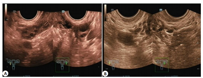
图 1 化疗前后ALN转移患者的卵巢超声情况
患者女,48岁; A: 化疗前卵巢超声图;B: 化疗后卵巢超声图.
Figure 1. Ovarian ultrasound of patients with AlN metastasis before and after chemotherapy.
表 1 两组患者的一般资料比较
Table 1. Comparison of baseline data between the two groups (Mean±SD)
组别 年龄(岁) BMI(kg/m2) 病程(月) TNM分期(1~2期/3~4期) ALN转移组(n=28) 44.02±2.06 23.58±2.36 6.23±2.69 15/14 未ALN转移组(n=34) 44.22±2.09 23.80±2.45 6.45±2.77 13/21 t 0.378 0.359 0.316 1.152 P 0.707 0.721 0.753 0.283  下载: 导出CSV
表 2 两组患者的皮质形态比较
Table 2. Comparison of cortical morphology between the two groups[n(%)]
组别 狭窄型 向心增厚型 偏心增厚型 无淋巴门型 ALN转移组(n=28) 4(14.29) 6(21.43) 18(64.29) 0(0.00) ALN未转移组(n=34) 19(55.88) 14(41.18) 1(2.94) 0(0.00) χ2 27.871 P < 0.001
下载: 导出CSV
表 3 两组患者的血流状况比较
Table 3. Comparison of blood flow between the two groups[n(%)]
组别 淋巴门型 散在型 周边型 混合型 ALN转移组(n=28) 5(15.15) 7(21.21) 19(57.58) 2(6.06) ALN未转移组(n=34) 26(76.47) 4(11.76) 2(5.88) 2(5.88) χ2 28.802 P < 0.001
下载: 导出CSV
表 4 两组患者的超声测量指标比较
Table 4. Comparison of ultrasonic measurement indexes between the two groups (Mean±SD)
组别 ALN纵径(cm) 皮质厚度(cm) D/W ALN转移组(n=28) 1.74±0.37 0.76±0.15 0.73±0.33 ALN未转移组(n=34) 0.40±0.21 0.26±0.09 0.27±0.07 t 17.037 15.491 7.243 P < 0.001 < 0.001 < 0.001
下载: 导出CSV
表 5 超声指标对ALN转移诊断效能分析
Table 5. Analysis of diagnostic efficiency of ultrasound parameters for AlN metastasis
诊断方法 真阳性
(n)假阳性
(n)真阴性
(n)假阴性
(n)准确率
(%)敏感度
(%)特异性
(%)阳性预测值
(%)阴性预测值
(%)纵径 > 1.0 cm 25 12 22 3 75.81 89.29 46.81 67.57 88.00 皮质偏心增厚型 18 13 21 10 62.90 64.29 53.85 58.06 67.74 周边型 19 15 19 9 61.29 67.86 50.00 55.88 67.86 联合检测 17 1 33 11 80.65 60.71 66.00 94.44 75.00
下载: 导出CSV
表 6 两组患者的卵巢功能比较
Table 6. Comparison of ovarian function between the two groups (Mean±SD)
组别 AMH(ng/mL) FSH(U/L) 化疗前 化疗后 化疗前 化疗后 ALN转移组(n=28) 2.74±1.37 1.21±0.25 7.33±1.33 8.25±0.45 ALN未转移组(n=34) 2.70±1.51 1.60±0.26 7.37±1.57 8.61±0.33 t 0.109 6.003 0.109 4.307 P 0.913 0.000 0.914 0.000
下载: 导出CSV
表 7 乳腺癌患者化疗前后的超声指标比较
Table 7. Comparison of ultrasound indexes of breast cancer patients before and after chemotherapy (Mean±SD)
超声指标 RI PI AMH r 0.365 0.441 P 0.002 0.039 FSH r 0.552 0.526 P 0.041 0.013 RI: 阻力指数; PI: 搏动指数.
下载: 导出CSV
表 8 相关性分析
Table 8. Correlation analysis (Mean±SD)
组别 RI PI 化疗前 化疗后 化疗前 化疗后 ALN转移组(n=28) 0.61±0.17 0.40±0.07 1.59±0.17 0.62±0.05 未ALN转移组(n=34) 0.66±0.15 0.26±0.09 1.55±0.11 1.26±0.13 t 1.215 6.887 1.074 26.430 P 0.229 0.000 0.287 0.000
下载: 导出CSV
-
[1] Chai R, Ma H, Xu M, et al. Differentiating axillary lymph node metastasis in invasive breast cancer patients: a comparison of radiomic signatures from multiparametric breast MR sequences[J]. J Magn Reson Imaging, 2019, 50(4): 1125-32. doi: 10.1002/jmri.26701 [2] Liu ZS, Feng B, Li CL, et al. Preoperative prediction of lymphovascular invasion in invasive breast cancer with dynamic contrast-enhanced-MRI-based radiomics[J]. J Magn Reson Imaging, 2019, 50(3): 847-57. doi: 10.1002/jmri.26688 [3] Yoo J, Kim BS, Yoon HJ. Predictive value of primary tumor parameters using 18F-FDG PET/CT for occult lymph node metastasis in breast cancer with clinically negative axillary lymph node[J]. Ann Nucl Med, 2018, 32(9): 642-8. doi: 10.1007/s12149-018-1288-2 [4] Tan WG, Xie XH, Huang ZY, et al. Construction of an immunerelated genes nomogram for the preoperative prediction of axillary lymph node metastasis in triple-negative breast cancer[J]. Artif Cells Nanomed Biotechnol, 2020, 48(1): 288-97. doi: 10.1080/21691401.2019.1703731 [5] Zhao YP, Ye WL, Liu DZ, et al. Redox and pH dual sensitive bone targeting nanoparticles to treat breast cancer bone metastases and inhibit bone resorption[J]. Nanoscale, 2017, 9(19): 6264-77. doi: 10.1039/C7NR00962C [6] Jiang X, Guo D, Li W, et al. Combination Twist1 and CA15-3 in axillary lymph nodes for breast cancer prognosis[J]. Mol Med Rep, 2017, 15(3): 1123-34. doi: 10.3892/mmr.2017.6138 [7] Zhou J, Guo FJ, Hao XP, et al. Use of breast magnetic resonance imaging and ultrasonography for identifying nonpalpable axillae metastases in newly diagnosed breast cancer patients[J]. Clin Breast Cancer, 2018, 18(1): e65-71. doi: 10.1016/j.clbc.2017.06.015 [8] Han C, Yang B, Zuo WS, et al. Prospective study found thatperipheral lymph node sampling reduced the false-negative rate ofsentinel lymph node biopsy forbreast cancer[J]. Chin J Cancer, 2016, 35(6): 327-32. [9] Rachel BM, Julia P, Julie O, et al. Real-time ex vivo perfusion of human lymph nodes invaded by cancer (REPLICANT): a feasibility study[J]. J Pathol, 2020, 250(3): 262-74. doi: 10.1002/path.5367 [10] Vanderburgh J, Hill JL, Gupta MK, et al. Tuning ligand density to optimize pharmacokinetics of targeted nanoparticles for dual protection against tumor-induced bone destruction[J]. ACS Nano, 2020, 14(1): 311-27. doi: 10.1021/acsnano.9b04571 [11] 金华, 罗伟权, 纪宗萍, 等. 乳腺癌超声影像组学图像特征logistic回归方程预测腋窝淋巴结转移风险[J]. 中国超声医学杂志, 2021, 37 (2): 139-42. doi: 10.3969/j.issn.1002-0101.2021.02.007 [12] 杨颛搏, 黄州, 王淑莲, 等. 腋窝前哨淋巴结阳性乳腺癌患者腋窝非前哨淋巴结转移风险预测[J]. 中华肿瘤杂志, 2020, 42(8): 653-9. doi: 10.3760/cma.j.cn112152-20190824-00545 [13] 林彩玲, 何毅辉, 林志武. 乳腺癌腋窝淋巴结转移危险因素的临床研究[J]. 中华实验外科杂志, 2020, 37(9): 1716-9. doi: 10.3760/cma.j.cn421213-20200512-00345 [14] 谭红娜, 武明辉, 周晶, 等. 乳腺X线影像组学方法预测乳腺癌腋窝淋巴结转移的价值[J]. 中华放射学杂志, 2020, 54(9): 859-63. doi: 10.3760/cma.j.cn112149-20190921-00437 [15] 马微妹, 李姣, 何妮, 等. 基于乳腺MRI及腋窝超声的列线图预测早期乳腺癌前哨淋巴结转移风险的价值[J]. 中华放射学杂志, 2020, 54 (7): 694-701. doi: 10.3760/cma.j.cn112149-20200420-00576 [16] 邢君, 马利军, 闫峥峥, 等. 乳腺癌非前哨淋巴结转移的危险因素研究[J]. 中华实验外科杂志, 2021, 38(3): 529-33. doi: 10.3760/cma.j.cn421213-20200801-01279 [17] 富泽龙, 田利, 张承玉, 等. 乳腺癌前哨淋巴结活检术中探索腋窝新分区的临床分析[J]. 中华医学杂志, 2020, 100(30): 2363-6. doi: 10.3760/cma.j.cn112137-20200303-00571 [18] 朱久俊, 焦得闯, 郭旭辉, 等. 新辅助化疗后锁骨上淋巴结病理完全缓解和乳腺癌患者预后的关系[J]. 中华普通外科杂志, 2020, 35(5): 366-70. doi: 10.3760/cma.j.cn113855-20190925-00567 [19] 陈相宏, 董福仁. DCE-MRI联合前哨淋巴结活检对腋窝淋巴结阳性乳腺癌患者新辅助化疗后状态转变的诊断价值[J]. 广东医学, 2020, 41(12): 1280-3. https://www.cnki.com.cn/Article/CJFDTOTAL-GAYX202012022.htm [20] 魏娜, 戴民, 杜葵英, 等. 纳米碳混悬液在乳腺癌新辅助化疗后前哨淋巴结活检中的临床应用[J]. 临床肿瘤学杂志, 2019, 24(4): 354-8. doi: 10.3969/j.issn.1009-0460.2019.04.014 [21] 周昊, 陈祖锦, 李云, 等. 腋窝淋巴结阳性乳腺癌新辅助化疗后腋窝治疗选择的研究[J]. 中国肿瘤临床, 2019, 46(5): 247-51. doi: 10.3969/j.issn.1000-8179.2019.05.050 [22] 彭惠阳, 武兆忠, 刘影, 等. 乳腺摄取99mTc-MDP与乳腺癌分子表达产物及预后的关系[J]. 中国老年学杂志, 2019, 39(18): 4437-42. doi: 10.3969/j.issn.1005-9202.2019.18.022 [23] 庞孟春, 钟凯, 俸跃波, 等. 乳腺癌动态增强MRI表现与腋窝淋巴结转移及ER、PR的关系[J]. 医学影像学杂志, 2021, 31(2): 255-8, 268. https://www.cnki.com.cn/Article/CJFDTOTAL-ZYXX201904009.htm [24] 纪晓惠, 石可心, 赵倩颖, 等. 高频超声对乳腺癌内乳淋巴结转移的诊断价值[J]. 中华超声影像学杂志, 2021, 30(1): 58-63. doi: 10.3760/cma.j.cn131148-20200528-00444 [25] 赵梦, 牛欢, 朱强, 等. 血清组织多肽特异性抗原表达水平在乳腺癌中的临床应用价值探讨[J]. 临床和实验医学杂志, 2021, 20(6): 594-8. doi: 10.3969/j.issn.1671-4695.2021.06.010 -

 点击查看大图
计量
- 文章访问数: 254
- HTML全文浏览量: 116
- PDF下载量: 3
- 被引次数: 0