


Value of color Doppler ultrasound combined with hepatic artery related index in predicting the effect and prognosis of interventional chemotherapy for elderly primary liver cancer
-
摘要:
目的探讨彩色多普勒超声联合肝动脉相关指数预测老年原发性肝癌(PLC)介入化疗疗效及预后的价值。 方法回顾性分析2017年3月~2018年12月本院收治的236例老年(>60岁)PLC患者的临床资料。统计老年PLC患者治疗情况;对比无效组和有效组患者的临床资料;Logistic回归分析影响老年PLC患者TACE治疗效果的因素;制作受试者工作特征曲线(ROC),分析肝动脉相关指数对老年PLC患者治疗效果的预测价值;术后随访1年,肝动脉相关指数分析老年PLC患者预后情况。 结果本研究老年PLC患者经TACE治疗后,治疗无效发生率为24.58%。Logistic回归分析结果显示,病理分型为低分化、肝动脉收缩期峰值(PSV)及肝动脉搏动指数(PI)均为影响老年PLC患者TACE治疗效果的因素(OR=4.011、3.340、2.807,P < 0.05)。ROC分析显示,PSV、PI预测老年PLC患者治疗无效的最佳截断点分别为82.42 cm/s、1.73,曲线下面积值分别为0.772、0.753。预后不良组PSV、PI均显著高于预后不良组(P < 0.05)。 结论彩色多普勒超声联合PSV、PI预测老年PLC介入化疗疗效及预后效能较高,可作为评估疗效及预后的重要指标。 Abstract:ObjectiveTo explore the value of color Doppler ultrasound combined with hepatic artery related index in predicting the efficacy and prognosis of interventional chemotherapy for elderly patients with primary liver cancer (PLC). MethodsThe clinical data of 236 elderly patients with PLC admitted to our hospital from March 2017 to December 2018 were retrospectively analyzed. Statistics of elderly patients with PLC were analyzed. The clinical data of patients in the invalid group and the effective group were compared. Logistic regression analysis was used to analyze the factors of TACE treatment in elderly patients with PLC. ROC was used to analyze the predictive value of hepatic artery correlation index for the treatment effect of elderly patients with PLC. After 1 year follow-up, the hepatic artery related index was used to analyze the prognosis of elderly patients with PLC. ResultsThe incidence of treatment failure in elderly PLC patients was 24.58% after TACE treatment. The pathological classification was poorly differentiated. Peak hepatic artery systole (PSV) and Hepatic artery pulsation index (PI) were all factors affecting the treatment effect of TACE in elderly patients with PLC (OR=4.011, 3.340, 2.807, all P < 0.05). ROC analysis showed that the best cut-off points for PSV and PI to predict treatment failure in elderly PLC patients were 82.42 cm/s and 1.73, respectively. The AUC values were 0.772 and 0.753, respectively. The PSV and PI of the poor prognosis group were significantly higher than those of the poor prognosis group (P < 0.05). ConclusionColor doppler ultrasound combined with PSV and PI has a higher efficacy in predicting the efficacy and prognosis of PLC interventional chemotherapy in the elderly. It can be used as an important indicator for evaluating the efficacy and prognosis. -
Key words:
- elderly /
- primary liver cancer /
- color Doppler ultrasound /
- hepatic artery index /
- chemotherapy /
- prognosis
-

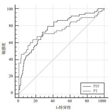
图 1 老年PLC彩色多普勒超声检查图
A:性别男,年龄72岁,BCLC分期IIa,child分级B,肿瘤数目3,PSV:67.76 cm/s,RI:0.86,DPI:0.39,PI:1.53;B:性别男,年龄68岁,BCLC分期IIIa,child分级B,肿瘤数目2,PSV:89.65 cm/s,RI:0.88,DPI:0.52,PI:1.59.
Figure 1. Color Doppler ultrasound examination of elderly PLC.

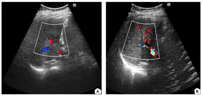
图 2 肝动脉相关指数对老年PLC患者治疗效果预测的ROC图
Figure 2. ROC diagram of hepatic artery related index for predicting the therapeutic effect of elderly patients with PLC.
表 1 对比无效组和有效组患者的临床资料
Table 1. Comparison of clinical data of patients in ineffective group and effective group[n(%)]
可能影响因素 无效组(n=58) 有效组(n=178) χ2/t P 性别(男) 48(82.76) 135(75.84) 1.202 0.273 年龄(岁, Mean±SD) 76.53±7.15 74.68±7.03 1.733 0.084 BMI(kg/m2, Mean±SD) 22.18±3.11 22.55±3.25 0.761 0.448 肝功能child分级 5.626 0.018 A级 28(48.28) 117(65.73) B级 30(51.72) 61(34.27) 肿瘤直径(cm, Mean±SD) 5.21±1.17 3.85±1.02 8.498 < 0.001 吸烟史 29(46.55) 68(38.20) 1.268 0.260 饮酒史 22(37.93) 54(30.34) 1.155 0.282 合并高血压 18(31.03) 43(24.16) 1.079 0.299 合并糖尿病 17(29.31) 42(23.60) 0.762 0.383 合并高血脂症 23(39.66) 57(32.02) 1.137 0.286 合并肝硬化 48(82.76) 137(76.97) 0.866 0.352 病理分型 10.068 0.002 中/高分化 19(32.76) 101(56.74) 低分化 39(67.24) 77(43.26) BCLC分期 7.783 0.005 A/B期 40(68.97) 152(85.39) C期 18(31.03) 26(14.61) 肿瘤数目 4.032 0.045 单发 40(68.97) 145(81.46) 多发 18(31.03) 33(18.54) TACE治疗次数(Mean±SD) 3.08±0.86 2.85±0.82 1.833 0.068 肝动脉直径(cm, Mean±SD) 0.37±0.09 0.35±0.08 1.602 0.110 RI(Mean±SD) 0.87±0.13 0.85±0.11 1.148 0.252 DPI(Mean±SD) 0.48±0.13 0.45±0.11 1.723 0.086 PSV(cm/s, Mean±SD) 91.32±14.35 78.21±12.87 6.546 < 0.001 PI(Mean±SD) 1.84±0.46 1.52±0.34 5.677 < 0.001 BCLC:巴塞罗那分期;TACE:肝动脉化疗栓塞;RI:阻力指数;DPI:血流灌注指数;PSV:肝动脉收缩期峰值;PI:搏动指数.  下载: 导出CSV
表 2 自变量赋值
Table 2. Assignment of independent variables
自变量 赋值 自变量 赋值 肝功能child分级 A级=0,B级=1 肿瘤数目 单发=0,多发=1 肿瘤直径 连续变量 PSV 连续变量 病理分型 中/高分化=0,低分化=1 PI 连续变量 BCLC分期 A/B期=0,C期=1
下载: 导出CSV
表 3 影响老年PLC患者TACE治疗效果的多因素Logistic回归分析
Table 3. Multivariate logistic regression analysis of the effects of TACE treatment in elderly patients with PLC
因素 β SE Wald χ2 P OR 95%CI 病理分型 1.389 0.368 14.247 < 0.001 4.011 2.150~7.481 PSV 1.206 0.415 8.445 0.011 3.340 1.254~6.084 PI 1.032 0.446 5.354 0.015 2.807 1.052~5.436
下载: 导出CSV
表 4 肝动脉相关指数对老年PLC患者治疗效果预测的ROC分析
Table 4. ROC analysis of hepatic artery related index for predicting the therapeutic effect of elderly patients with PLC
检测指标 最佳截断点 敏感度(%) 特异性(%) AUC 95%CI PSV(cm/s) 82.42 79.31(46/58) 67.42(120/178) 0.772 0.713~0.824 PI 1.73 63.79(37/58) 83.71(149/178) 0.753 0.693~0.807
下载: 导出CSV
表 5 预后良好组和预后不良组肝动脉相关指数对比
Table 5. Comparison of hepatic artery related indexes between the good prognosis group and the poor prognosis group (Mean±SD)
指标 预后不良组(n=72) 预后良好组(n=164) χ2/t P 肝动脉直径(cm) 0.38±0.17 0.34±0.15 1.810 0.072 RI 0.75±0.16 0.73±0.13 1.012 0.313 DPI 0.48±0.13 0.45±0.11 1.823 0.070 PSV(cm/s) 94.36±13.25 75.76±10.25 11.436 < 0.001 PI 2.26±0.63 1.77±0.38 7.373 < 0.001
下载: 导出CSV
-
[1] Broutier L, Mastrogiovanni G, Verstegen MM, et al. Human primary liver cancer-derived organoid cultures for disease modeling and drug screening[J]. Nat Med, 2017, 23(12): 1424-35. doi: 10.1038/nm.4438 [2] Kim EK, Song MJ, Jung Y, et al. Proteomic analysis of primary colon cancer and synchronous solitary liver metastasis[J]. Cancer Genomics Proteomics, 2019, 16(6): 583-92. doi: 10.21873/cgp.20161 [3] Chen J, Zhang Y, Cai H, et al. Comparison of the effects of postoperative prophylactic transcatheter arterial chemoembolization (TACE) and transhepatic arterial infusion (TAI) after hepatectomy for primary liver cancer[J]. J Buon, 2018, 23(3): 629-34. http://www.ncbi.nlm.nih.gov/pubmed/30003729 [4] Rennert J, Wiesinger I, Schicho A, et al. Color coded perfusion imaging with contrast enhanced ultrasound (CEUS) for post-interventional success control following irreversible electroporation (IRE) of primary and secondary malignant liver lesions[J]. J Gastrointestin Liver Dis, 2019, 28(3): 311-8. doi: 10.15403/jgld-254 [5] Schwarze V, Marschner C, Völckers W, et al. The diagnostic performance of contrast-enhanced ultrasound (CEUS) for evaluating hepatocellular carcinoma (HCC) juxtaposed to MRI findings; a retrospective single-center analysis of 292 patients[J]. Clin Hemorheol Microcirc, 2020, 76(2): 155-60. doi: 10.3233/CH-209213 [6] 中华人民共和国卫生和计划生育委员会医政医管局. 原发性肝癌诊疗规范(2017年版)[J]. 中华肝脏病杂志, 2017, 25(12): 886-95. doi: 10.3760/cma.j.issn.1007-3418.2017.12.002 [7] 张百红, 岳红云. 实体瘤疗效评价标准简介[J]. 国际肿瘤学杂志, 2016, 43(11): 845-7. doi: 10.3760/cma.j.issn.1673-422X.2016.11.011 [8] Jeong S, Zheng B, Wang H, et al. Nervous system and primary liver cancer[J]. Biochim Biophys Acta Rev Cancer, 2018, 1869(2): 286-92. doi: 10.1016/j.bbcan.2018.04.002 [9] 王华利, 熊清芳, 杨永峰. 279例老年原发性肝癌临床特点及预后影响因素分析[J]. 实用老年医学, 2017, 31(12): 1135-7. https://www.cnki.com.cn/Article/CJFDTOTAL-SYLA201712011.htm [10] Swaid F, Geller DA. Minimally invasive primary liver cancer surgery[J]. Surg Oncol Clin N Am, 2019, 28(2): 215-27. doi: 10.1016/j.soc.2018.11.002 [11] Chen X, Chang Z, Liu Z. D-dimer increase: an unfavorable factor for patients with primary liver cancer treated with TACE[J]. Cancer Chemother Pharmacol, 2019, 83(4): 797-802. doi: 10.1007/s00280-019-03778-6 [12] Luo YJ, Jiang Y. Comparison of efficiency of TACE plus HIFU and TACE alone on patients with primary liver cancer[J]. J Coll Physicians Surg Pak, 2019, 29(5): 414-7. doi: 10.29271/jcpsp.2019.05.414 [13] 赵剑, 吴涯昆, 田银生. CT联合MRI评估原发性肝癌患者的介入及分子靶向治疗效果[J]. 分子影像学杂志, 2021, 44(1): 41-6. doi: 10.12122/j.issn.1674-4500.2021.01.08 [14] Rubini A, Guiban O, Cantisani V, et al. Multiparametric ultrasound evaluation of parotid gland tumors: B-mode and color Doppler in comparison and in combination with contrast-enhanced ultrasound and elastography. A case report of a misleading diagnosis[J]. J Ultrasound, 2020, 15(4): 136-42. [15] Iliescu L, Toma L, Mercan-Stanciu A, et al. Contrast-enhanced ultrasonography in the diagnosis of portal vein thrombosis: a pictorial review[J]. Ultrasound Q, 2019, 35(4): 311-5. doi: 10.1097/RUQ.0000000000000451 [16] 诸巧红, 王颖. 彩色多普勒超声在原发性肝癌与肝血管瘤鉴别诊断中的效果分析[J]. 健康大视野, 2019, 10(7): 229-30. https://www.cnki.com.cn/Article/CJFDTOTAL-YYXK201832027.htm [17] 钟景云, 梁满球, 聂悦富, 等. 肝硬化并发原发性肝癌合并肝囊肿患者的影像学特征[J]. 分子影像学杂志, 2018, 41(2): 165-8. doi: 10.3969/j.issn.1674-4500.2018.02.07 [18] Dubinsky TJ, Revels J, Wang S, et al. Comparison of superb microvascular imaging with color flow and power Doppler imaging of small hepatocellular carcinomas[J]. J Ultrasound Med, 2018, 37 (12): 2915-24. doi: 10.1002/jum.14654 [19] Blackburn H, West S. Management of postembolization syndrome following hepatic transarterial chemoembolization for primary or metastatic liver cancer[J]. Cancer Nurs, 2016, 39(5): . http://www.ncbi.nlm.nih.gov/pubmed/26484962 [20] Taylor AC, Maddirela D, White SB. Role of radioembolization for biliary tract and primary liver cancer[J]. Surg Oncol Clin N Am, 2019, 28(4): 731-43. doi: 10.1016/j.soc.2019.07.001 [21] 李栩聪, 林谦益, 陶克奇, 等. 不同Child-Pugh分级的原发性肝癌并发食管静脉曲张破裂出血套扎术后患者的生存分析[J]. 分子影像学杂志, 2018, 41(1): 77-80. doi: 10.3969/j.issn.1674-4500.2018.01.17 [22] He FJ, Zhang P, Wang MJ, et al. Left armpit subcutaneous metastasis of gastric cancer: a case report[J]. World J Clin Cases, 2019, 7(23): 4137-43. doi: 10.12998/wjcc.v7.i23.4137 -

 点击查看大图
计量
- 文章访问数: 256
- HTML全文浏览量: 84
- PDF下载量: 2
- 被引次数: 0