


Blood flow characteristics and complications of diabetic patients with high frequency color Doppler ultrasound
-
摘要:
目的探讨乳腺癌患者采用高频彩色多普勒超声检查的血流阻力指数(RI)、血流特点、声像特征及其诊断价值。 方法选取2019年1月~2021年1月安徽省肿瘤医院甲乳外科经病理学确诊的96例乳腺癌患者(病例组)、同期经病理学检查确诊的乳腺良性疾病患者100例(良性组),对两组患者的高频彩色多普勒超声检查资料进行分析,对比两组的RI、血流特点、声像特征差异,并以病理学结果作为金标准计算各项指标在诊断乳腺癌中的价值。 结果病例组的肿块呈不规则形状、边缘毛刺征、微钙化病灶、边界模糊的检出率均高于良性组(P < 0.05);肿块形状特征诊断乳腺癌与乳腺良性疾病的敏感度为66.67%,特异性为60.00%,ROC曲线下面积为0.633;边缘毛刺征诊断乳腺癌与乳腺良性疾病的敏感度为73.96%,特异性为58.00%,ROC曲线下面积为0.660;微钙化病灶诊断乳腺癌与乳腺良性疾病的敏感度为31.25%,特异性为91.00%,ROC曲线下面积为0.611;边界模糊诊断乳腺癌与乳腺良性疾病的敏感度为27.08%,特异性为89.00%,ROC曲线下面积为0.580;病例组的血流分级主要为Ⅱ级(48.96%)、Ⅲ级(27.08%),良性组的血流分级主要为0级(53.00%)、Ⅰ级(24.00%),两组差异有统计学意义(P < 0.05);血流分级诊断乳腺癌与乳腺良性疾病的敏感度为76.04%,特异性为77.00%,ROC曲线下面积为0.765;病例组的RI值低于良性组(P < 0.05);病灶RI值诊断乳腺癌与乳腺良性疾病的敏感度为77.03%,特异性为55.17%,ROC曲线下面积为0.681。 结论根据高频彩色多普勒超声检查的声像特征、血流分级、RI参数鉴别诊断乳腺癌及乳腺良性疾病均具有一定的临床价值,临床上可以综合几种指标分析,提高临床的诊断效率。 Abstract:ObjectiveTo explore the blood flow resistance index (RI), blood flow characteristics, sound image characteristics and diagnostic value of high-frequency color Doppler ultrasound in breast cancer patients. MethodsFrom January 2019 to January 2021, 96 breast cancer patients (case group) diagnosed by pathology in the Department of Thyroid Breast Surgery of Anhui Cancer Hospital and 100 patients with benign breast disease diagnosed by pathological examination during the same period (benign group). The high-frequency color Doppler ultrasound examination data of the patients in the two groups were analyzed. The differences in RI, blood flow characteristics, and sound image characteristics of the two groups were compared. The pathological results were used as the gold standard to calculate the value of various indicators in the diagnosis of breast cancer. ResultsThe detection rates of irregular shape, edge burr sign, microcalcification lesions, and blurred borders in the case group were higher than those in the benign group(P < 0.05). The breast was diagnosed by drawing the ROC curve and the shape of the tumor. The sensitivity of cancer and benign breast diseases was 66.67%, the specificity was 60.00%, and the area under the ROC curve was AUC value of 0.633. The marginal burr sign for the diagnosis of breast cancer and benign breast diseases had a sensitivity of 73.96%, specificity of 58.00%.The area under the AUC value was 0.660. The sensitivity of microcalcified lesions in the diagnosis of breast cancer and benign breast diseases was 31.25%, the specificity was 91.00%, and the AUC value of the area under the ROC curve was 0.611. The sensitivity of the fuzzy boundary in the diagnosis of breast cancer and benign breast diseases was 27.08%, specificity 89.00%, area under the ROC curve AUC value of 0.580. The blood flow classification of the case group were mainly grade II (48.96%) and grade III (27.08%). The blood flow classification of the benign group was mainly grade 0 (53.00%), grade I (24.00%). The difference between the two groups was significant (P < 0.05). The sensitivity of blood flow classification in the diagnosis of breast cancer and benign breast diseases was 76.04%, the specificity was 77.00%, and the ROC curve was below. The area AUC value was 0.765. The RI value of the case group was lower than that of the benign group (P < 0.05). The sensitivity of the RI value of the lesion to diagnose breast cancer and benign breast diseases was 77.03%, and the specificity was 55.17%, the AUC value of the area under the ROC curve was 0.681. ConclusionAccording to the characteristics of high-frequency color Doppler ultrasonography, blood flow classification, and RI parameters, the differential diagnosis of breast cancer and benign breast diseases has certain clinical value. Several indicators can be analyzed clinically to improve clinical diagnosis efficiency. -
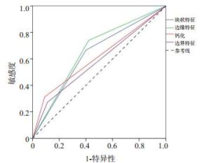
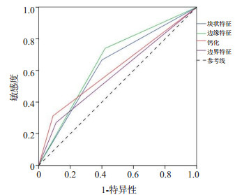
图 1 声像特征鉴别诊断乳腺癌的ROC曲线
Figure 1. ROC curve for differential diagnosis of breast cancer with sound image characteristics.

图 2 血流分级诊断乳腺癌与乳腺良性疾病的ROC曲线
Figure 2. The ROC curve of blood flow classification in the diagnosis of breast cancer and benign breast diseases.
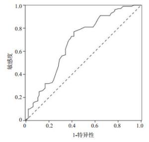
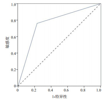
图 3 病灶RI值诊断乳腺癌与乳腺良性疾病的ROC曲线
Figure 3. ROC curve of lesion RI value in diagnosis of breast cancer and benign breast diseases.

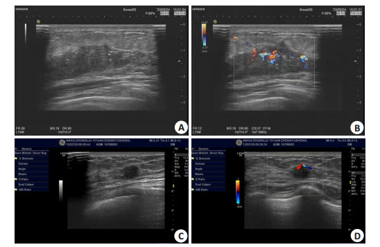
图 4 患者女,48岁,因右侧乳腺包块就诊
A: 化疗前肿块的二维灰阶; B: 化疗前肿块的彩色多普勒血流信号丰富; C: 化疗后包块体积明显缩小; D: 彩色多普勒提示血流信号减少.
Figure 4. A48-year-old female patient was diagnosed with right breast mass.
表 1 两组患者的一般资料分析
Table 1. Analysis of visitors' purchase information (Mean±SD)
组別 年龄(岁) BMl(kg/m2) 患侧分布[n(%)] 肿块直径(cm) 左侧 右侧 病例组(n=96) 46.2±7.5 23.8±2.2 52(54.17) 44(45.83) 1.66±0.74 良性组(n=100) 44.8±6.0 23.6±2.4 46(46.00) 54(54.00) 1.50±0.62 t/χ2 1.446 0.607 1.307 1.643 P 0.150 0.544 0.253 0.102  下载: 导出CSV
表 2 病例组和良性组的声像特征比较
Table 2. Comparison of sound image characteristics between case group and benign group [n(%)]
声像特征 病例组(n=96) 良性组(n=100) χ2 P 肿块形状 13.984 0.000 类圆形、微分叶状 32(33.33) 60(60.00) 不规则性 64(66.67) 40(40.00) 边缘特征 20.490 0.000 光整 25(26.04) 58(58.00) 毛刺 71(73.96) 42(42.00) 钙化灶 15.213 0.000 无 66(68.75) 91(91.00) 微钙化灶 30(31.25) 9(9.00) 边界 8.273 0.004 清晰 70(72.92) 89(89.00) 模糊 26(27.08) 11(11.00) 后方回声 1.595 0.207 增强或无改变 76(79.17) 86(86.00) 衰减 20(20.83) 14(14.00)
下载: 导出CSV
表 3 声像特征鉴别诊断乳腺癌的价值
Table 3. The value of sound image characteristics in differential diagnosis of breast cancer
特征 敏感度(%) 特异性(%) 漏诊率(%) 误诊率(%) AUC 肿块形状 66.67 60.00 33.33 40.00 0.633 边缘 73.96 58.00 26.04 42.00 0.660 钙化灶 31.25 91.00 68.75 9.00 0.611 边界 27.08 89.00 72.92 11.00 0.580
下载: 导出CSV
表 4 病例组和良性组的血流分级特征比较
Table 4. Comparison of blood flow classification characteristics between case group and benign group[n(%)]
组别 0级 Ⅰ级 Ⅱ级 Ⅲ级 病例组(n=96) 9(9.38) 14(14.58) 47(48.96) 26(27.08) 良性组(n=100) 53(53.00) 24(24.00) 11(11.00) 12(12.00) Z -6.982 P 0.000
下载: 导出CSV
-
[1] Marmot MG, Altman DG, Cameron DA, et al. The benefits and harms of breast cancer screening: an independent review[J]. Br J Cancer, 2013, 108(11): 2205-40. doi: 10.1038/bjc.2013.177 [2] Luo WQ, Huang QX, Huang XW, et al. Predicting breast cancer in breast imaging reporting and data system (BI-RADS) ultrasound category 4 or 5 lesions: a nomogram combining radiomics and BI-RADS[J]. Sci Rep, 2019, 9(1): 11921. doi: 10.1038/s41598-019-48488-4 [3] Chen T, Gao H, Guo W, et al. A novel application of the Automated Breast Volume Scanner (ABVS) in the diagnosis of soft tissue tumors[J]. Clin Imaging, 2015, 39(3): 401-7. doi: 10.1016/j.clinimag.2015.01.005 [4] Yeo SH, Kim GR, Lee SH, et al. Comparison of ultrasound elastography and color Doppler ultrasonography for distinguishing small triple-negative breast cancer from fibroadenoma[J]. J Ultrasound Med, 2018, 37(9): 2135-46. doi: 10.1002/jum.14564 [5] Zhu YC, Zu DM, Zhang Y, et al. A comparative study on superb microvascular imaging and conventional ultrasonography in differentiating BI-RADS 4 breast lesions[J]. Oncol Lett, 2019, 18 (3): 3202-10. http://www.ingentaconnect.com/content/sp/ol/2019/00000018/00000003/art00126 [6] 刘海燕, 郑慧. 高频彩色多普勒超声鉴别乳腺良恶性肿瘤: 基于其灰阶声像及血流特征性分析[J]. 中华全科医学, 2019, 17(9): 1551-3. https://www.cnki.com.cn/Article/CJFDTOTAL-SYQY201909034.htm [7] 周笔峰, 邹婧, 杨凤玲. 超声血流及MRI参数与乳腺癌病理特征关联性分析[J]. 新疆医科大学学报, 2020, 43(4): 484-7, 490. doi: 10.3969/j.issn.1009-5551.2020.04.024 [8] Niu JC, Ma JX, Guan XZ, et al. Correlation between Doppler ultrasound blood flow parameters and angiogenesis and proliferation activity in breast cancer[J]. Med Sci Monit, 2019, 25: 7035-41. doi: 10.12659/MSM.914395 [9] 徐敏涛, 李鸿超, 毛爱弟. 彩色多普勒超声检查对女性乳腺恶性病变的诊断价值[J]. 海南医学, 2019, 30(11): 1433-5. doi: 10.3969/j.issn.1003-6350.2019.11.022 [10] 中国抗癌协会乳腺癌专业委员会. 中国抗癌协会乳腺癌诊治指南与规范(2017年版)[J]. 中国癌症杂志, 2017, 27(9): 695-759. https://www.cnki.com.cn/Article/CJFDTOTAL-ZGAZ201709004.htm [11] 马燕, 李晶, 任卫东, 等. 超微血管成像与乳腺肿瘤微血管密度的相关性研究[J]. 临床与病理杂志, 2016, 36(4): 364-9. https://www.cnki.com.cn/Article/CJFDTOTAL-WYSB201604003.htm [12] Fukui K, Masumoto N, Shiroma N, et al. Novel tumor-infiltrating lymphocytes ultrasonography score based on ultrasonic tissue findings predicts tumor-infiltrating lymphocytes in breast cancer [J]. Breast Cancer, 2019, 26(5): 573-80. doi: 10.1007/s12282-019-00958-3 [13] Guo Q, Dong ZW, Zhang L, et al. Ultrasound features of breast cancer for predicting axillary lymph node metastasis[J]. J Ultrasound Med, 2018, 37(6): 1354-3. doi: 10.1002/jum.14469 [14] Wu T, Sultan LR, Tian JW, et al. Machine learning for diagnostic ultrasound of triple-negative breast cancer[J]. Breast Cancer Res Treat, 2019, 173(2): 365-73. doi: 10.1007/s10549-018-4984-7 [15] 刘瑜. 高频彩色多普勒超声检查在乳腺癌患者诊断中的应用价值[J]. 医学临床研究, 2019, 36(2): 324-5, 326. https://www.cnki.com.cn/Article/CJFDTOTAL-ZMYX202002039.htm [16] 雷蕾, 王琪琪, 薛秀秀. 高频彩色多普勒超声在乳腺肿瘤疾病中的诊断价值[J]. 海南医学, 2018, 29(22): 3187-9. doi: 10.3969/j.issn.1003-6350.2018.22.024 [17] 张润, 刘双艳, 李伶俐, 等. 乳腺超声造影与彩色多普勒超声在乳腺肿瘤良恶性诊断中应用[J]. 临床军医杂志, 2018, 46(6): 648-9. https://www.cnki.com.cn/Article/CJFDTOTAL-JYGZ201806012.htm [18] 刘畅, 徐新雅. 高频彩色多普勒超声在早期乳腺癌患者中的表现及诊断价值[J]. 影像研究与医学应用, 2018, 2(13): 113-4. doi: 10.3969/j.issn.2096-3807.2018.13.073 [19] 赵晓乐. 应用高频彩色多普勒超声诊断乳腺癌对降低漏诊误诊的作用分析[J]. 影像研究与医学应用, 2020, 4(22): 123-5. doi: 10.3969/j.issn.2096-3807.2020.22.061 [20] 欧阳燕, 杨腾坚. 探讨乳腺钙化的声像图特征及对乳腺良恶性病变的诊断价值[J]. 养生保健指南, 2019(49): 272. [21] Watanabe T, Kaoku S, Yamaguchi T, et al. Multicenter prospective study of color Doppler ultrasound for breast masses: utility of our color Doppler method[J]. Ultrasound Med Biol, 2019, 45(6): 1367-79. doi: 10.1016/j.ultrasmedbio.2019.01.021 -

 点击查看大图
计量
- 文章访问数: 326
- HTML全文浏览量: 239
- PDF下载量: 6
- 被引次数: 0