


Clinical value of MRI combined with CSF detection in the diagnosis and differential diagnosis of central nervous system infection
-
摘要:
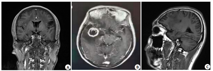
目的探讨MRI联合脑脊液(CSF)检查在诊断和鉴别中枢神经感染中的临床价值。 方法选取2019年1月~2020年5月在我院治疗的中枢神经系统感染性疾病患者151例,其中病毒性脑膜炎84例,化脓性脑膜炎35例,结核性脑膜炎32例,均给予MRI检查,检查CSF中的乳酸脱氢酶(LDH)、蛋白、乳酸和白细胞计数(WBC)。 结果结核性脑膜炎MRI异常检出率高于病毒性脑膜炎和化脓性脑膜炎(P < 0.05);病毒性脑膜炎和化脓性脑膜炎MRI异常检出率比较差异无统计学意义(P > 0.05);病毒性脑膜炎病灶区域脑膜强化检出率高于化脓性脑膜炎和结核性脑膜炎(P < 0.05);化脓性脑膜炎病灶呈环形强化检出率高于病毒性脑膜炎(P < 0.05);结核性脑膜炎颅底脑膜强化检出率高于病毒性脑膜炎和化脓性脑膜炎(P < 0.05);病毒性脑膜炎CSF中LDH、蛋白、乳酸和WBC明显低于化脓性脑膜炎和结核性脑膜炎(P < 0.05);结核性脑膜炎CSF中LDH高于化脓性脑膜炎(P < 0.05),而蛋白和WBC低于化脓性脑膜炎(P < 0.05);筛选出LDH(X1)、蛋白(X2)和WBC(X3)3个指标建立判别函数,即:Y病脑=-0.056X1- 0.065X2-0.062X3+1.168,Y化脑=-0.041X1+0.102X2+0.089X3-1.102,Y结脑=0.112X1-0.057X2-0.078+1.032;MRI联合CSF参数函数诊断病毒性脑膜炎的敏感度、特异性和准确率分别为79.76%、74.63%和77.48%,诊断化脓性脑膜炎的敏感度、特异性和准确率分别为80.00%、81.03%和80.79%,诊断结核性脑膜炎的敏感度、特异性和准确率分别为78.13%、84.87%和83.44%。 结论MRI联合CSF检测在诊断和鉴别中枢神经感染中有较高应用价值,值得临床使用。 Abstract:ObjectiveTo investigate the clinical value of MRI combined with cerebrospinal fluid (CSF) in the diagnosis and differential diagnosis of central nervous system infection. MethodsOne hundred and fifty-one patients with central nervous system infectious diseases treated in our hospital from January 2019 to may 2020 were selected, including 84 cases of viral meningitis, 35 cases of purulent meningitis and 32 cases of tuberculous meningitis. All patients were examined by MRI. The lactate dehydrogenase (LDH), protein, lactic acid (LA) and white blood cell count (WBC) in CSF were examined. ResultsThe abnormal detection rate of MRI in tuberculous meningitis was significantly higher than that in viral meningitis and purulent meningitis (P < 0.05). There was no significant difference in abnormal detection rate of MRI between viral meningitis and purulent meningitis (P > 0.05). The detection rate of meningeal enhancement in viral meningitis was significantly higher than that in purulent meningitis and tuberculous meningitis (P < 0.05). The detection rate of ring enhancement in purulent meningitis was significantly higher than that of viral meningitis (P < 0.05). The detection rate of meningeal enhancement in tuberculous meningitis was significantly higher than that in viral meningitis and purulent meningitis (P < 0.05). The LDH, protein, LA and WBC in CSF of viral meningitis were significantly lower than those in purulent meningitis and tuberculous meningitis (P < 0.05). The LDH in CSF of tuberculous meningitis was significantly higher than that in purulent meningitis (P < 0.05), while the protein and WBC were significantly lower than those in purulent meningitis (P < 0.05). Three indicators, LDH (X1), protein (X2) and WBC (X3), were screened out to establish discriminant functions, namely: Yviral meningitis=-0.056X1-0.065X2-0.062X3+ 1.168, Ypurulent meningitis=-0.041X1 + 0.102X2 + 0.089X3-1.102, Ytuberculous meningitis =0.112X1-0.057X2-0.078 + 1.032. The sensitivity, specificity and accuracy of MRI combined with CSF parameter function in the diagnosis of viral meningitis were 79.76%, 74.63% and 77.48%, the sensitivity, specificity and accuracy of diagnosing purulent meningitis were 80.00%, 81.03% and 80.79%, the sensitivity, specificity and accuracy rate of diagnosing tuberculous meningitis were 78.13%, 84.87% and 83.44%. ConclusionMRI combined with CSF detection has high application value in the diagnosis and differential diagnosis of central nervous system infection. -
表 1 不同疾病类型患者一般资料比较
Table 1. Comparison of general data of patients with different disease types (Mean±SD)
组別 男/女 年龄(岁) BM(kg/m2) 病毒性脑膜炎(n=84) 51/33 32.15±7.32 22.65±2.13 化脓性脑膜炎(n=35) 22/13 34.65±8.03 22.80±3.01 结核性脑膜炎(n=32) 19/13 31.88±8.43 22.50±2.87 F/χ2 0.089 1.203 0.846 P 0.957 0.422 0.911  下载: 导出CSV
表 2 不同疾病类型MRI异常检出率比较
Table 2. Comparison of abnormal detection rate of MRI in different disease types[n (%)]
组別 异常检出率 χ2 P 病毒性脑膜炎(n=84) 33(39.29) 化脓性脑膜炎(n=35) 13(37.14) 7.477 0.024 结核性脑膜炎(n=32) 21(65.63)ab aP < 0.05 vs病毒性脑膜炎;bP < 0.05 vs化脓性脑膜炎.
下载: 导出CSV
表 3 不同疾病类型MRI征象比较
Table 3. Comparison of MRI signs of different disease types[n(%)]
组別 病灶区域脑膜强化 病灶呈环形强化 颅底脑膜强化 病毒性脑膜炎(n=84) 29(34.52) 2(2.38) 2(2.38) 化脓性脑膜炎(n=35) 4(11.43)a 8(22.86)a 1(2.86) 结核性脑膜炎(n=32) 4(12.50)a 2(6.25) 15(46.88)ab χ2 10.286 14.32 47.257 aP < 0.05 vs病毒性脑膜炎;bP < 0.05 vs化脓性脑膜炎.
下载: 导出CSV
表 4 不同疾病类型CSF参数比较
Table 4. Comparison of CSF parameters of different disease types (Mean±SD)
组別 LDH(U/L) 蛋白(g/L) LA(U/L) WBC(×109/L) 病毒性脑膜炎(n=84) 16.68±4.22 1.12±0.32 11.45±2.03 0.22±0.09 化脓性脑膜炎(n=35) 75.51±11.80a 3.65±1.00a 51.15±9.44a 4.49±1.10a 结核性脑膜炎(n=32) 87.62±12.26ab 3.01±0.94ab 52.21±8.94a 1.78±0.72ab F 21.106 10.541 32.211 13.544 P 0.000 0.000 0.000 0.000 aP < 0.05 vs病毒性脑膜炎;bP < 0.05 vs化脓性脑膜炎;LDH:乳酸脱氢酶;LA:乳酸;WBC:白细胞计数.
下载: 导出CSV
表 5 MRI联合CSF诊断价值
Table 5. Diagnostic value of MRI combined with CSF(%)
组別 敏感度 特异性 准确率 病毒性脑膜炎 79.76(67/84) 74.63(50/67) 77.48(117/151) 化脓性脑膜炎 80.00(28/35) 81.03(94/116) 80.79(112/151) 结核性脑膜炎 78.13(25/32) 84.87(101/119) 83.44(126/151) χ2 0.046 2.941 3.932 P 0.977 0.230 0.140 注:联合方法采用并联方式.
下载: 导出CSV
-
[1] Hwang M, Bergmann CC. Intercellular communication is key for protective IFNα/β signaling during viral central nervous system infection[J]. Viral Immunol, 2019, 32(1): 1-6. doi: 10.1089/vim.2018.0101 [2] Agnihotri SP. Central nervous system opportunistic infections[J]. Semin Neurol, 2019, 39(3): 383-90. doi: 10.1055/s-0039-1687842 [3] Zida S, Kolia-Diafouka P, Kania D, et al. Combined testing for Herpes simplex virus and Mycobacterium tuberculosis DNA in cerebrospinal fluid of patients with aseptic meningitis in Burkina Faso, West Africa[J]. J Clin Lab Anal, 2019, 33(3): e22719. DOI: 10.1002/jcla.22719. [4] Tsai WC, Lien CY, Lee JJ, et al. The prognostic factors of HIV-negative adult cryptococcal meningitis with a focus on cranial MRI-based neuroimaging findings[J]. J Clin Neurosci, 2018, 55: 57-61. doi: 10.1016/j.jocn.2018.06.044 [5] Wen L, Zhu YR, Yan M. Clinical presentations and outcomes of post-operative central nervous system infection caused by multidrug- resistant/extensively drug-resistant Acinetobacter baumannii: a retrospective study[J]. Surg Infect, 2019, 20(6): 460-4. doi: 10.1089/sur.2018.286 [6] Tenforde MW, Mokomane M, Leeme TB, et al. Mortality in adult patients with culture-positive and culture-negative meningitis in the Botswana national meningitis survey: a prevalent cohort study[J]. Lancet Infect Dis, 2019, 19(7): 740-9. doi: 10.1016/S1473-3099(19)30066-0 [7] 徐佳佳, 赵年, 刘四斌. CT联合磁共振诊断中枢神经系统感染的临床研究[J]. 中西医结合心脑血管病杂志, 2019, 17(14): 2199-202. doi: 10.12102/j.issn.1672-1349.2019.14.037 [8] 刘兵, 姚长青, 赵树军, 等. 磁共振成像联合脑脊液检测对中枢神经感染的鉴别诊断效果[J]. 中华医院感染学杂志, 2017, 27(13): 2943-6. https://www.cnki.com.cn/Article/CJFDTOTAL-ZHYY201713017.htm [9] Viswanathan S. Scrub typhus meningitis versus acute bacterial meningitis and tuberculous meningitis[J]. Indian Pediatr, 2018, 55 (1): 25-6. http://www.ncbi.nlm.nih.gov/pubmed/29396931 [10] 向旭, 殷洁, 崔莹, 等. 脑脊液可溶性髓细胞触发受体-1与硫化氢在中枢神经系统感染患儿不同发病时期的含量变化及临床意义研究[J]. 中华医院感染学杂志, 2018, 28(9): 1407-10. https://www.cnki.com.cn/Article/CJFDTOTAL-ZHYY201809036.htm [11] 阿迪力·阿布来提, 帕尔哈提·苏里坦, 艾买提江·阿不力米提. 磁共振成像联合脑脊液检测对中枢神经感染的鉴别诊断效果分析[J]. 影像研究与医学应用, 2019, 3(19): 77-8. https://www.cnki.com.cn/Article/CJFDTOTAL-YXYY201919038.htm [12] 赵露, 姜文雯, 张馨, 等. 结核性脑膜炎MRI脑膜病变形态和信号特点及颅内继发性改变特征分析[J]. 脑与神经疾病杂志, 2019, 27(1): 33-7. https://www.cnki.com.cn/Article/CJFDTOTAL-LYSJ201901008.htm [13] van Laarhoven A, Dian S, Aguirre-Gamboa R, et al. Cerebral tryptophan metabolism and outcome of tuberculous meningitis: an observational cohort study[J]. Lancet Infect Dis, 2018, 18(5): 526-35. doi: 10.1016/S1473-3099(18)30053-7 [14] McCarron EP, Sreenivasan S. Importance of CSF lactate concentration in the diagnosis of acute bacterial meningitis[J]. Clin Med: Lond, 2018, 18(4): 351. [15] 伊洁, 关鸿志, 倪安平, 等. 成人疑似病毒性脑炎、脑膜炎患者xTAG脑脊液病毒多重检测结果分析[J]. 现代检验医学杂志, 2019, 34(1): 14-6, 21. https://www.cnki.com.cn/Article/CJFDTOTAL-SXYN201901004.htm [16] 刘乐, 李朋朋, 马萍. 3项指标检测对中枢神经系统感染的鉴别诊断价值[J]. 检验医学与临床, 2018, 15(21): 3200-2. doi: 10.3969/j.issn.1672-9455.2018.21.011 [17] 王玖红. 脑脊液生化指标对中枢神经系统性疾病患者病情严重程度的评估价值[J]. 检验医学与临床, 2019, 16(20): 3043-5. doi: 10.3969/j.issn.1672-9455.2019.20.040 [18] 李轲, 谢付静, 杨亚培, 等. 单独及联合检测脑脊液中3种标志物对儿童病毒性脑炎与不典型化脓性脑炎的鉴别诊断价值[J]. 中华实用诊断与治疗杂志, 2018, 32(4): 375-7. https://www.cnki.com.cn/Article/CJFDTOTAL-HNZD201804020.htm [19] 张勤, 张帆, 崔凯, 等. LDH、CSFβ2-MG及中性粒细胞CD64对老年患者中枢神经系统感染诊断价值[J]. 中国病原生物学杂志, 2018, 13(10): 1155-7, 1165. https://www.cnki.com.cn/Article/CJFDTOTAL-ZISC201810022.htm [20] 吴长金, 陈兵. 脑脊液生化指标对结核性脑膜炎诊断效能评价[J]. 中国临床医生杂志, 2019, 47(8): 935-7. https://www.cnki.com.cn/Article/CJFDTOTAL-ZLYS201908019.htm [21] 曲慧. 小儿急性中枢神经系统病毒感染临床特征及影响预后的相关因素分析[J]. 陕西医学杂志, 2017, 46(1): 17-9. https://www.cnki.com.cn/Article/CJFDTOTAL-SXYZ201701007.htm [22] 任会丽, 方伟军, 韩远远. 头颅MRI增强扫描与脑脊液检查对婴幼儿颅内结核的早期诊断价值[J]. 分子影像学杂志, 2020, 43 (2): 304-8. doi: 10.12122/j.issn.1674-4500.2020.02.26 -

 点击查看大图
计量
- 文章访问数: 253
- HTML全文浏览量: 81
- PDF下载量: 3
- 被引次数: 0