


Epicardial adipose tissue measured by computerized tomography is a risk factor for troponin elevation in patients with paroxysmal supraventricular tachycardia
-
摘要:
目的探讨CT测量心外膜脂肪组织(EAT)体积是否为阵发性室上速(PSVT)患者肌钙蛋白I(cTnI)阳性的危险因素。 方法选取2014年6月~2020年8月因“阵发性室上速”就诊且在院期间有检测cTnI及心脏CT的患者为研究对象(n=30),根据cTnI水平将患者分为cTnI阳性组(n=10)和cTnI阴性组(n=20),比较其临床和人口学特征、EAT体积、冠心病的发生率。分析EAT体积是否为PSVT患者cTnI阳性的相关危险因素。将没有冠心病的PSVT患者分为cTnI阳性组和cTnI阴性组,比较其EAT体积。 结果对比于发作PSVT时cTnI阴性的患者,cTnI阳性的患者EAT体积更大(P=0.032)。EAT体积是PSVT患者cTnI阳性的相关危险因素(r=0.351,P=0.028)。没有确诊冠心病的PSVT患者中cTnI阳性组EAT体积与cTnI阴性组差异无统计学意义(P=0.062)。 结论发作PSVT时cTnI阳性的患者EAT体积更大,临床上应警惕发作PSVT时cTnI阳性的患者。且EAT体积是PSVT患者cTnI阳性的相关危险因素,这为改善cTnI阳性PSVT患者的预后提供参考资料。cTnI阳性的PSVT患者即使暂未确诊冠心病,但需更高频次的随访,警惕心血管疾病的发生。 -
关键词:
- 心外膜脂肪组织 /
- 阵发性室上性心动过速 /
- 肌钙蛋白I /
- 冠心病
Abstract:ObjectiveTo investigate whether the volume of Epicardial Adipose Tissue (EAT) measured by CT is a risk factor of Cardiac Troponin I (cTnI) elevation in patients with acute Paroxysmal Supraventricular Tachycardia (PSVT). MethodsThe objecters were patients with acute PSVT received treatment in Peking University Shenzhen Hospital from June 2014 to August 2020(n=30). The cTnI and CT Scan of the Heart were detected during the hospitalization. All the patients were divided into the cTnI positive group and the cTnI negative group. Their clinical and demographic characteristics, EAT volume, the incidence of CHD were compared. PSVT patients without CHD were divided into cTnI positive group and cTnI negative group, and their EAT volume was compared. ResultsCompared with the patients with cTnI negative at the onset of PSVT, the patients with cTnI elevation had a larger EAT volume (P=0.032). EAT volume was a related risk factor for cTnI elevation in patients with acute PSVT(r=0.351, P=0.028). In PSVT patients without diagnosed CHD, the EAT volume of cTnI positive group made no difference with that of cTnI negative group (P=0.062). ConclusionThough patients with cTnI elevation during the attack of PSVT had lager EAT volume, they should be on the alert. In addition, EAT volume is a related risk factor for cTnI elevation in PSVT patients, which provides reference information for improving the prognosis. PSVT patients with cTnI positive need more frequent follow-up to object the occurrence of cardiovascular diseases, even if CHD has not been confirmed for the time being. -

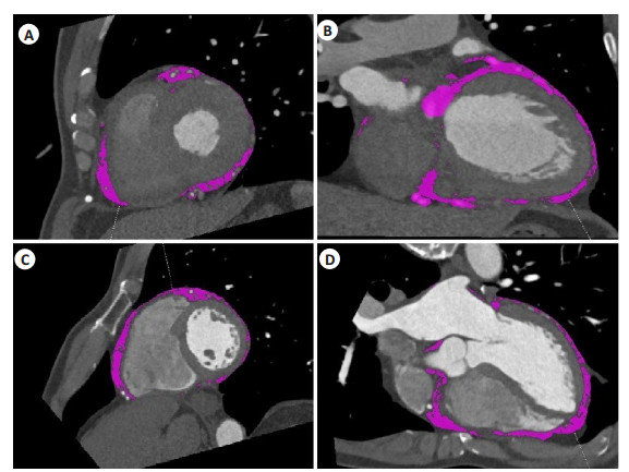
图 1 CT测量发作PSVT时cTnI阳性和cTnI阴性患者EAT体积
A~B: 发作PSVT时cTnI阳性患者EAT体积测量图;C~D: 发作PSVT时cTnI阴性患者EAT体积测量图.
Figure 1. The EAT volume measured by CT in cTnI positive and cTnI negative patients with Paroxysmal Supraventricular Tachycardia.
表 1 发作PSVT时cTnI阳性和cTnI阴性患者临床和人口学特征比较
Table 1. Comparation of clinical and demographic characteristics in cTnI positive/negative patients with Paroxysmal Supraventricular Tachycardia (Mean±SD)
变量 cTnI阳性(n=10) cTnI阴性(n=20) P 年龄(岁) 60.00±19.00 55.95±10.81 0.544 女性[n(%)] 4(40) 11(55) 1 高血压[n(%)] 6(60) 10(50) 0.715 血红蛋白浓度(g/L) 125.80±18.26 133.35±13.76 0.215 肌酐(μmol/L) 151.10±157.13 70.40±16.21 0.139 糖化血红蛋白(%) 5.89±0.89 5.54±0.32 0.126 尿酸(μmol/L) 368.30±109.73 302.35±76.36 0.064 促甲状腺激素(mU/L) 2.64±2.45 2.39±1.30 0.716 高密度脂蛋白(mmol/L) 1.10±0.22 1.19±0.32 0.402 低密度脂蛋白(mmol/L) 2.84±0.75 2.84±1.03 0.998 冠心病发病率[n(%)] 4(40) 6(30) 0.068 心外膜脂肪组织(cm2) 67.67±13.89 57.36±10.66 0.032  下载: 导出CSV
-
[1] 黄卫斌. 窄QRS心动过速的诊断思考[J]. 内科急危重症杂志, 2016, 22(6): 418-20. https://www.cnki.com.cn/Article/CJFDTOTAL-NKJW201606007.htm [2] Zellweger MJ, Schaer BA, Cron TA, et al. Elevated troponin levels in absence of coronary artery disease after supraventricular tachycardia[J]. Swiss Med Wkly, 2003, 133(31/32): 439-41. http://europepmc.org/abstract/MED/14562187 [3] Ben Yedder N, Roux JF, Paredes FA. Troponin elevation in supraventricular tachycardia: primary dependence on heart rate[J]. Can J Cardiol, 2011, 27(1): 105-9. doi: 10.1016/j.cjca.2010.12.004 [4] Carlberg DJ, Tsuchitani S, Barlotta KS, et al. Serum troponin testing in patients with paroxysmal supraventricular tachycardia: outcome after ED care[J]. Am J Emerg Med, 2011, 29(5): 545-8. doi: 10.1016/j.ajem.2010.01.041 [5] Chow GV, Hirsch GA, Spragg DD, et al. Prognostic significance of cardiac troponin I levels in hospitalized patients presenting with supraventricular tachycardia[J]. Medicine, 2010, 89(3): 141-8. doi: 10.1097/MD.0b013e3181dddb3b [6] Gaborit B, Sengenes C, Ancel P, et al. Role of epicardial adipose tissue in health and disease: a matter of fat?[J]. Compr Physiol, 2017, 7(3): 1051-82. http://www.tandfonline.com/servlet/linkout?suffix=CIT0026&dbid=8&doi=10.1080%2F17446651.2018.1500894&key=28640452 [7] Mazurek T, Zhang L, Zalewski A, et al. Human epicardial adipose tissue is a source of inflammatory mediators[J]. Circulation, 2003, 108(20): 2460-6. doi: 10.1161/01.CIR.0000099542.57313.C5 [8] Sacks HS, Fain JN. Human epicardial fat: what is new and what is missing?[J]. Clin Exp Pharmacol Physiol, 2011, 38(12): 879-87. doi: 10.1111/j.1440-1681.2011.05601.x [9] Nagy E, Jermendy AL, Merkely B, et al. Clinical importance of epicardial adipose tissue[J]. Arch Med Sci, 2017, 13(4): 864-74. http://europepmc.org/abstract/MED/28721155 [10] Page RL, Joglar JA, Caldwell MA, et al. 2015 ACC/AHA/HRS guideline for the management of adult patients with supraventricular tachycardia: a report of the American college of cardiology/ American heart association task force on clinical practice guidelines and the heart rhythm society[J]. J Am Coll Cardiol, 2016, 67(13): e27-e115. http://www.sciencedirect.com/science/article/pii/S0735109717307933 [11] Fox DJ, Tischenko A, Krahn AD, et al. Supraventricular tachycardia: diagnosis and management[J]. Mayo Clin Proc, 2008, 83(12): 1400-11. doi: 10.4065/83.12.1400 [12] Kanjwal K, Imran N, Grubb B, et al. Troponin elevation in patients with various tachycardias and normal epicardial coronaries[J]. Indian Pacing Electrophysiol J, 2008, 8(3): 172-4. http://onlinelibrary.wiley.com/resolve/reference/PMED?id=18679521 [13] Schueler M, Vafaie M, Becker R, et al. Prevalence, kinetic changes and possible reasons of elevated cardiac troponin T in patients with AV nodal re-entrant tachycardia[J]. Acute Cardiac Care, 2012, 14 (4): 131-7. doi: 10.3109/17482941.2012.741246 [14] Xue F, Jiang TB, Jiang B, et al. Cardiac troponin I elevation with supraventricular tachycardia: two case reports and review of the literature[J]. BMC Res Notes, 2014, 7: 136. doi: 10.1186/1756-0500-7-136 [15] Sayadnik M, Shafiee A, Jenab Y, et al. Predictors of high-sensitivity cardiac troponin T elevation in patients with acute paroxysmal supraventricular tachycardia and ischemic heart disease[J]. Tex Heart Inst J, 2017, 44(5): 306-11. doi: 10.14503/THIJ-15-5338 [16] Murer M, Cuculi F, Toggweiler S, et al. Elevated high-sensitivity troponin does not indicate the presence of coronary artery disease in patients presenting with supraventricular tachycardia[J]. Cardiol J, 2017, 24(6): 642-8. doi: 10.5603/CJ.a2017.0058 [17] Kozinski M, Krintus M, Kubica J, et al. High-sensitivity cardiac troponin assays: From improved analytical performance to enhanced risk stratification[J]. Crit Rev Clin Lab Sci, 2017, 54(3): 143-72. doi: 10.1080/10408363.2017.1285268 [18] Vrsalovic M. Prognostic effect of cardiac troponin elevation in acute aortic dissection: a meta-analysis[J]. Int J Cardiol, 2016, 214: 277-8. doi: 10.1016/j.ijcard.2016.03.230 [19] Vallabhajosyula S, Sakhuja A, Geske JB, et al. Role of admission troponin-T and serial troponin-T testing in predicting outcomes in severe Sepsis and septic shock[J]. J Am Heart Assoc, 2017, 6(9): 005930. http://europepmc.org/articles/PMC5634261/ [20] Ghersin I, Zahran M, Azzam ZS, et al. Prognostic value of cardiac troponin levels in patients presenting with supraventricular tachycardias[J]. J Electrocardiol, 2020, 62: 200-3. doi: 10.1016/j.jelectrocard.2020.09.001 [21] Sacks HS, Fain JN. Human epicardial adipose tissue: a review[J]. Am Heart J, 2007, 153(6): 907-17. doi: 10.1016/j.ahj.2007.03.019 [22] Rosito GA, Massaro JM, Hoffmann U, et al. Pericardial fat, visceral abdominal fat, cardiovascular disease risk factors, and vascular calcification in a community-based sample: the Framingham Heart Study[J]. Circulation, 2008, 117(5): 605-13. doi: 10.1161/CIRCULATIONAHA.107.743062 [23] Russo R, Di Iorio B, Di Lullo L, et al. Epicardial adipose tissue: new parameter for cardiovascular risk assessment in high risk populations[J]. J Nephrol, 2018, 31(6): 847-53. doi: 10.1007/s40620-018-0491-5 [24] Fitzgibbons TP, Czech MP. Epicardial and perivascular adipose tissues and their influence on cardiovascular disease: basic mechanisms and clinical associations[J]. J Am Heart Assoc, 2014, 3: e000582. http://europepmc.org/articles/PMC4187500/ [25] Ghersin I, Zahran M, Azzam ZS, et al. Prognostic value of cardiac troponin levels in patients presenting with supraventricular tachycardias[J]. J Electrocardiol, 2020, 62: 200-3. doi: 10.1016/j.jelectrocard.2020.09.001 [26] Nishio S, Kusunose K, Yamada H, et al. Echocardiographic epicardial adipose tissue thickness is associated with symptomatic coronary vasospasm during provocative testing[J]. J Am Soc Echocardiogr, 2017, 30(10): 1021-7. e1. doi: 10.1016/j.echo.2017.06.024 [27] Kim MN, Kim HL, Park SM, et al. Association of epicardial adipose tissue with coronary spasm and coronary atherosclerosis in patients with chest pain: analysis of data collated by the KoRean wOmen'S chest pain rEgistry (koROSE)[J]. Heart Vessels, 2018, 33 (1): 17-24. doi: 10.1007/s00380-017-1029-9 -

 点击查看大图
图(1) / 表(1)
计量
- 文章访问数: 495
- HTML全文浏览量: 216
- PDF下载量: 11
- 被引次数: 0