


Value of macular morphological changes in early diagnosis of diabetic retinopathy
-
摘要:
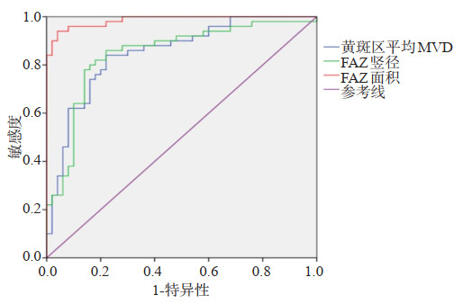
目的探讨黄斑区形态改变在糖尿病视网膜病变(DR)早期诊断中的价值。 方法选取2018年1月~2019年6月在我院治疗的2型DR患者108例,其中非增殖性DR(NPDR组)68例,增殖性DR(PDR组)40例,同时选取单纯2型糖尿病患者50例作为对照,各组均行光学相干断层扫描血流成像(OCTA)检查,比较各组黄斑区血流密度(MVD)、黄斑中心凹无血管区(FAZ)及黄斑视网膜厚度差异。 结果NPDR组和PDR组黄斑区MVD分别为(0.48±0.02)%和(0.47±0.03)%,低于对照组(P < 0.05);NPDR组和PDR组FAZ竖径分别为0.37±0.03 mm和0.38±0.02 mm,高于对照组(P < 0.05);PDR组FAZ面积为0.49±0.04 mm2,高于对照组和NPDR组(P < 0.05);对照组、NPDR组和PDR组黄斑区平均视网膜厚度的差异无统计学意义(P>0.05);黄斑区MVD、FAZ竖径、FAZ面积诊断NPDR和PDR的ROC曲线下面积分别为0.845、0.840和0.922(P < 0.05),截断值分别为0.50%、0.37 mm和0.43 mm2,敏感度分别为80.50%、79.80%和88.50%,特异性分别为78.00%、75.00%和84.00%。 结论DR患者黄斑区MVD降低,而FAZ竖径及面积扩大,在诊断中有一定应用价值。 -
关键词:
- 糖尿病视网膜病变 /
- 黄斑区 /
- 光学相干断层扫描血流成像 /
- 诊断价值
Abstract:ObjectiveTo investigate the value of macular morphological changes in the early diagnosis of diabetic retinopathy (DR). MethodsA total of 108 patients with type 2 DR treated in our hospital from January 2018 to June 2019 were selected. There were 68 cases of non proliferative DR (NPDR group), 40 cases of proliferative DR (PDR group) and 50 cases of simple type 2 diabetes mellitus (DM group). The optical coherence tomography blood flow imaging (OCTA) was performed in each group, the blood flow density (MVD), macular fovea avascular area (FAZ) and macular retinal thickness were compared among the groups. ResultsThe MVD of macular area in NPDR group and PDR group were (0.48±0.02)% and (0.47±0.03)%, which were significantly lower than those in DM group (P < 0.05). The vertical diameter of FAZ in NPDR group and PDR group were 0.37±0.03 mm and 0.38±0.02 mm respectively, which were significantly higher than those in DM group (P < 0.05). The area of FAZ in PDR group was 0.49±0.04 mm2, which was significantly higher than that in DM group and NPDR group (P < 0.05). There was no significant difference of mean macular retinal thickness between DM group, NPDR group and PDR group (P>0.05). The area under ROC curve of MVD, FAZ vertical diameter and FAZ area in the diagnosis of NPDR and PDR were 0.845, 0.840 and 0.922, respectively (P < 0.05). The cut-off values were 0.50%, 0.37 mm and 0.43 mm2, respectively. The sensitivity were 80.50%, 79.80% and 88.50%, and the specificity were 78.00%, 75.00% and 84.00%, respectively. ConclusionIn DR patients, the MVD of macular area decreases, while the vertical diameter and area of FAZ are enlarged, which has certain application value in the diagnosis. -
Key words:
- diabetic retinopathy /
- macular area /
- optical coherence tomography /
- diagnostic value
-

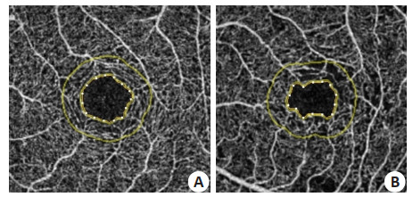
图 1 黄斑区图像
A: 单纯2型糖尿病患者, 可见黄斑区血管密度稀疏, FAZ边缘较规则; B: DR患者, 可见黄斑区血管密度更加稀疏, FAZ边缘不规则.
Figure 1. Macular area image.
表 1 临床资料比较
Table 1. Comparison of clinical data of each group (Mean±SD)
组别 男/女(n) 年龄(岁) 糖尿病病程(年) 空腹血糖(mmol/L) DM组(n=50) 30/20 64.40±5.59 6.39±1.10 6.10±1.04 NPDR组(n=68) 39/29 65.10±6.60 10.20±1.05a 6.03±1.00 PDR组(n=40) 22/18 65.03±6.12 11.05±1.43a 6.15±1.01 F/χ2 0.230 1.022 8.281 0.672 P 0.891 0.562 0.000 0.722 aP < 0.05 vs DM组.  下载: 导出CSV
表 2 各组黄斑区MVD比较
Table 2. Comparison of macular MVD in each group
组别 黄斑区平均MVD(Mean±SD, %) F P DM组(n=50) 0.52±0.03 12.281 0.000 NPDR组(n=68) 0.48±0.02a PDR组(n=40) 0.47±0.03a aP < 0.05 vs DM组; MVD: 黄斑区血流密度.
下载: 导出CSV
表 3 各组FAZ竖径、横径及面积比较
Table 3. Comparison of vertical diameter, transverse diameter and area of FAZ in each group (Mean±SD)
组别 竖径(mm) 横径(mm) 面积(mm2) DM组(n=50) 0.35±0.02 0.35±0.03 0.40±0.02 NPDR组(n=68) 0.37±0.03a 0.36±0.03 0.44±0.03a PDR组(n=40) 0.38±0.02a 0.36±0.02 0.49±0.04ab F 7.271 0.998 8.203 P 0.000 0.601 0.000 aP < 0.05 vs DM组; bP < 0.05 vs NPDR组.
下载: 导出CSV
表 4 各组黄斑区视网膜厚度比较
Table 4. Comparison of macular retinal thickness in each group (Mean±SD)
组别 黄斑区平均视网膜厚度(mm) F P DM组(n=50) 244.19±11.26 1.442 0.343 NPDR组(n=68) 245.50±12.14 PDR组(n=40) 244.80±12.03
下载: 导出CSV
-
[1] Das R, Spence G, Hogg RE, et al. Disorganization of inner Retina and outer retinal morphology in diabetic macular edema[J]. J Am MedAssco Ophthalmol, 2018, 136(2): 202-8. http://www.ncbi.nlm.nih.gov/pubmed/29327033 [2] Mercuri S, Davis B, Yap T, et al. Longitudinal morphological assessment of macular changes in Glaucoma[J]. Acta Ophthalmol, 2019, 97(S263): 76-7. doi: 10.1111/j.1755-3768.2019.5211 [3] Mete M, Alfano A, Maggio E, et al. Inverted ILM flap for the treatment of myopic macular holes: healing processes and morphological changes in comparison with complete ILM removal [J]. J Ophthalmol, 2019(8): 1-8. http://www.researchgate.net/publication/333576994_Inverted_ILM_Flap_for_the_Treatment_of_Myopic_Macular_Holes_Healing_Processes_and_Morphological_Changes_in_Comparison_with_Complete_ILM_Removal [4] Yang QH, Zhang Y, Zhang XM, et al. Prevalence of diabetic retinopathy, proliferative diabetic retinopathy and non-proliferative diabetic retinopathy in Asian T2DM patients: a systematic review and Metaanalysis[J]. Int J Ophthalmol, 2019, 12(2): 302-11. http://kns.cnki.net/KCMS/detail/detail.aspx?dbcode=CJFD&filename=GYZZ201902019 [5] Brondani LA, Crispim D, Pisco J, et al. The G allele of the rs12050217 polymorphism in the BDKRB1 gene is associated with protection for diabetic retinopathy[J]. Curr Eye Res, 2019, 44(9): 994-9. doi: 10.1080/02713683.2019.1610178 [6] Lu Y, Simonett JM, Wang J, et al. Evaluation of automatically quantified foveal avascular zone metrics for diagnosis of diabetic retinopathy using optical coherence tomography angiography[J]. Invest Ophthalmol Vis Sci, 2018, 59(6): 2212-21. doi: 10.1167/iovs.17-23498 [7] 王茜, 陈冰. 糖尿病视网膜病变黄斑区血流密度和黄斑中心凹无血管区面积的变化及其意义[J]. 医学临床研究, 2020, 37(2): 255-8. http://www.wanfangdata.com.cn/details/detail.do?_type=perio&id=hnyx202002030 [8] 程泉. OCTA对T2MD患者FAZ形态学、视网膜血流密度及厚度的研究[D]. 昆明: 昆明医科大学, 2018. [9] Sahlsten J, Jaskari J, Kivinen J, et al. Deep learning fundus image analysis for diabetic retinopathy and macular edema grading[J]. Sci Rep, 2019, 9(1): 10750-2. doi: 10.1038/s41598-019-47181-w [10] 中华医学会糖尿病学分会. 中国2型糖尿病防治指南(2013年版[) J]. 中国糖尿病杂志, 2014, 22(8): 2-42. https://www.cnki.com.cn/Article/CJFDTOTAL-ZGTL201408030.htm [11] 许迅. 糖尿病性视网膜病变新的国际临床分型[J]. 上海医学, 2005, 28(1): 8-9. doi: 10.3969/j.issn.0253-9934.2005.01.003 [12] 韩秀香. 眼科影像学检查在早期诊断及治疗糖尿病视网膜病变中的作用价值[J]. 影像研究与医学应用, 2020, 4(18): 85-6. https://www.cnki.com.cn/Article/CJFDTOTAL-YXYY202018041.htm [13] Liu L, JianGao, Bao WL, et al. Analysis of foveal microvascular abnormalities in diabetic retinopathy using optical coherence tomography angiography with projection artifact removal[J]. J Ophthalmol, 2018(1): 1-9. http://www.researchgate.net/publication/327736099_Analysis_of_Foveal_Microvascular_Abnormalities_in_Diabetic_Retinopathy_Using_Optical_Coherence_Tomography_Angiography_with_Projection_Artifact_Removal [14] Han W, Wei H, Kong W, et al. Association between retinol binding protein 4 and diabetic retinopathy among type 2 diabetic patients: a meta-analysis[J]. Acta Diabetol, 2020, 57(10): 1203-18. doi: 10.1007/s00592-020-01535-3 [15] Lynch G, Romo JSA, Linderman R, et al. Within-subject assessment of foveal avascular zone enlargement in different stages of diabetic retinopathy using en face OCT reflectance and OCT angiography[J]. Biomed Opt Express, 2018, 9(12): 5982-96. doi: 10.1364/BOE.9.005982 [16] Bates NM, Tian J, Smiddy WE, et al. Relationship between the morphology of the foveal avascular zone, retinal structure, and macular circulation in patients with diabetes mellitus[J]. Sci Rep, 2018, 8(1): 5355-6. doi: 10.1038/s41598-018-23604-y [17] Thompson IA, Durrani AK, Patel S. Optical coherence tomography angiography characteristics in diabetic patients without clinical diabetic retinopathy[J]. Eye (Lond), 2019, 33(4): 648-52. doi: 10.1038/s41433-018-0286-x [18] 王健, 陈松. 非增生期糖尿病视网膜病变黄斑微血管形态学改变的相干光断层扫描血管成像观察[J]. 中国医师杂志, 2018, 20(8): 1132- 4, 1138. doi: 10.3760/cma.j.issn.1008-1372.2018.08.004 [19] AttaAllah HR, Mohamed AAM, Ali MA. Macular vessels density in diabetic retinopathy: quantitative assessment using optical coherence tomography angiography[J]. Int Ophthalmol, 2019, 39 (8): 1845-59. doi: 10.1007/s10792-018-1013-0 [20] 邬嘉蔚, 柯晓云, 符敏. 光相干断层扫描血管成像在糖尿病视网膜病变诊断中的应用研究进展[J]. 中华眼底病杂志, 2018, 34(1): 86-9. doi: 10.3760/cma.j.issn.1005-1015.2018.01.026 [21] 李艳, 叶舒, 罗雪英, 等. 眼底血管荧光造影与光学相干断层扫描在糖尿病性黄斑水肿临床诊断中的应用价值比较[J]. 广西医学, 2020, 42(5): 548-50. https://www.cnki.com.cn/Article/CJFDTOTAL-GYYX202005007.htm [22] 雷颖庆, 周琦, 段成霞, 等. OCTA定量分析糖尿病视网膜病变患者黄斑区微血管改变[J]. 眼科新进展, 2020, 40(2): 161-4. https://www.cnki.com.cn/Article/CJFDTOTAL-XKJZ202002015.htm -

 点击查看大图
计量
- 文章访问数: 591
- HTML全文浏览量: 382
- PDF下载量: 5
- 被引次数: 0