


Value of spiral CT in the diagnosis of preoperative staging in elderly patients with gastric cancer
-
摘要:
目的探讨螺旋CT诊断老年胃癌术前分期的价值。 方法选取2017年1月~2020年1月在我院治疗的老年胃癌患者110例,在我院行螺旋CT、胃镜检查,分析螺旋CT、胃镜胃癌检出率差异,并检验螺旋CT术前T、N分期与病理 结果一致性。 结果螺旋CT和胃镜胃癌检出率分别为92.73%和95.45%,差异无统计学意义(P>0.05);螺旋CT术前T分期与术后病理诊断一致性Kappa值为0.893(P < 0.05),准确率92.73%;螺旋CT术前N分期与术后病理诊断一致性Kappa值为0.927(P < 0.05),准确率95.45%。 结论螺旋CT与胃镜诊断老年胃癌均有较好的效果,其中螺旋CT术前T、N分期判断有较好的价值。 Abstract:ObjectiveTo explore the value of spiral CT in the diagnosis of preoperative staging of senile gastric cancer. MethodsA total 110 elderly patients with gastric cancer treated in our hospital from January 2017 to January 2020 were selected. Spiral CT and gastroscopy were performed in our hospital, the difference of spiral CT, upper gastric cancer detection rates were analyzed. And the consistency of spiral CT preoperative T and N staging and pathologic results was examined. ResultsThe detection rates of gastric cancer by spiral CT and gastroscopy were 92.73% and 95.45% respectively and the difference was not statistically significant (P>0.05). The Kappa value of consistent with the preoperative T staging of spiral CT and postoperative pathological diagnosis was 0.893 (P < 0.05), the accuracy rate was 92.73%. The Kappa value of consistent with the preoperative N staging of spiral CT and postoperative pathological diagnosis was 0.927 (P < 0.05), the accuracy rate was 95.45%. ConclusionSpiral CT and gastroscopy have good results in the diagnosis of gastric cancer in the elderly, among which spiral CT has a good value in preoperative T and N stage. -
Key words:
- spiral CT /
- gastroscope /
- gastric cancer /
- old age /
- diagnostic value
-

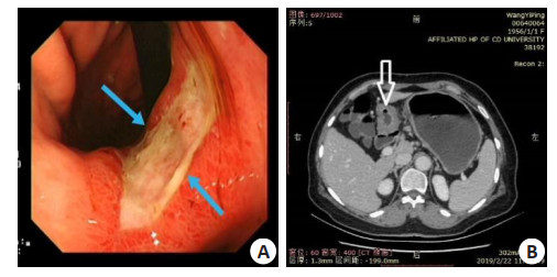
图 1 某患者胃镜及螺旋CT影像学表现
A: 胃镜; B: 螺旋CT; 图中箭头为病灶.
Figure 1. Gastroscopy and spiral CT image of a patient.
表 1 螺旋CT和胃镜检出情况比较[n(%)]
Table 1. Comparison between spiral CT and gastroscope
检查方法 检出率 χ2 P 螺旋CT 102(92.73) 0.736 0.391 胃镜 105(95.45)  下载: 导出CSV
表 2 螺旋CT术前T分期判断情况
Table 2. Preoperative T staging of spiral CT
螺旋CT 病理诊断 Kappa P T1 T2 T3 T4 T1 7 1 0 0 0.893 0.000 T2 1 32 3 0 T3 0 1 43 2 T4 0 0 0 20
下载: 导出CSV
表 3 螺旋CT术前N分期判断情况
Table 3. Preoperative N staging of spiral CT
螺旋CT 病理诊断 Kappa P N0 N1 N2 N0 24 3 0 0.927 0.000 N1 2 25 0 N2 0 0 56
下载: 导出CSV
-
[1] 温晓媛. 胃癌术后病理分析对早诊断的警示作用[J]. 中国药物与临床, 2019, 19(10): 1677-8. https://www.cnki.com.cn/Article/CJFDTOTAL-YWLC201910044.htm [2] Areia M, Spaander MC, Kuipers EJ, et al. Endoscopic screening for gastric cancer: a cost-utility analysis for countries with an intermediate gastric cancer risk[J]. United European Gastroenterol J, 2018, 6(2): 192-202. doi: 10.1177/2050640617722902 [3] Hirasawa T, Aoyama K, Tanimoto T, et al. Application of artificial intelligence using a convolutional neural network for detecting gastric cancer in endoscopic images[J]. Gastric Cancer, 2018, 21(4): 653-60. doi: 10.1007/s10120-018-0793-2 [4] 李文娟, 张霁雯, 罗酩, 等. C-13和胃蛋白酶原及肿瘤标志物联合检测在胃癌早期诊断中的作用研究[J]. 中国肿瘤临床与康复, 2018, 25 (9): 1069-72. https://www.cnki.com.cn/Article/CJFDTOTAL-ZGZK201809013.htm [5] 朱文杰, 孙晓林, 刘颖. 窄带成像技术联合高清放大胃镜在诊断早期胃癌中的价值分析[J]. 现代消化及介入诊疗, 2019, 24(2): 214-5, 218. https://www.cnki.com.cn/Article/CJFDTOTAL-XDXH201902029.htm [6] 谭冬梅, 成小蓉. 胃镜下活体组织检查与外科手术病理诊断胃癌的价值对比分析[J]. 结直肠肛门外科, 2018, 24(S2): 19-20. https://www.cnki.com.cn/Article/CJFDTOTAL-DCGM2018S2015.htm [7] Virgilio E, Giarnieri E, Giovagnoli MR, et al. Gastric juice MicroRNAs as potential biomarkers for screening gastric cancer: a systematic review[J]. Anticancer Res, 2018, 38(2): 613-6. http://www.researchgate.net/publication/322863781_Gastric_Juice_MicroRNAs_as_Potential_Biomarkers_for_Screening_Gastric_Cancer_A_Systematic_Review [8] 李航, 吴锷, 刘盛, 等. 胃癌患者术前胃镜活检病理与外科术后病理诊断对照分析[J]. 中国实验诊断学, 2018, 22(9): 1541-2. doi: 10.3969/j.issn.1007-4287.2018.09.016 [9] Pérez-Antón E, Egui A, Thomas MC, et al. Impact of benznidazole treatment on the functional response of Trypanosoma cruzi antigenspecific CD4+CD8+ T cells in chronic Chagas disease patients[J]. PLoS Negl Trop Dis, 2018, 12(5): e0006480. doi: 10.1371/journal.pntd.0006480 [10] Brenkman HJF, van Putten M, Visser E, et al. Timing of postoperative chemotherapy in patients undergoing perioperative chemotherapy and gastrectomy for gastric cancer[J]. Surg Oncol, 2018, 27(3): 421-7. doi: 10.1016/j.suronc.2018.05.026 [11] Honda S, Furukawa K, Nishiwaki N, et al. Risk factors for postoperative delirium after gastrectomy in gastric cancer patients [J]. World J Surg, 2018, 42(11): 3669-75. doi: 10.1007/s00268-018-4682-y [12] Egbe TO, Kobenge FM, Arlette MMJ, et al. Pyosalpinges after hysterosalpingography in a patient with lower genital tract infection and managed by laparoscopic surgery in a resource low tertiary hospital case report and literature review[J]. Fertil Res Pract, 2018, 4 (1): 1-8. doi: 10.1186/s40738-018-0046-4 [13] Cho BJ, Bang CS, Park SW, et al. Automated classification of gastric neoplasms in endoscopic images using a convolutional neural network. Endoscopy[J]. 2019, 51(12): 1121-9. [14] Alvarado-Cabrero I, Gil-Hernández S, Ruelas-Perea A, et al. Immunohistochemical assessment of HER2 expression in gastric cancer. A clinicopathologic study of 93 cases[J]. Cir Cir, 2017, 85 (6): 504-9. [15] Saito H, Kono Y, Murakami Y, et al. Highly activated PD-1/PD-L1 pathway In gastric cancer with PD-L1 expression[J]. Anticancer Res, 2018, 38(1): 107-12. http://smartsearch.nstl.gov.cn/paper_detail.html?id=bf0e8fc033a5a44b21ce7351aa0a844d [16] Sumer F, Karakas S, Gundogan E, et al. Totally laparoscopic resection and extraction of specimens via transanal route in synchronous colon and gastric cancer[J]. G Chir, 2018, 39(2): 82-6. http://europepmc.org/abstract/MED/29694306 [17] Imamura T, Komatsu S, Ichikawa D, et al. Reconstruction method as an independent risk factor for postoperative bone mineral density loss in gastric cancer[J]. J Gastroenterol Hepatol, 2018, 33(2): 418- 25. doi: 10.1111/jgh.13910 [18] 刘红芬, 王征, 王莉, 等. 肿瘤标记物对胃癌诊断和预后判断的价值以及与免疫炎性反应指标的相关性[J]. 河北医科大学学报, 2019, 40 (7): 855-8. doi: 10.3969/j.issn.1007-3205.2019.07.026 [19] 祖明立. 胃镜下活体组织检查与外科手术病理诊断胃癌的效果比较[J]. 河北医药, 2019, 41(4): 557-9, 563. https://www.cnki.com.cn/Article/CJFDTOTAL-HBYZ201904019.htm [20] 王川予, 陈秀华, 刘媛, 等. 口服胃肠超声造影在老年人胃肿瘤诊断中的应用价值[J]. 中华老年医学杂志, 2019, 38(11): 1262-5. doi: 10.3760/cma.j.issn.0254-9026.2019.11.017 [21] 李长健, 朱广辉. 能谱CT在胃癌诊断应用中的新进展[J]. 医学综述, 2020, 26(6): 1209-13. doi: 10.3969/j.issn.1006-2084.2020.06.032 [22] 王凯瑞, 赵多文, 王嘉彤, 等. 18F-FDG PET/CT在胃癌诊断中应用价值[J]. 中华肿瘤防治杂志, 2020, 27(7): 554-8. https://www.cnki.com.cn/Article/CJFDTOTAL-QLZL202007011.htm [23] 沈美铖, 何承勇, 文铁. 128层螺旋CT对胃癌可切除状况的评估价值[J]. 实用癌症杂志, 2020, 35(3): 448-50. https://www.cnki.com.cn/Article/CJFDTOTAL-SYAZ202003029.htm [24] 余佑高, 操焰林, 曹雪松. 胃癌应用气钡双重造影与多排螺旋CT临床诊断价值对比分析[J]. 河北医学, 2020, 26(5): 829-32. https://www.cnki.com.cn/Article/CJFDTOTAL-HCYX202005031.htm [25] 曹征, 肖刚, 兰军. 64排螺旋CT对进展期胃癌患者术前分型和分期的临床研究[J]. 解放军医药杂志, 2018, 30(2): 106-9. https://www.cnki.com.cn/Article/CJFDTOTAL-HBGF201802030.htm -

 点击查看大图
计量
- 文章访问数: 410
- HTML全文浏览量: 204
- PDF下载量: 5
- 被引次数: 0