


Value of SWE in the diagnosis of HBV liver fibrosis and its influencing factors
-
摘要:
目的探讨实时剪切波弹性成像(SWE)诊断乙肝病毒(HBV)感染肝纤维化的价值及其影响因素。 方法选取我院既往确诊的HBV感染肝纤维化患者137例作为纤维化组,选取同期HBV感染未出现肝纤维化的患者60例作为对照组,对比两组患者的SWE测定的肝脏弹性模量值,并根据不同的病理学纤维化程度分期进行分层分析;采用受试者工作曲线(ROC)分析SWE检测鉴别诊断肝纤维化患者的临床价值;采用Logistic回归方法分析影响SWE检测诊断肝纤维化的影响因素。 结果纤维化组患者的肝脏弹性模量测定值高于对照组,差异具有统计学意义(P < 0.05);不同病理学分期的肝纤维化患者肝脏弹性模量组间比较,差异具有统计学意义(P < 0.05),S1~S4期肝脏弹性模量测定值逐渐增大;SWE测定肝脏纤维化值诊断肝纤维化的灵敏度为86.13%,特异度为85.00%,漏诊率为13.87%,误诊率为15.00%,ROC曲线下面积AUC值为0.889;Logistic回归模型分析显示,肝纤维化病理学分期越高、炎症分级越高与肝脏弹性模量值正确诊断肝纤维化呈正相关(P < 0.05)。 结论SWE诊断HBV感染肝纤维化作为一种无创手段具有较高的灵敏度和特异度,但其诊断效能受到纤维化程度及炎症分级的影响。 Abstract:ObjectiveTo explore the value of real-time shear wave elastography (SWE) in the diagnosis of hepatitis B virus (HBV) infection and its influencing factors. MethodsA total of 137 patients with liver fibrosis diagnosed with HBV infection in our hospital were selected as the fibrosis group. Sixty patients with HBV infection without liver fibrosis during the same period were selected as the control group. The liver elastic modulus values measured by SWE of the two groups were compared. According to different pathological fibrosis stages, stratified analysis, receiver operating curve (ROC) was used to analyze the clinical value of SWE detection in differential diagnosis of liver fibrosis; logistic regression method was used to analyze the influence of SWE detection in diagnosis of liver fibrosis Influencing factors. ResultsThe measured value of hepatic elastic modulus of patients in the fibrosis group was higher than that of the control group (P < 0.05). The difference in liver elastic modulus of patients with different pathological stages of liver fibrosis was significant (P < 0.05). The measured values of hepatic elastic modulus from stage S1 to S4 gradually increased. The ROC curve was drawn and the sensitivity of SWE to determine the hepatic fibrosis value in the diagnosis of liver fibrosis was 86.13%, the specificity was 85.00%, and the missed diagnosis rate was 13.87%, the misdiagnosis rate was 15.00%. The area under the ROC curve AUC value was 0.889. Logistic regression model analysis showed the higher the pathological stage of liver fibrosis. The higher the inflammation grade was positively correlated with the correct diagnosis of hepatic elastic modulus (P < 0.05). ConclusionSWE diagnosis of HBV infection liver fibrosis as a non-invasive method has high sensitivity and specificity. Its diagnostic performance is affected by the degree of fibrosis and inflammation grade. -

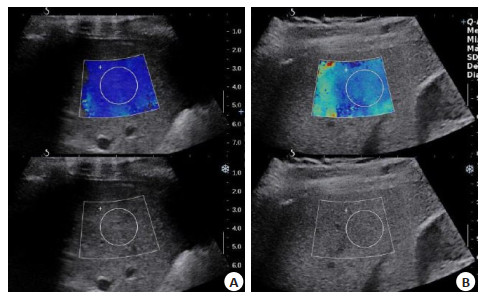
图 1 两组患者SWE影像学示例
A: 对照组某患者SWE检测结果, 病理学分期S0期; B: 纤维化组某患者SWE检测结果,病理学分期S3期.
Figure 1. SWE results of the two groups.

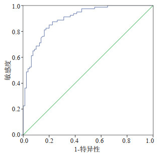
图 2 SWE鉴别诊断肝纤维化的ROC曲线
Figure 2. SWE ROC curve for differential diagnosis of hepatic fibrosis.
表 1 不同病理学分期的肝纤维化患者肝脏弹性模量比较
Table 1. Comparison of liver elastic modulus in patients with different pathological stages (kPa, Mean±SD)
纤维化程度 肝脏弹性模量 F P S1期(n=26) 5.95±1.03 48.209 0.000 S2期(n=35) 8.85±1.58 S3期(n=56) 10.67±1.72 S4期(n=20) 14.31±1.98  下载: 导出CSV
表 2 SWE诊断肝纤维化患者的一般资料对比
Table 2. SWE Comparison of general data on diagnosis of hepatic fibrosis
因素 正确诊断(n=118) 漏诊(n=19) t/χ2 P 年龄(岁, Mean±SD) 58.8±7.7 57.6±7.8 0.629 0.530 BMI(kg/m2, Mean±SD) 23.8±2.2 24.5±2.4 -1.271 0.206 ALT(U/L, Mean±SD) 44.2±6.9 43.1±7.1 0.642 0.522 AST(U/L, Mean±SD) 55.4±9.8 53.7±10.2 0.698 0.486 性别[n(%)] 2.319 0.128 男 65 (55.08) 14(11.86) 女 53 (44.92) 5(4.24) 肝纤维化程度[n(%)] 25.596 0.000 S1 16 (13.56) 10 (8.47) S2 27 (22.88) 8(6.78) S3 55 (46.61) 1(0.85) S4 20 (16.95) 0(0.00) HBeAg[n(%)] 1.665 0.197 阳性 62 (52.54) 13(11.02) 阴性 56 (47.46) 6(5.08) 炎症分级[n(%)] 5.291 0.021 G1~G2 82 (69.49) 18(15.25) G3~G4 36 (30.51) 1(0.85) SWE: 剪切波弹性成像.
下载: 导出CSV
表 3 SWE诊断肝纤维化患者的影响因素研究
Table 3. SWE Factors affecting the diagnosis of hepatic fibrosis
参数 β SE Walds P OR 95%CI 肝纤维化程度 0.884 0.304 8.456 0.000 2.421 1.334-4.392 炎症分级 0.775 0.315 6.053 0.002 2.171 1.171-4.024 常数项 1.048 0.487 4.631 0.045 2.852 1.098-7.408
下载: 导出CSV
-
[1] Shim KY, Eom YW, Kim MY, et al. Role of the renin-angiotensin system in hepatic fibrosis and portal hypertension[J]. Korean J Intern Med, 2018, 33(3): 453-61. doi: 10.3904/kjim.2017.317 [2] 陶国运, 张丽, 孙昳, 等. HBV感染者肝脏弹性模量值及血清肝纤维化指标与肝纤维化的相关研究[J]. 中华临床医师杂志: 电子版, 2015, 9(11): 2075-9. https://www.cnki.com.cn/Article/CJFDTOTAL-ZLYD201511013.htm [3] Yamamura S, Kawaguchi T, Nakano D, et al. Profiles of advanced hepatic fibrosis evaluated by FIB-4 index and shear wave elastography in health checkup examinees[J]. Hepatol Res, 2020, 50(2): 199-213. doi: 10.1111/hepr.13436 [4] Li Y, Huang YS, Wang ZZ, et al. Systematic review with metaanalysis: the diagnostic accuracy of transient elastography for the staging of liver fibrosis in patients with chronic hepatitis B[J]. Aliment Pharmacol Ther, 2016, 43(4): 458-69. doi: 10.1111/apt.13488 [5] Yamamura S, Kawaguchi T, Nakano D, et al. Profiles of advanced hepatic fibrosis evaluated by FIB-4 index and shear wave elastography in health checkup examinees[J]. Hepatol Res, 2020, 50(2): 199-213. doi: 10.1111/hepr.13436 [6] Jiang WX, Huang SR, Teng H, et al. Diagnostic accuracy of point shear wave elastography and transient elastography for staging hepatic fibrosis in patients with non-alcoholic fatty liver disease: a meta-analysis [J]. BMJ Open, 2018, 8(8): e021787. doi: 10.1136/bmjopen-2018-021787 [7] Park DW, Lee YJ, Chang W, et al. Diagnostic performance of a point shear wave elastography (pSWE) for hepatic fibrosis in patients with autoimmune liver disease[J]. PLoS One, 2019, 14(3): e0212771. doi: 10.1371/journal.pone.0212771 [8] 徐朝阳, 牛高华. 实时剪切波弹性成像技术评估肝纤维化程度及其分期的临床意义[J]. 实用医院临床杂志, 2020, 17(2): 123-6. https://www.cnki.com.cn/Article/CJFDTOTAL-YYLC202002037.htm [9] 中华医学会肝病学分会, 中华医学会感染病学分会. 慢性乙型肝炎防治指南2010年更新版[J]. 中华实验和临床感染病杂志: 电子版, 2011, 5(1): . https://www.cnki.com.cn/Article/CJFDTOTAL-ZSGR201101015.htm [10] Abe T, Kuroda H, Fujiwara Y, et al. Accuracy of 2D shear wave elastography in the diagnosis of liver fibrosis in patients with chronic hepatitis C[J]. J Clin Ultrasound, 2018, 46(5): 319-27. doi: 10.1002/jcu.22592 [11] Dubois M, Ronot M, Houssel-Debry P, et al. Performance of B-mode ratio and 2D shear wave elastography for the detection and quantification of hepatic steatosis and fibrosis after liver transplantation[J]. Eur J Gastroenterol Hepatol, 2020, 32(2): 222-30. doi: 10.1097/MEG.0000000000001500 [12] Kohla MAS, Fayoumi AE, Akl M, et al. Early fibrosis regression by shear wave elastography after successful direct-acting anti-HCV therapy[J]. Clin Exp Med, 2020, 20(1): 143-8. doi: 10.1007/s10238-019-00597-0 [13] Wang HW, Shi HN, Cheng J, et al. Real-time shear wave elastography (SWE) assessment of short- and long-term treatment outcome in Budd-Chiari syndrome: a pilot study[J]. PLoS One, 2018, 13(5): e0197550. doi: 10.1371/journal.pone.0197550 [14] 刘博儒, 董雪, 黄丽萍. 剪切波弹性成像评估慢性乙型肝炎肝纤维化的价值及影响因素[J]. 临床肝胆病杂志, 2018, 34(11): 2329-33. https://www.cnki.com.cn/Article/CJFDTOTAL-LCGD201811019.htm [15] 周哲, 周璐, 张玉洁, 等. 瞬时弹性成像在自身免疫性肝炎纤维化早期诊断中的价值及影响因素[J]. 中华消化杂志, 2018, 38(5): 344-6. doi: 10.3760/cma.j.issn.0254-1432.2018.05.011 [16] 崔艾琳, 王佳冰, 徐莉力, 等. 二维剪切波弹性成像与瞬时弹性成像对慢性乙型肝炎患者肝纤维化诊断效能的探讨[J]. 临床超声医学杂志, 2018, 20(12): 819-22. https://www.cnki.com.cn/Article/CJFDTOTAL-LCCY201812009.htm [17] 刘洪伟, 黄颖. 实时二维剪切波弹性成像技术在成人肝脏硬度诊断方面的应用研究[J]. 影像研究与医学应用, 2019, 3(5): 115-6. https://www.cnki.com.cn/Article/CJFDTOTAL-YXYY201905076.htm [18] 卢超政. 实时剪切波弹性成像对慢性乙型肝炎患者早期肝纤维化的诊断价值[J]. 现代诊断与治疗, 2016, 27(19): 3656-7. https://www.cnki.com.cn/Article/CJFDTOTAL-XDZD201619081.htm [19] 王坤, 张文晓, 王明辉, 等. 声辐射脉冲成像及实时剪切波弹性成像对慢性乙型肝炎肝纤维化诊断的对比分析[J]. 中国医师进修杂志, 2018, 41(2): 133-6. doi: 10.3760/cma.j.issn.1673-4904.2018.02.010 [20] 殷珊娱. 实时剪切波超声弹性成像技术对腓肠肌舒张及收缩功能研究[C]. 2018海峡两岸医药卫生交流与合作会议暨第十届海峡两岸超声医学高端论坛论文集, 2018: 1075-6. [21] 李向珍, 房秀霞. 实时剪切波弹性成像对乙型肝病诊断价值及其与肝纤维化分期关系的研究[J]. 内蒙古医科大学学报, 2019, 41(4): 365-7. https://www.cnki.com.cn/Article/CJFDTOTAL-NMYX201904010.htm [22] 朱学苹, 李开林, 梁志超, 等. 定点剪切波弹性成像联合血清学指标无创评估肝纤维化临床研究[J]. 现代医用影像学, 2020, 29(1): 139-41. https://www.cnki.com.cn/Article/CJFDTOTAL-XDYY202001058.htm [23] 陈丽, 高枫, 张贺彬, 等. 实时剪切波弹性成像技术评估低病毒载量慢性乙型肝炎患者肝脏病理改变的临床意义[J]. 国际流行病学传染病学杂志, 2019, 46(6): 481-4. doi: 10.3760/cma.j.issn.1673-4149.2019.06.006 [24] 沈萍, 龚兆萍, 汪敏. 超微血管成像联合剪切波弹性成像在乳腺肿瘤患者良恶性鉴别诊断中的应用及其对诊断价值的影响[J]. 生物医学工程与临床, 2018, 22(4): 418-22. https://www.cnki.com.cn/Article/CJFDTOTAL-SGLC201804012.htm [25] 徐华勤, 郭建锋, 顾斌. 剪切波弹性成像技术对小肝癌和肝硬化结节的鉴别诊断价[J]. 医学美学美容, 2018, 27(23): 89. http://med.wanfangdata.com.cn/Paper/Detail?id=PeriodicalPaper_yxmxmr18201823084 [26] 叶俊钊, 王伟, 廖冰, 等. 实时剪切波弹性成像诊断慢性乙型肝炎肝纤维化的影响因素[J]. 实用医学杂志, 2016, 32(15): 2454-8. https://www.cnki.com.cn/Article/CJFDTOTAL-SYYZ201615011.htm -

 点击查看大图
计量
- 文章访问数: 544
- HTML全文浏览量: 218
- PDF下载量: 4
- 被引次数: 0