


Early MRI findings of seromuscular-layer injury after ultrasound ablation for uterine fibroids and their clinical significance
-
摘要:
目的探究子宫肌瘤超声消融后子宫浆肌层损伤早期MRI影像学特征及其临床意义。 方法回顾性分析2019年9月~2020年9月在本院接受高强度聚焦超声(HIFU)消融术治疗的150例子宫肌瘤患者的临床资料,所有患者均于手术前后接受MR检查,影像学资料完整。比较手术前后患者的MRI信号改变情况,根据MRI图像特征确定术后患者子宫浆肌层损伤情况,将患者分为损伤组(n=43)和无损伤组(n=107),观察损伤组患者的MRI形态特征,比较两组患者的超声消融情况及术后不良反应发生情况。 结果术前,子宫肌瘤在T1WI上表现为低、等或高信号,在T2WI上表现为低或高低混杂信号、等信号或高信号,增强扫描均显示强化;术后肌瘤T1WI信号增高,T2WI信号变化无明显规律,增强扫描均显示无强化。超声消融后,子宫浆肌层损伤率为28.67%(43/150),子宫浆肌层损伤的MRI表现为:T2WI序列显示肌瘤周边肌层信号连续,边界清晰;动态增强扫描显示子宫浆肌层呈环状强化,局部灌注缺损,前壁肌瘤的宫浆肌层损伤率最高为31.40%。损伤组的辐照时间、消融总剂量和肌瘤体积消融率均高于无损伤组(P < 0.05),术后两组患者均出现一定不良反应,且损伤组患者阴道排液发生率高于无损伤组(P < 0.05)。 结论子宫肌瘤患者接受HIFU消融术治疗后,早期会出现子宫浆肌层损伤,治疗期间的辐照时间长、消融总剂量及肌瘤体积消融率高会增加患者子宫浆肌层损伤的风险,使患者术后出现不良反应,通过MRI检查可有效评估患者的子宫浆肌层损伤情况。 -
关键词:
- 子宫肌瘤 /
- 高强度聚焦超声消融术 /
- 浆肌层损伤 /
- 磁共振成像
Abstract:ObjectiveTo explore the early MRI findings of seromuscular-layer injury after ultrasound ablation for uterine fibroids, and analyze their clinical significance. MethodsWe retrospectively analyzed clinical data of 150 patients with uterine fibroids received high-intensity focused ultrasound (HIFU) ablation between September 2019 and September 2020. All patients received MR examination before and after operation and had complete imaging data. Changes in MRI signals before and after operation were analyzed, and the condition of seromuscular-layer injury after operation was determined according to the MRI images. The subjects were divided into the injury group (n=43) and the non-injury group (n=107). MRI results of the injury group were observed. The situation of ultrasound ablation and postoperative adverse reactions were compared between 2 groups. ResultsBefore operation, uterine fibroids showed low, equal, or high signals on T1WI, low, mixed, equal or high signals on T2WI, and enhanced scan showed enhancement. After operation, fibroids showed increased signals on T1WI, but no apparent rule of signal changes on T2WI. The enhanced scan showed no enhancement. After ultrasound ablation, the incidence of seromuscular-layer injury was 28.67% (43/150). T2WI sequence showed continuous signals of peripheral muscle layer of the fibroids, with clear boundaries. The dynamic enhanced scan showed ring enhancement of the seromuscular-layer, and local perfusion defect. The rate of seromuscular-layer injury in patients with anterior wall fibroids reached 31.40%. The irradiation time, total ablation dose, and ablation rate of fibroids were longer and higher in the injury group than in the non-injury group (P < 0.05). Adverse reactions were found in both groups after operation, and the incidence of vaginal discharge was significantly higher in the injury group than in the non-injury group (P < 0.05). ConclusionAfter HIFU ablation, patients with uterine fibroids will have seromuscular-layer injury in the early stage. Long irradiation time, high total ablation dose and high ablation rate of fibroids will increase the risk of seromuscular-layer injury, causing adverse reactions after operation. MR examination can effectively assess the condition of seromuscular-layer injury. -

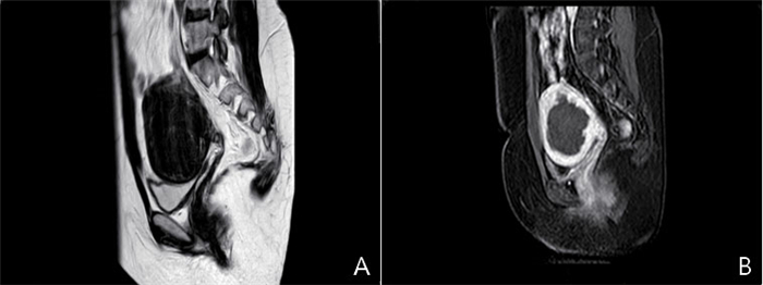
图 1 超声消融前后子宫肌瘤MRI表现
A: 消融前, T2WI显示子宫后壁肌层内有一圆形病灶, 凸向浆膜; B: T2WI序列显示肌瘤周边肌层信号连续, 边界清晰; 动态增强扫描显示子宫浆肌层呈环状强化, 局部灌注缺损.
Figure 1. MRI images of uterine fibroids before and after ultrasound ablation
表 1 两组患者肌瘤位置及消融情况比较
Table 1. Comparison of the location and ablation of fibroids between the two groups (Mean±SD)
组别 子宫肌瘤体积(cm3) 声源功率(W) 辐照时间(s) 消融总剂量(kJ) 肌瘤体积消融率(%) 损伤组(n=43) 85.31±12.68 376.51±28.47 576.25±91.87 317.66±36.75 87.24±5.26 无损伤组(n=107) 82.74±13.11 381.34±15.33 542.35±87.64 286.54±42.17 82.76±6.35 t 1.096 1.340 2.112 4.492 4.094 P 0.275 0.182 0.036 <0.001 <0.001  下载: 导出CSV
表 2 两组患者术后不良反应发生情况比较
Table 2. Comparison of postoperative adverse reactions between the two groups [n(%)]
组别 发热 下腹部疼痛 骶尾部痛 阴道排液 损伤组(n=43) 1(2.33) 16(37.21) 8(18.60) 8(18.60) 无损伤组(n=107) 4(3.74) 50(46.73) 26(24.30) 7(6.54) χ2 - 1.128 1.567 - P 0.613 0.288 0.451 0.036
下载: 导出CSV
-
[1] 刘玉婷, 谢晓绘, 官孝萍, 等. HIFU消融MR T2WI不同信号子宫肌瘤的临床疗效及安全性分析[J]. 中国介入影像与治疗学, 2019, 16(6): 349-53. https://www.cnki.com.cn/Article/CJFDTOTAL-JRYX201906008.htm [2] Yang SH, Kong FJ, Hou RJ, et al. Ultrasound guided high-intensity focused ultrasound combined with gonadotropin releasing hormone analogue (GnRHa) ablating uterine leiomyoma with homogeneous hyperintensity on T2 weighted MR imaging[J]. Br J Radiol, 2017, 90(1073): 20160760. doi: 10.1259/bjr.20160760 [3] Yin N, Hu L, Xiao ZB, et al. Factors influencing thermal injury to skin and abdominal wall structures in HIFU ablation of uterine fibroids [J]. Int J Hyperthermia, 2018, 34(8): 1298-303. doi: 10.1080/02656736.2018.1433880 [4] 寸江平, 赵卫, 范宏杰, 等. MRI在高强度聚焦超声消融子宫肌瘤中的应用进展[J]. 中国医学影像技术, 2019, 35(6): 946-9. https://www.cnki.com.cn/Article/CJFDTOTAL-ZYXX201906054.htm [5] Zhao WP, Zhang J, Han ZY, et al. A clinical investigation treating different types of fibroids identified by MRI-T2WI imaging with ultrasound guided high intensity focused ultrasound[J]. Sci Rep, 2017, 7(1): 10812. doi: 10.1038/s41598-017-11486-5 [6] 魏庆, 陈锦云, 刘一诺, 等. 子宫肌瘤超声消融后早期子宫浆肌层损伤的MRI评价[J]. 第三军医大学学报, 2019, 41(12): 1161-6. https://www.cnki.com.cn/Article/CJFDTOTAL-DSDX201912011.htm [7] Chen J, Li Y, Wang Z, et al. Evaluation of high-intensity focused ultrasound ablation for uterine fibroids: an IDEAL prospective exploration study[J]. BJOG, 2018, 125(3): 354-64. doi: 10.1111/1471-0528.14689 [8] Peng S, Zhang L, Hu L, et al. Factors influencing the dosimetry for high-intensity focused ultrasound ablation of uterine fibroids: a retrospective study[J]. Medicine (Baltimore), 2015, 94(13): e650. doi: 10.1097/MD.0000000000000650 [9] 江昭颖, 朱小刚, 薛敏. 高强度聚焦超声消融治疗子宫肌瘤后妊娠结局及影响因素分析[J]. 中国实用妇科与产科杂志, 2020, 36(2): 168-72. https://www.cnki.com.cn/Article/CJFDTOTAL-ZGSF202002019.htm [10] Liu YC, Zhang WW, He M, et al. Adverse effect analysis of highintensity focused ultrasound in the treatment of benign uterine diseases [J]. Int J Hyperthermia, 2018, 35(1): 56-61. doi: 10.1080/02656736.2018.1473894 [11] 魏庆, 陈锦云, 刘一诺, 等. HIFU消融子宫肌瘤后浆肌层损伤对消融治疗有效性及安全性的影响[J]. 中国介入影像与治疗学, 2019, 16(8): 455-9. https://www.cnki.com.cn/Article/CJFDTOTAL-JRYX201908003.htm [12] 范宏杰, 寸江平, 姚瑞红, 等. 高强度聚焦超声治疗子宫肌瘤的消融率的多因素分析[J]. 实用放射学杂志, 2018, 34(9): 1427-9, 1474. doi: 10.3969/j.issn.1002-1671.2018.09.029 [13] Zhang YJ, Xiao ZB, Lv FR, et al. MRI evaluation of endopelvic fascial swelling and analysis of influencing factors in patients with uterine fibroids after high-intensity focused ultrasound ablation[J]. Int J Hyperthermia, 2020, 37(1): 175-81. doi: 10.1080/02656736.2019.1701100 [14] 张学花, 翟昭华, 董国礼, 等. MRI评价子宫肌瘤高强度聚焦超声消融术后盆底筋膜改变[J]. 中国医学影像技术, 2017, 33(10): 1540-4. http://www.cnki.com.cn/Article/CJFDTotal-ZYXX201710035.htm [15] Zhao WP, Chen JY, Chen WZ. Dynamic contrast-enhanced MRI serves as a predictor of HIFU treatment outcome for uterine fibroids with hyperintensity in T2-weighted images[J]. Exp Ther Med, 2016, 11(1): 328-34. doi: 10.3892/etm.2015.2879 [16] 尹娜, 王玲, 胡亮, 等. HIFU治疗超声衰减宽度 < 10 mm声通道腹壁瘢痕子宫肌瘤患者的有效性及安全性[J]. 中国介入影像与治疗学, 2018, 15(4): 221-5. [17] Tempest N, Hapangama D. Should we be putting our scalpels down? Is HIFU the answer to fertility-sparing fibroid treatment?[J]. BJOG: Int J Obstet Gy, 2018, 125(3): 366. doi: 10.1111/1471-0528.14691 [18] 崔运能, 田橄, 陈向东, 等. 高强度聚焦超声治疗子宫肌瘤及子宫腺肌瘤术后早期并发症的MR表现[J]. 临床放射学杂志, 2018, 37(3): 461-5. https://www.cnki.com.cn/Article/CJFDTOTAL-LCFS201803026.htm [19] Cun JP, Fan HJ, Zhao W, et al. Factors influencing MR changes associated with sacral injury after high-intensity focused ultrasound ablation of uterine fibroids [J]. Int J Hyperthermia, 2019, 36(1): 21-8. doi: 10.1080/02656736.2018.1528391 [20] Chen JY, Chen WZ, Zhang L, et al. Safety of ultrasound-guided ultrasound ablation for uterine fibroids and adenomyosis: a review of 9988 cases [J]. Ultrason Sonochem, 2015, 27: 671-6. doi: 10.1016/j.ultsonch.2015.05.031 -

 点击查看大图
计量
- 文章访问数: 855
- HTML全文浏览量: 380
- PDF下载量: 8
- 被引次数: 0