


Application value of MRI diffusion-weighted imaging in the preoperative diagnosis of renal space-occupying lesions
-
摘要:
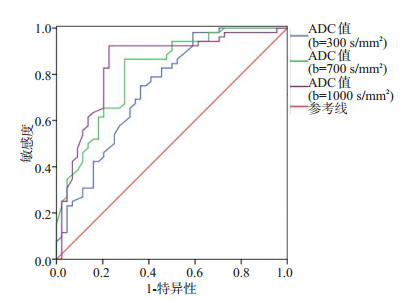
目的分析MRI弥散加权成像(DWI)在肾脏占位性病变术前诊断中的应用价值。 方法选取2019年1~12月我院收治的96例肾脏占位性病变患者,病理证实其中肾脏恶性肿瘤患者52例(肾脏恶性病变组)和肾脏良性肿瘤患者44例(肾脏良性病变组),同时选取96例健康志愿者作为对照组,均进行DWI检查,比较不同弥散敏感系数b值下各组表观扩散系数(ADC)值,并分析不同弥散敏感系数b值下ADC值对肾脏占位性病变的诊断价值。 结果肾脏恶性病变组表现为T1WI、T2WI呈现稍高信号,其中9例信号均匀,43例表现为不同程度高低混杂信号,肿瘤实质部分DWI表现为高信号,肿瘤坏死部分呈现低信号;肾脏良性病变组表现为T1WI低信号,T2WI高信号,DWI表现为均匀稍低信号;肾脏恶性病变磁共振弥散加权成像在肾脏占位性病变术前诊断中的应用组病灶最长直径明显大于肾脏良性病变组(P < 0.05);弥散敏感系数b值分别为300、700、1000 s/mm2时,肾脏恶性病变组、肾脏良性病变组以及对照组所对应的ADC值差异具有统计学意义(P < 0.05),表现为肾脏良性病变组 > 对照组 > 肾脏恶性病变组;以病理活检结果为金标准,弥散敏感系数b值分别为300、700、1000 s/mm2时,ADC值分别取最佳截断值3.86×103、2.50×103、1.71×103 mm2/s时,诊断肾脏占位性病变的AUC分别为0.742(95%CI: 0.642~0.842)、0.811(95%CI: 0.725~0.897)、0.842(95%CI: 0.758~0.927),敏感度分别为98.10%、86.50%、92.30%,特异度分别为40.90%、70.50%、77.30%。 结论DWI在肾脏占位性病变术前诊断中具有良好的应用价值,可用于区别良恶性肾脏占位性病变,不同弥散敏感系数b值下ADC值对肾脏占位性病变都具有较好的诊断效能。 Abstract:ObjectiveTo analyze the application value of magnetic resonance (MRI) diffusion weighted imaging (DWI) in the preoperative diagnosis of renal space- occupying lesions. MethodsNinty-six patients with renal space-occupying lesions admitted to our hospital from January 2019 to December 2019 were selected. Fifty-two patients with renal malignant tumors (renal malignant lesion group) and 44 patients with benign renal tumors (benign renal lesion group) were pathologically confirmed, and 96 healthy volunteers were selected as controls (normal group). They were given DWI examination. The apparent diffusion coefficient (ADC) values of each group under different diffusion sensitivity coefficient b values were compared, and the diagnostic value of ADC values under different diffusion sensitivity coefficient b values on renal spaceoccupying lesions was analyzed. ResultsThe longest diameter of lesions in renal malignant lesion group was larger than that in benign renal lesion group. In renal malignant lesion group, T1WI and T2WI showed slightly high signals, including 9 cases of uniform signals and 43 cases of high-low mixed signals of different degrees, and DWI showed high signal in tumor parenchyma and showed low signal in necrotic tumor. In renal benign lesion group, T1WI showed low signals and T2WI showed high signals, and DWI showed uniform slightly low signals. The longest diameter of the lesion in renal malignant lesion group was significantly larger than that in benign renal lesion group (P < 0.05). When the diffuse sensitivity coefficient b value was 300, 700 or 1000 s/mm2 respectively, the ADC values in malignant lesion group, benign renal lesion group and normal group were significantly different (P < 0.05), showing benign renal lesion group > normal group > renal malignant lesion group. Taking pathological biopsy results as the gold standards, the AUC values were 0.742 (95%CI: 0.642-0.842), 0.811 (95%CI: 0.725-0.897) and 0.842 (95%CI: 0.758-0.927), and sensitivities were 98.10%, 86.50% and 92.30% and specificities were 40. 90%, 70.50% and 77.30% when diffuse sensitivity coefficient b values were 300, 700 and 1000 s/mm2. And the the best cutoff values of ADC were 3.86×10³, 2.50×10³ and 1.71×103 mm2/s respectively. ConclusionDWI has good application value in the preoperative diagnosis of renal space-occupying lesions. It can be used to distinguish benign and malignant renal space-occupying lesions. ADC value has good diagnostic efficacy on renal space- occupying lesions under different diffusion sensitivity coefficient b values. -

图 2 不同弥散敏感系数b值下ADC值诊断肾脏占位性病变的ROC曲线图
Figure 2. ROC curves of ADC values in diagnosing renal space-occupying lesions under different diffusion sensitivity coefficient b values.
表 1 肾脏占位性病变组和对照组ADC值比较
Table 1. Comparison of ADC values between renal space-occupying lesion group and normal group (Mean± SD, ×103 mm2/s)
b值 肾脏恶性病变组(n=52) 肾脏良性病变组(n=44) 对照组(n=96) F P 300 s/mm2 1.74±0.22 3.78±0.60 2.76±0.41 277.309 0.000 700 s/mm2 1.50±0.18 3.45±0.57 2.33±0.33 330.500 0.000 1000 s/mm2 1.31±0.15 3.06±0.44 2.09±0.26 434.101 0.000  下载: 导出CSV
-
[1] Yu CJ, Wu SD. A 4-year-old boy with right renal space-occupying lesion diagnosed with inflammatory pseudotumor[J]. Urology, 2020: S0090-S4295(20)31362-5. [2] López Zúñiga MÁ, Vallejo Palomino T, Martin Toro MA, et al. Diagnostic capacity of pocket-sized ultrasound devices at point of care by a non-radiologist resident in patients with suspected abdominal pathology[J]. Ultrasound Med Biol, 2020, 46(2): 263-8. doi: 10.1016/j.ultrasmedbio.2019.10.019 [3] Baloi P, Del Chicca F, Ruetten M, et al. The human Bosniak model applied to a cat with renal cystadenoma. A classification to differentiate benign and malignant cystic renal masses via computed tomography and ultrasound[J]. Tierarztl Prax Ausg K Kleintiere Heimtiere, 2015, 43(1): 45-9. doi: 10.15654/TPK-140268 [4] 张梁, 周志斌. 高分辨率磁共振血管壁成像在颅内动脉粥样硬化性疾病中的应用[J]. 分子影像学杂志, 2020, 43(1): 45-8. doi: 10.12122/j.issn.1674-4500.2020.01.10 [5] Pitra T, Pivovarcikova K, Tupy R, et al. Magnetic resonance imaging as an adjunct diagnostic tool in computed tomography defined Bosniak ⅡF- Ⅲ renal cysts: a multicenter study[J]. World J Urol, 2018, 36(6): 905-11. doi: 10.1007/s00345-018-2176-z [6] Döring A, Adalid V, Boesch C, et al. Diffusion-weighted magnetic resonance spectroscopy boosted by simultaneously acquired water reference signals[J]. Magn Reson Med, 2018, 80(6): 2326-38. doi: 10.1002/mrm.27222 [7] 刘玉荣. 磁共振动态增强检查与弥散加权成像在肝结节性病变与小肝癌诊断中的比较[J]. 中国医药指南, 2018, 16(14): 83. https://www.cnki.com.cn/Article/CJFDTOTAL-YYXK201814065.htm [8] 李晓青, 文明, 路彦斌, 等. 腹部磁共振弥散加权成像(DWI)技术在对肝脏良、恶性肿瘤鉴别中的应用价值分析[J]. 影像研究与医学应用, 2019, 3(6): 96-7. https://www.cnki.com.cn/Article/CJFDTOTAL-YXYY201906061.htm [9] Li R, Wu G, Wang R. Application values of 3.0T magnetic resonance diffusion weighted imaging for distinguishing liver malignant tumors and benign lesions[J]. Oncol Lett, 2018, 15(2): 2091-6. http://europepmc.org/abstract/MED/29434910 [10] 姚文君, 张涛, 王龙胜, 等. Bosniak分级系统在肾脏囊性病变MSCT诊断中的应用价值[J]. 中华全科医学, 2018, 16(9): 1515-7, 1529. https://www.cnki.com.cn/Article/CJFDTOTAL-SYQY201809032.htm [11] 马海鸿, 潘家洁, 王建新. 双源CT双能量在肾脏占位性病变检查中的应用研究[J]. 临床和实验医学杂志, 2019, 18(4), 18: 423-6. doi: 10.3969/j.issn.1671-4695.2019.04.025 [12] Ballard DH, De Alba L, Migliaro M, et al. CT imaging spectrum of infiltrative renal diseases[J]. Abdom Radiol, 2017, 42(11): 2700-9. doi: 10.1007/s00261-017-1193-5 [13] Del Chicca F, Salesov E, Joerger F, et al. Perfusion-weighted and diffusion-weighted magnetic resonance imaging of the liver, spleen, and kidneys of healthy adult male cats[J]. Am J Vet Res, 2019, 80(2): 159-67. doi: 10.2460/ajvr.80.2.159 [14] Li YM, Lin DD, Weng YL, et al. Early diffusion-weighted imaging and proton magnetic resonance spectroscopy features of liver transplanted tumors treated with radiation in rabbits: correlation with histopathology[J]. Radiat Res, 2019, 191(1): 52-9. http://www.ncbi.nlm.nih.gov/pubmed/30376410 [15] 周蕾蕾, 张作恒, 陈宇辰, 等. 基于卷积神经网络的肾脏占位CT图像的良恶性分类研究[J]. 国际生物医学工程杂志, 2018, 41(5): 417-22. doi: 10.3760/cma.j.issn.1673-4181.2018.05.008 [16] 王娇, 胡彩虹, 马许静, 等. 扩散张量成像在原发性肾小球肾炎早期病理损害的临床应用价值[J]. 临床放射学杂志, 2019, 38(5): 868-73. https://www.cnki.com.cn/Article/CJFDTOTAL-LCFS201905030.htm [17] 彭泳涵, 刘敏, 王振, 等. 磁共振扩散加权成像在肾积水和肾积脓鉴别诊断中的应用[J]. 中华泌尿外科杂志, 2019, 40(2): 122-6. doi: 10.3760/cma.j.issn.1000-6702.2019.02.009 [18] Tian W, Lu JB, Jiao D, et al. An evaluation of the clinical diagnostic value of contrast-enhanced ultrasound combined with contrastenhanced computed tomography in space-occupying lesions of the kidney[J]. Onco Targets Ther, 2017, 10: 3493-9. doi: 10.2147/OTT.S135500 [19] 蔡恩明. 磁共振弥散加权成像技术在肝脏良、恶性肿瘤中的鉴别诊断价值[J]. 实用临床医药杂志, 2018, 22(1): 71-3. https://www.cnki.com.cn/Article/CJFDTOTAL-XYZL201801022.htm [20] 席建平, 马新. 磁共振弥散加权成像诊断联合多b值检查对前列腺癌的诊断价值[J]. 实用癌症杂志, 2018, 33(11): 164-7. https://www.cnki.com.cn/Article/CJFDTOTAL-SYAZ201811047.htm [21] 陆蓉, 胥常云, 林璐, 等. 3.0T磁共振弥散加权成像表观弥散系数对肾脏肿瘤性病变的诊断价值[J]. 中国临床医学, 2018, 25(4): 601-5. https://www.cnki.com.cn/Article/CJFDTOTAL-LCYX201804020.htm [22] 王淼, 白亚飞. 增强磁共振成像和扩散加权成像鉴别肾脏良、恶性病变的诊断效能[J]. 临床医学研究与实践, 2019, 4(20): 134-5, 141. https://www.cnki.com.cn/Article/CJFDTOTAL-YLYS201920057.htm -

 点击查看大图
图(2) / 表(1)
计量
- 文章访问数: 527
- HTML全文浏览量: 285
- PDF下载量: 8
- 被引次数: 0