


Clinical application of 3D ASL in the evaluation of cerebral watershed perfusion in patients with different degrees of carotid stenosis
-
摘要:
目的探讨三维动脉自旋标记(3D ASL)技术在评估不同程度颈动脉狭窄后大脑分水岭灌注状态方面的临床应用价值。 方法收集2018年1月~2020年3月于脑病科初诊为缺血性脑血管病患者60例,经颈部彩色多普勒超声分别评估双侧颈动脉狭窄度。同时行3D ASL成像检查,使用Functool软件将原始数据自动生成脑血流量(CBF)伪彩图,分别于皮质分水岭前角及后角、放射冠及半卵圆中心选取兴趣区(ROI=200±20 mm2),测量并分析不同程度颈动脉狭窄患者大脑分水岭区CBF值差异。 结果共采集颈动脉120根,其中颈动脉无狭窄30根,轻度狭窄49根,中度狭窄26根,重度狭窄15根,颈动脉轻度狭窄患者皮质分水岭前、后角CBF值与颈动脉无狭窄患者差异无统计学意义(P > 0.05),内分水岭区CBF值低于无狭窄患者,差异有统计学意义(P < 0.05);颈动脉中度、重度狭窄患者皮质分水岭及内分水岭CBF值均明显低于无狭窄患者,差异有统计学意义(P < 0.05)。 结论3D ASL成像技术可敏感评估不同程度颈动脉狭窄患者大脑分水岭区脑灌注状态,对于患者治疗方案的选择以及预后评估具有重要应用价值。 Abstract:ObjectiveTo investigate the clinical value of 3D ASL in assessing the perfusion status of cerebral watershed after different degrees of carotid stenosis. MethodsSixty patients with ischemic cerebrovascular disease who were initially diagnosed in the Department of encephalopathy from January 2018 to March 2020 were collected. The degree of bilateral carotid artery stenosis was evaluated by CDFI. Meanwhile, 3D ASL imaging was performed, and the original data were automatically generated into a pseudo color map of cerebral blood flow (CBF) using functool software, and three-dimensional arterial spin labeling (3D ASL) imaging examination. The interest areas (ROI=200±20 mm2) were selected from the front and back corners of the cortical watershed, the radial crown and the center of the semicircular circle. The CBF values of the cerebral watershed in patients with different degrees of carotid stenosis were measured and analyzed. ResultsThere were 120 cases of carotid artery, 30 cases of no stenosis, 49 cases of mild stenosis, 26 cases of moderate stenosis, 15 cases of severe stenosis. There was no significant difference in CBF between the patients with mild stenosis and the patients without stenosis (P > 0.05). The CBF value in the inner watershed was lower than that in the patients without stenosis (P < 0.05), The CBF values of cortical watershed and internal watershed in patients with moderate and severe carotid stenosis were significantly lower than those in patients without stenosis (P < 0.05). Conclusion3D ASL imaging technology can sensitively evaluate the cerebral perfusion in the watershed area of patients with different degrees of carotid stenosis. It has important application value for the selection of treatment plan and prognosis evaluation of patients. -
Key words:
- carotid artery /
- hemodynamics /
- arterial spin labeling /
- cerebral blood flow
-

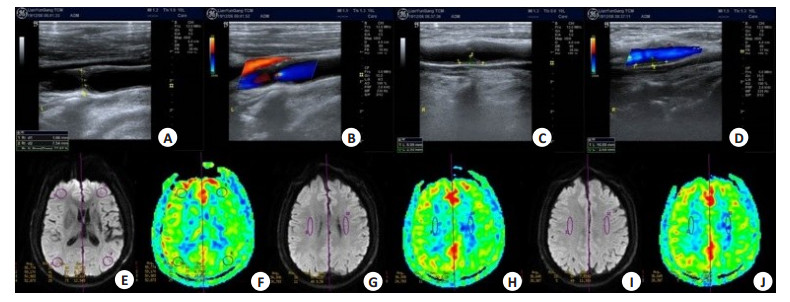
图 1 患者男,56岁,右肢活动不利1年余
A~D: 颈部CDFI提示左侧颈内动脉起始部重度狭窄(狭窄率77%),右侧颈总动脉分叉处轻度狭窄; E~J: 3DASL(PLD=1525 ms)提示左侧皮质分水岭及内分水岭区CBF值明显低于右侧.
Figure 1. A56-year-old Male patient, right limb movement is not good for more than one year.
表 1 不同程度颈动脉狭窄后分水岭区CBF值比较
Table 1. Comparison of CBF values in watershed area after different degrees of carotid stenosis[mL/(min·100 g), Mean±SD]
颈动脉狭窄程度 皮质分水岭 内分水岭 前角 后角 放射冠 半卵圆中心 无狭窄(n=30) 54.25±4.57 53.34±4.65 37.55±4.87 36.86±4.81 轻度狭窄(n=49) 53.12±4.61 52.78±4.82 32.64±4.83 32.83±4.78 中度狭窄(n=26) 43.64±5.42 42.17±5.26 23.87±5.81 24.21±5.77 重度狭窄(n=15) 28.58±5.86 27.35±6.08 20.51±5.43 19.85±5.24  下载: 导出CSV
-
[1] Derex L, Cho TH. Mechanical thrombectomy in acute ischemic stroke[J]. Rev Neurol, 2017, 173(3): 106. doi: 10.1016/j.neurol.2016.06.008 [2] 赵仁军, 刘英, 杨加惠. TCD检测颈内动脉颅外段重度狭窄或闭塞后颅内血流动力学改变的临床价值[J]. 中西医结合心脑血管病杂志, 2017, 15(13): 1653-5. doi: 10.3969/j.issn.1672-1349.2017.13.037 [3] Weill C, Suissa L, Darcourt J, et al. The pathophysiology of watershed infarction: a three-dimensional time-of-flight magnetic resonance angiography study[J]. J Stroke Cerebrovasc Dis, 2017, 26(9): 1966- 73. doi: 10.1016/j.jstrokecerebrovasdis.2017.06.016 [4] 范晓媛, 冯逢. 动脉自旋标记MRI技术在烟雾病中的应用[J]. 国际医学放射学杂志, 2019, 42(6): 668-72. https://www.cnki.com.cn/Article/CJFDTOTAL-GWLC201906009.htm [5] 金可鑫, 丁佳悦, 孟然. 磁共振灌注成像在预警和预后缺血性脑卒中的应用[J]. 中华老年心脑血管病杂志, 2020, 22(2): 216-8. https://www.cnki.com.cn/Article/CJFDTOTAL-LNXG202002034.htm [6] 张帆, 徐缓, 李运祥, 等. 320排CT全脑灌注成像结合4D-CTA在大脑中动脉闭塞患者侧支循环评估中的应用[J]. 中国CT和MRI杂志, 2019, 17(8): 36-8. https://www.cnki.com.cn/Article/CJFDTOTAL-CTMR201908011.htm [7] Kaczmarz S, Griese V, Preibisch C, et al. Increased variability of watershed areas in patients with high-grade carotid Stenosis[J]. Neuroradiology, 2018, 60(3): 311-23. doi: 10.1007/s00234-017-1970-4 [8] 程令刚, 何文. 超声造影技术在脑灌注成像中的应用及进展[J]. 首都医科大学学报, 2019, 40(6): 813-7. doi: 10.3969/j.issn.1006-7795.2019.06.001 [9] 郑园园, 惠品晶, 韩佳霖, 等. 经颅多普勒量化评估单侧颈内动脉重度狭窄或闭塞侧支循环的可行性[J]. 中风与神经疾病杂志, 2018, 35 (9): 782-6. https://www.cnki.com.cn/Article/CJFDTOTAL-ZFSJ201809004.htm [10] 李婷, 林雁潮, 王瑶, 等. 颈部血管超声、CT血管成像及磁共振成像在急性缺血性脑卒中患者颈动脉狭窄诊断中的应用比较[J]. 实用医学杂志, 2019, 35(12): 2005-9. doi: 10.3969/j.issn.1006-5725.2019.12.032 [11] 吴秀美, 严江志, 蔡俊秀, 等. 不同程度颈内动脉狭窄患者脑分水岭区灌注状态研究[J]. 中华老年心脑血管病杂志, 2019, 21(4): 407-10. https://www.cnki.com.cn/Article/CJFDTOTAL-LNXG201904020.htm [12] 伍满燕, 梁文卿, 陈江天, 等. 颈动脉粥样硬化性疾病的诊治进展[J]. 中华老年心脑血管病杂志, 2019, 21(11): 1223-6. doi: 10.3969/j.issn.1009-0126.2019.11.028 [13] 黄崇闲. 彩色多普勒超声诊断颈动脉狭窄的准确性评价[J]. 深圳中西医结合杂志, 2019, 29(17): 152-3. https://www.cnki.com.cn/Article/CJFDTOTAL-SZZX201917076.htm [14] 李晓, 赵辉林, 孙贝贝, 等. MR测定颈动脉易损斑块特征与急性缺血性脑卒中的关系[J]. 实用放射学杂志, 2017, 33(3): 373-7. doi: 10.3969/j.issn.1002-1671.2017.03.013 [15] 雷少阳, 张淑倩. ASL技术在中枢神经系统中的应用进展[J]. 国际医学放射学杂志, 2020, 43(1): 73-7. https://www.cnki.com.cn/Article/CJFDTOTAL-GWLC202001019.htm [16] 时彤彤, 徐加焕, 谢元忠, 等. 动脉自旋标记成像评估颈内动脉狭窄患者脑血流量[J]. 中国介入影像与治疗学, 2020, 17(6): 364-8. https://www.cnki.com.cn/Article/CJFDTOTAL-JRYX202006015.htm [17] Madhok DY, Vitt JR, Nguyen AT. Overview of neurovascular physiology[J]. Curr Neurol Neurosci Rep, 2018, 18(12): 99. doi: 10.1007/s11910-018-0905-8 [18] 周建国, 符大勇, 卢明聪, 等. 三维动脉自旋标记在单侧颈内动脉闭塞后侧支循环评估中的价值[J]. 分子影像学杂志, 2020, 43(1): 130-3. doi: 10.12122/j.issn.1674-4500.2020.01.27 [19] 杨玉婵, 段庆红, 李小宝, 等. 颈动脉狭窄脑血流动力学变化与动脉狭窄程度相关性的CT研究[J]. 实用放射学杂志, 2015(5): 726-9, 734. [20] 林天烨, 有慧, 冯逢, 等. 动脉自旋标记MR技术进展及应用[J]. 中华放射学杂志, 2019, 53(5): 431-4. [21] 周建国, 符大勇, 马先军, 等. ASL对大脑中动脉M1段闭塞后侧支循环建立显示的临床应用[J]. 实用放射学杂志, 2018, 34(8): 1164-6, 1171. [22] Ma H, Wang ZZ, Xu K, et al. Three-dimensional arterial spin labeling imaging and dynamic susceptibility contrast perfusionweighted imaging value in diagnosing glioma grade prior to surgery [J]. Exp Ther Med, 2017, 13(6): 2691-8. http://europepmc.org/abstract/MED/28587332 [23] 吕洪伟. 三维准连续动脉自旋标记在脑缺血性疾病中的应用进展[J]. 实用放射学杂志, 2019, 35(12): 2033-6. -

 点击查看大图
图(1) / 表(1)
计量
- 文章访问数: 457
- HTML全文浏览量: 203
- PDF下载量: 8
- 被引次数: 0