


Value of CT and MRI in evaluating the effects of intervention and molecular targeted therapy on patients with primary hepatocellular carcinoma
-
摘要:
目的探讨CT及MRI评估原发性肝癌(HCC)患者介入及分子靶向治疗效果的价值。 方法回顾性选取我院于2017年1月~2019年1月收治的80例中晚期HCC患者作为研究对象,患者均行肝动脉化疗栓塞术(TACE)治疗+索拉非尼治疗,3月后均行CT、MRI和数字减影血管造影(DSA)检查。比较CT联合MRI与单独CT检查HCC患者治疗后3月的临床有效例数、有效率,并以DSA检查结果为金标准,计算两种检查方法的敏感度、特异度、准确率和kappa值,并比较两种检查方法对患者治疗后的残余和复发病灶检出情况。 结果经TACE+索拉非尼治疗后3月,DSA显示总目标病灶数由98个减少至86个,病灶明显缩小,临床疾病控制患者64例,无效患者16例,疾病控制率80.00%;共检出残留和复发病灶共82个,其中Ⅰ型、Ⅱ型、Ⅲ型、Ⅳ型碘油沉积残留和复发病灶数分别为9、18、42、17个。CT联合MRI检出患者治疗后Ⅱ型以及总碘油沉积残留、复发病灶个数均大于CT检查(P < 0.05); CT联合MRI评估TACE+索拉非尼治疗HCC患者疗效的灵敏度95.31%高于单独CT检查的84.38%(P < 0.05),且CT联合MRI评估临床疗效与DSA检查的Kappa值为0.809,且大于单独CT评估与DSA检查的Kappa值(0.605)。结论TACE+索拉非尼治疗HCC患者3月后具有较好的临床疗效,且相比单独CT检查,CT联合MRI更有利于患者治疗后残留和复发灶的检出,同时对于患者治疗后3月疗效的评估具有更高的灵敏度和应用价值。 Abstract:ObjectiveTo explore the value of CT and MRI in evaluating the effects of intervention and molecular targeted therapy on patients with primary hepatocellular carcinoma (HCC). MethodsA total of 80 patients with advanced HCC admitted to the hospital from January 2017 to January 2019 were retrospectively enrolled as the research objects. All patients underwent hepatic artery chemoembolization (TACE) and sorafenib therapy, and CT, MRI and digital subtraction angiography (DSA) were performed after 3 months. The number of clinically effective cases and response rate were compared after 3 months treatment by CT combined MRI and CT alone. Taking DSA results as the golden standard, the sensitivity, specificity, accuracy and kappa values of the two methods were calculated. The detection situations of residual and recurrence lesions were compared after treatment by the two methods. ResultsAfter 3 months of TACE and sorafenib treatment, DSA showed that the total number of target lesions decreased from 98 to 86, the lesions significantly reduced There were 64 clinical disease control patients and 16 ineffective patients, with 80.00% disease control rate. There were 82 residual and recurrence lesions. Among them, there were 9, 18, 42 and 17 residual and recurrence lesions of type I, II, III and IV iodized oil deposition, respectively. The detected number of type II iodized oil deposition and recurrence lesions by CT combined with MRI was more than that by CT (P < 0.05). The sensitivity of CT combined with MRI for evaluating the curative effect of TACE and sorafenib on HCC patients was higher than that of CT examination alone (95.31% vs 84.38%, P < 0.05). Kappa value of CT combined with MRI for evaluating clinical curative effect and DSA examination was 0.809, which was greater than that of CT alone (0.605). ConclusionThe clinical curative effect of TACE and sorafenib is good on HCC patients 3 months later. Compared with CT alone, CT combined with MRI is more conducive to detecting residual and recurrent lesions after treatment, which has of higher sensitivity and application value in evaluating curative effect after 3 months of treatment. -

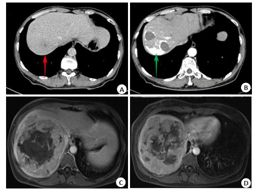
图 1 HCC患者行TACE+索拉非尼治疗前后的CT与MRI影像学检查
A: 肝右叶低密度团块影, 增强后呈“快进快出”表现; B: 病灶无明显强化, 无新病灶出现; C, D: 肝右叶团块状混杂信号影, 病灶部分弥散受限, 增强后病灶周围不均匀强化, 中央无强化, 无新病灶出现.
Figure 1. CT and MRI examination in HCC patients before and after TACE combined with sorafenib treatment.
表 1 两种检查方法对不同碘油沉积检出残余、复发病灶的比较
Table 1. Comparison on the detected number of different iodized oil deposition and recurrence lesions by two methods (n)
影像学方法 不同碘油沉积检出残余、复发病灶 合计 Ⅰ型 Ⅱ型 Ⅲ型 Ⅳ型 DSA 9 18 42 17 86 CT联合MRI 8 17 40 17 82 CT 7 11 39 17 74 χ2* 4.018 4.410 P* > 0.05 0.045 > 0.05 > 0.05 0.036 *χ2值及P值为CT联合MRI与CT检杳之间的比较; DSA: 数字减影血管造影.  下载: 导出CSV
表 2 两种检查方法对治疗后3月的疗效评估结果与金标准比
Table 2. Comparison of curative effect at 3 months after treatment and golden standard by the two methods (n)
DSA(金标准) n CT联合MRI CT 完全缓解 部分缓解 疾病稳定 疾病进展 完全缓解 部分缓解 疾病稳定 疾病进展 完全缓解 8 7 1 0 0 5 3 0 0 部分缓解 16 0 15 0 1 0 12 1 3 疾病稳定 40 0 0 38 2 0 0 33 7 疾病进展 16 0 0 2 14 0 1 1 14 合计 80 7 16 40 17 5 16 35 24
下载: 导出CSV
表 3 两种检查方法对TACE+索拉非尼治疗后疗效的评估效能比较
Table 3. Comparison of the evaluation efficiency on the curative effect of TACE combined with sorafenib by the two methods (%, n=80)
检查方法 灵敏度 特异度 准确率 阳性预测值 阴性预测值 kappa值 CT联合MRI 95.31(61/64) 87.50(14/16) 93.75(75/80) 96.83(61/63) 82.35(14/17) 0.809 CT 84.38(54/64) 87.50(14/16) 85.00(68/80) 96.43(54/56) 58.33(14/24) 0.605 χ2 4.195 0.286 3.225 0.152 2.651 P 0.041 0.593 0.073 0.697 0.103
下载: 导出CSV
-
[1] Lee DH, Lee JM. Recent advances in the image-guided tumor ablation of liver malignancies: radiofrequency ablation with multiple electrodes, real- time multimodality fusion imaging, and new energy sources[J]. Korean J Radiol, 2018, 19(4): 545-59. doi: 10.3348/kjr.2018.19.4.545 [2] Yoon IS, Shin JH, Han K, et al. Ultrasound- guided intraoperative radiofrequency ablation and surgical resection for liver metastasis from malignant gastrointestinal stromal tumors[J]. Korean J Radiol, 2018, 19(1): 54-62. doi: 10.3348/kjr.2018.19.1.54 [3] 杨青松. CT扫描联合磁共振诊断原发性肝癌及评估其介入治疗术后的临床效果评价[J]. 当代医学, 2019, 25(30): 110-2. https://www.cnki.com.cn/Article/CJFDTOTAL-DDYI201930047.htm [4] 樊建朝, 赵香田. 核磁共振、增强CT及超声造影对肝癌介入治疗疗效评估的对比研究[J]. 中西医结合肝病杂志, 2019, 29(5): 463-5. https://www.cnki.com.cn/Article/CJFDTOTAL-ZXGB201905025.htm [5] 杨德顺, 甘椿银. CT扫描联合磁共振诊断原发性肝癌及评估其介入治疗术后的临床效果[J]. 影像研究与医学应用, 2019, 3(2): 11-3. https://www.cnki.com.cn/Article/CJFDTOTAL-YXYY201902005.htm [6] 中华人民共和国卫生部. 原发性肝癌诊疗规范(2011年版[) J]. 临床肝胆病杂志, 2011, 20(11): 929-46. https://www.cnki.com.cn/Article/CJFDTOTAL-LCZL201110017.htm [7] Lencioni R, Llovet JM. Modified RECIST (mRECIST) assessment for hepatocellular carcinoma[J]. Semin Liver Dis, 2010, 30(1): 52- 60. doi: 10.1055/s-0030-1247132 [8] Ichigo S, Takagi H, Matsunami K, et al. A large ovarian leiomyoma discovered incidentally in a 76-year-old woman: case report[J]. Eur J Gynaecol Oncol, 2015, 36(2): 203-5. http://europepmc.org/abstract/MED/26050361 [9] Han Y, Shao N, Xi XM, et al. Use of microwave ablation in the treatment of patients with multiple primary malignant tumors[J]. Thorac Cancer, 2017, 8(4): 365-71. doi: 10.1111/1759-7714.12445 [10] 金丹, 周烈, 吴明勇, 等. 肝动脉化疗栓塞术联合索拉菲尼治疗原发性肝癌的疗效观察[J]. 中国临床研究, 2017, 30(3): 339-41. https://www.cnki.com.cn/Article/CJFDTOTAL-ZGCK201703015.htm [11] 吴林波, 刘秋宏, 王勇强. 增强CT扫描及MRI在评估肝癌介入治疗疗效中的应用对比分析[J]. 影像研究与医学应用, 2019, 3(13): 91-2. https://www.cnki.com.cn/Article/CJFDTOTAL-YXYY201913055.htm [12] Hou WJ, Zhu XL. Extra vascular interventional treatment of liver cancer, present and future[J]. Drug Discov Ther, 2015, 9(5): 335-41. doi: 10.5582/ddt.2015.01049 [13] Liu YM, Qin H, Wang CB, et al. Comparison of therapeutic effectiveness of combined interventional therapy for 1126 cases of primary liver cancer[J]. World J Gastroenterol, 2006, 12(31): 5060-3. doi: 10.3748/wjg.v12.i31.5060 [14] 郑新闻, 刘丹, 李振平, 等. 3.0T MRI与64排CT评价原发性肝癌介入治疗后疗效的价值[J]. 中国CT和MRI杂志, 2019, 17(2): 32-4, 56. https://www.cnki.com.cn/Article/CJFDTOTAL-CTMR201902010.htm [15] 周占文. CT扫描联合磁共振诊断原发性肝癌及评估其介入治疗术后的临床效果[J]. 胃肠病学和肝病学杂志, 2017, 26(8): 926-9. https://www.cnki.com.cn/Article/CJFDTOTAL-WCBX201708023.htm [16] 朱芳成, 陈鸿光, 郎清. CT增强扫描在评估原发性肝癌介入治疗近期疗效中的应用价值分析[J]. 中国CT和MRI杂志, 2019, 17(2): 29- 31, 97. https://www.cnki.com.cn/Article/CJFDTOTAL-CTMR201902009.htm [17] Wang LX, Liu K, Lin GW, et al. Solitary necrotic nodules of the liver: histology and diagnosis with CT and MRI[J]. Hepat Mon, 2012, 12 (8): e6212. DOI: 10.5812/hepatmon.6212. [18] 刘佩. MRI检查在原发性肝癌患者TACE术后疗效评估中的应用价值[J]. 黑龙江中医药, 2019, 48(6): 154-5. https://www.cnki.com.cn/Article/CJFDTOTAL-HLZY201906113.htm [19] Zhang DW, Xu AX. Application of dual-source CT perfusion imaging and MRI for the diagnosis of primary liver cancer[J]. Oncol Lett, 2017, 14(5): 5753-8. http://www.ncbi.nlm.nih.gov/pmc/articles/PMC5661360/ [20] Valls C, Cos M, Figueras J, et al. Pretransplantation diagnosis and staging of hepatocellular carcinoma in patients with cirrhosis: value of dual- phase helical CT[J]. AJR Am J Roentgenol, 2004, 182(4): 1011-7. doi: 10.2214/ajr.182.4.1821011 [21] 吴绍腾, 钟胜, 王有枝, 等. CT增强扫描联合AFP检测在评估原发性肝癌介入治疗近期疗效中的应用价值分析[J]. 现代医用影像学, 2020, 29(5): 883-5. https://www.cnki.com.cn/Article/CJFDTOTAL-XDYY202005026.htm -

 点击查看大图
计量
- 文章访问数: 473
- HTML全文浏览量: 246
- PDF下载量: 8
- 被引次数: 0