


Emergency management strategy of medical imaging department under the spread of COVID-19
-
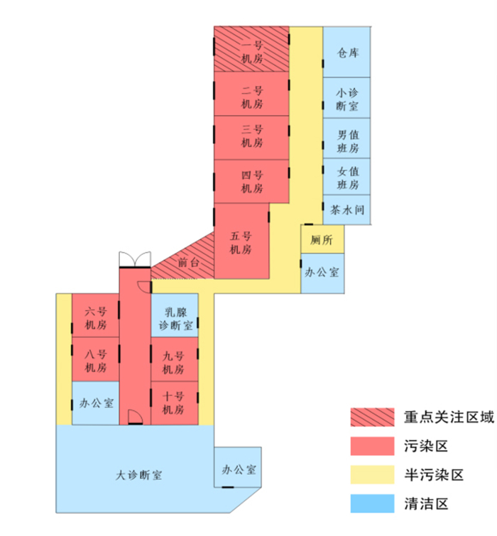
摘要: COVID-19疫情传播速度快、波及范围广,严重危害人民的健康,早期发现并隔离疑似或确诊患者对控制疫情至关重要。COVID-19的筛查离不开影像检查。本文总结了影像学在COVID-19疫情管理中的作用,并分析了影像检查流程中可能发生的院内感染因素,提出COVID-19疫情期间影像科室的应急管理策略,包括成立疫情防控管理小组、感染控制区域划分及严格管理、防护级别管理和用品配置、放射检查分类防控管理、提高诊断医师对疾病的鉴别能力、规范受检患者、场地环境、仪器设备和工作人员的管理等,以期对疫情防控有所帮助。Abstract: The epidemic of COVID-19 spreads quickly and widely, which seriously endangers people's health. Early detection and isolation of suspected or confirmed patients is essential to control the development of the epidemic. The screening of COVID-19 is inseparable from diagnostic imaging. This paper analyzes the possible infection factors in the process of examination. We put forward the emergency management strategy of medical imaging department during the epidemic situation of COVID-19, including establishment of epidemic control management team, division and strict administration of infection control area, protection level management and supplies configuration, classification management of radiation examination, enhancement of the disease differentiation ability, standardized management of patients, environment, devices and staff, and hoping to be helpful to the prevention and control of the epidemic.
-
[1] Huang C, Wang Y, Li X, et al. Clinical features of patients infected with 2019 novel coronavirus in Wuhan, China[J]. Lancet, 2020, 395 (10223): 497-506. doi: 10.1016/S0140-6736(20)30183-5 [2] 国家卫生健康委办公厅, 国家中医药管理局办公室.新型冠状病毒感染的肺炎诊疗方案(试行第五版)[EB/OL].[2020- 02-14]. http://www.gov.cn/zhengce/zhengceku/2020-02/09/5476407/files/7651e65b7d1443081053c29ad37fb07.pdf. [3] 霍力, 杨治, 等.新冠肺炎非定点救治医院核医学影像检查中的防控措施[J].中华放射医学与防护杂志, 2020, 40(3): 168-72. doi: 10.3760/cma.j.issn.0254-5098.2020.03.002 [4] 中华医学会影像技术分会.新型冠状病毒肺炎影像学检查院内感染防控管理:中华医学会影像技术分会推荐意见(第一版)[J].中华放射学杂志, 2020, 54(4): 286-91. doi: 10.3760/cma.j.cn112149-20200209-00115 [5] Lanaspa M, Annamalay AA, LeSouëf P, et al. Epidemiology, etiology, X-ray features, importance of co-infections and clinical features of viral pneumonia in developing countries[J]. Expert Rev Anti Infect Ther, 2014, 12(1): 31-47. doi: 10.1586/14787210.2014.866517 [6] 陈淮, 邹玉坚, 蓝博文, 等.重型和危重型新型冠状病毒肺炎患者床边胸部X线平片表现及其在随访中的作用[J/OL].中华放射学杂志, 2020, 54. http://rs.yiigle.com/yufabiao/1184724.htm. [7] Fang YC, Zhang HQ, Xie JC, et al. Sensitivity of chest CT for COVID-19: comparison to RT-PCR[J]. Radiology, 2020, 76: 200432-43. http://cn.bing.com/academic/profile?id=3628e86c5f809573c97537dda9e6df80&encoded=0&v=paper_preview&mkt=zh-cn [8] Ai T, Yang ZL, Hou HY, et al. Correlation of chest CT and RT-PCR testing in coronavirus disease 2019 (COVID-19) in China: a report of 1014 cases[J]. Radiology, 2020, 76: 200642-9. http://cn.bing.com/academic/profile?id=75dacdd44e5dda0149ec532800d839c3&encoded=0&v=paper_preview&mkt=zh-cn [9] Lei JQ, Li JF, Li X, et al. CT Imaging of the 2019 Novel Coronavirus (2019-nCoV) Pneumonia[J]. Radiology, 2020, 295(1): 18-27. doi: 10.1148/radiol.2020200236 [10] Chung M, Bernheim A, Mei XY, et al. CT Imaging Features of 2019 Novel Coronavirus (2019-nCoV)[J]. Radiology, 2020, 295(1): 202-7. doi: 10.1148/radiol.2020200230 [11] Xu X, Yu CC, Qu J, et al. Imaging and clinical features of patients with 2019 novel coronavirus SARS-CoV-2[J]. Eur J Nucl Med Mol Imaging, 2020, 47(5): 1275-80. doi: 10.1007/s00259-020-04735-9 [12] Shi HS, Han XY, Zheng CS. Evolution of CT manifestations in a patient recovered from 2019 novel coronavirus (2019-nCoV) pneumonia in Wuhan, China[J]. Radiology, 2020, 295(1): 20-31. doi: 10.1148/radiol.2020200269 [13] Duan YN, Qin J. Pre- and posttreatment chest CT findings: 2019 novel coronavirus (2019-nCoV) pneumonia[J]. Radiology, 2020, 295(1): 21-9. doi: 10.1148/radiol.2020200323 [14] Zhang K, Liu XH, Shen J, et al. Clinically applicable AI system for accurate diagnosis, quantitative measurements, and prognosis of COVID-19 pneumonia using computed tomography[J]. Cell, 2020, S8674(20): 30551-61. https://www.cell.com/cell/fulltext/S0092-8674(20)30551-1?rss=yes [15] Phan LT, Nguyen TV, Luong QC, et al. Importation and human- to-human transmission of a novel coronavirus in Vietnam[J]. N Engl J Med, 2020, 382(9): 872-4. doi: 10.1056/NEJMc2001272 [16] 秦耿耿, 蔡裕兴.南方医科大学南方医院新型冠状病毒放射检查防控指南(2020年1月24日修订稿)[EB/OL].[2020-01-24]. http://www.nfyy.com/ks/fzk/yxzdk. [17] 朱宏, 史文钊, 刘莉, 等.广东驰援湖北洪湖市医疗队的抗疫实践及思考[J/OL].中华医学杂志, 2020: 100. http://rs.yiigle.com/yufabiao/1184395.htm. -

 下载:
下载:
 点击查看大图
图(1)
计量
- 文章访问数: 703
- HTML全文浏览量: 364
- PDF下载量: 8
- 被引次数: 0