


Intelligent assisted diagnosis of COVID-19 based on CT images
-
摘要:
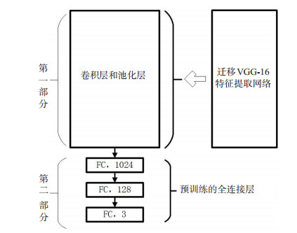
目的结合COVID-19患者肺部CT影像学特征,探讨深度学习技术在COVID-19辅助诊断上的价值。 方法搜集武汉大学中南医院和华中科技大学同济医学院确诊为COVID-19患者的部分CT影像资料构建小样本COVID-19数据集,将VGG-16具有提取高层抽象特征部分与设计的全连接层共同构成初步的基于迁移学习的COVID-19智能辅助诊断模型,使用COVID-19训练集迭代训练诊断模型,不断优化全连接层网络参数,最后训练出一个基于VGG-16卷积神经网络迁移学习的COVID-19智能辅助诊断模型。 结果在COVID-19测试集中早期、进展期和重症期3个类别的样本上,COVID-19智能辅助诊断模型测试的敏感度分别为0.95、0.93和0.96,F1 Score分别为0.98、0.95和0.92,综合的诊断准确率达到94.59%。 结论小样本数据集上采用迁移学习技术训练的COVID-19辅助诊断模型具有较高的可靠性,在防控疫情的关键时期,能快速地为医生提供诊断的参考意见,提高医生的工作效率。 Abstract:ObjectiveTo investigate the value of deep learning technology in the diagnosis of pneumonitis associated with new coronavirus infection in combination with CT imaging features of patients with new coronavirus infected pneumonia (COVID- 19). MethodsWe collected CT image data of patients diagnosed as COVID-19 in Central South Hospital of Wuhan University and Tongji Medical College of Huazhong University of Science and Technology to construct a small sample COVID-19 data set. VGG-16 was taken to extract high- level abstract features and design the full The connection layer together constitutes the preliminary COVID-19 intelligent auxiliary diagnosis model based on transfer learning. The COVID-19 training set was used to iteratively train the diagnosis model, continuously optimize the parameters of the fully connected layer network. We finally trained a migration based on the VGG-16 convolutional neural network. Learning COVID-19 intelligent assistant diagnosis model. ResultsOn the samples of the three categories of early, advanced and severe stages in the COVID-19 test set, the sensitivities of the COVID-19 intelligent auxiliary diagnostic model test were 0.95, 0.93 and 0.96, and the F1 Scores were 0.98, 0.95 and 0.92, respectively. The comprehensive diagnostic accuracy rate reached 94.59%. ConclusionThe COVID-19 assisted diagnosis model trained with transfer learning technology on a small sample data set has high reliability. It can provide doctors with reference opinions for diagnosis and improve doctors' work efficiency in the critical period of epidemic prevention and control. -
Key words:
- COVID-19 /
- CT imaging /
- assisted diagnosis /
- transfer learning
-
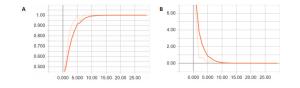
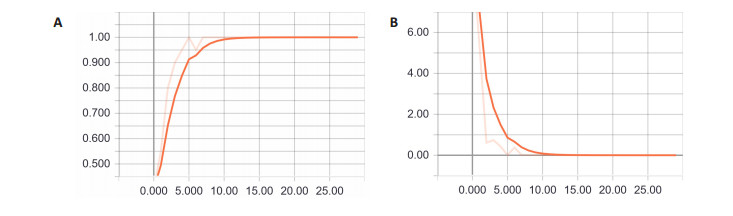
图 2 准确率和损失函数随迭代次数变化的曲线
A:准确率随迭代次数的变化曲线; B:损失函数随迭代次数的收敛曲线
Figure 2. Curves of accuracy and loss function as a function of the number of iterations

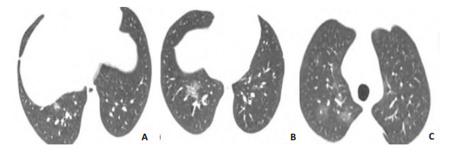
图 3 早期CT影像
COVID-19患者, 女, 32岁, 无明显症状, 胸部CT影像显示双肺下叶可见多发磨玻璃结节(A), 其中双肺下叶外侧基底段结节(B, C)边缘模糊[21]
Figure 3. Early CT images
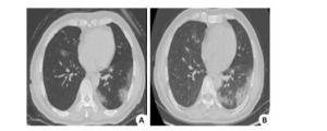
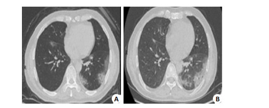
图 4 进展期CT影像
COVID-19确诊患者, 男, 57岁, 发热2 d. A: CT示左肺下叶胸膜下及右叶间裂附近有多发团、片状磨玻璃密度影; B: 4 d后复查, CT示病变进展, 左肺下叶病变范围增大, 内部密度增高[18]
Figure 4. CT images in advanced stage
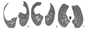
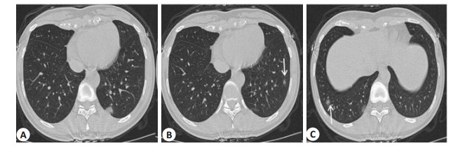
图 5 重症期CT影像
COVID-19确诊患者, 男, 30岁, 出现反复发热症状, COVID-19核酸检测阳性.A:出现发热症状后次日行胸部CT平扫,仅见右肺下叶有小片状密度增高影; B: 3 d后复查CT影像可见新增病灶,呈不规则GGO; C: 6 d后复查CT示双肺内多发淡薄GGO,较前明显进展, 右下肺病灶呈纤维条索影[22].
Figure 5. CT images of severe disease
表 1 COVID-19 CT数据集的划分
Table 1. Partitioning of the COVID-19 CT dataset
数据集 样本数(n) 样本张量 训练集 292 292×224×224 测试集 74 74×224×224  下载: 导出CSV
表 2 COVID-19 CT数据集在CT表现的分布情况
Table 2. Distribution of COVID-19 CT dataset in CT performance
CT表现 训练集 测试集 标签 早期 84 21 1 进展期 120 30 2 重症期 88 23 3
下载: 导出CSV
表 3 准确率和损失函数随迭代次数增加的变化情况
Table 3. Changes in accuracy and loss function with increasing number of iterations
迭代次数 准确率 损失函数值 迭代次数 准确率 损失函数值 迭代次数 准确率 损失函数值 1 0.4 9.9078 11 1 0.0033 21 1 0.0029 2 0.55 5.3854 12 1 0.0053 22 1 0.0016 3 0.8 0.6144 13 1 0.0020 23 1 0.0012 4 0.9 0.7374 14 1 0.0021 24 1 0.0015 5 0.95 0.4342 15 1 0.0026 25 1 0.0013 6 1 0.0110 16 1 0.0009 26 1 0.0016 7 0.95 0.3804 17 1 0.0047 27 1 0.0016 8 1 0.0182 18 1 0.0018 28 1 0.0012 9 1 0.0062 19 1 0.0025 29 1 0.0013 10 1 0.0047 20 1 0.0028 30 1 0.0012
下载: 导出CSV
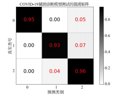
表 4 三分类混淆矩阵
Table 4. Three-class confusion matrix
混淆矩阵(n) 预测类别 0 1 2 0 20 0 1 真实类别 1 0 28 2 2 0 1 22
下载: 导出CSV
表 5 评估模型的方法
Table 5. Methods for evaluating models
诊断结果 敏感度 精确度 F1 Score 类别 0 0.95 1.00 0.98 1 0.93 0.97 0.95 2 0.96 0.88 0.92
下载: 导出CSV
-
[1] Xu X, Chen P, Wang J, et al. Evolution of the novel coronavirus from the ongoing Wuhan outbreak and modeling of its spike protein for risk of human transmission[J]. Sci China Life Sci, 2020, 63(3): 457- 60. http://www.wanfangdata.com.cn/details/detail.do?_type=perio&id=zgkx-ec202003015 [2] Zhu N, Zhang D, Wang W, et al. A Novel Coronavirus from Patients with Pneumonia in China, 2019[J]. N Engl J Med, 2020, 382(8): 727- 33 http://d.old.wanfangdata.com.cn/Periodical/zhjhhhx202003016 [3] Cheng VCC, Wong SC, To KKW, et al. Preparedness and proactive infection control measures against the emerging Wuhan coronavirus pneumonia in China[J]. J Hosp Infect, 2020, 104(3): 254-5. [4] Fang Y, Zhang H, Xie J, et al. Sensitivity of Chest CT for COVID-19: Comparison to RT-PCR[J]. Radiology, 2020, 2: 200432-8. [5] 谭鸣, 冯晓源, 刘士远, 等.新型冠状病毒肺炎影像检查诊断与感染控制指导意见[J/OL].中国医学计算机成像杂志, 2020: 1-19[2020- 04-19]. 10.19627/j.cnki.cn31-1700/th.20200309.001. [6] 王继元, 李真林, 蒲立新, 等.基于人工智能的正位DR胸片质控体系研究与应用[J].生物医学工程学杂志, 2020, 37(1): 158-68. http://www.wanfangdata.com.cn/details/detail.do?_type=perio&id=swyxgcx202001020 [7] 刘珍娟, 傅迎霞, 张羽, 等.不同CT图像重建算法下基于深度学习的肺结节检测算法效能[J].中国医学影像技术, 2019, 35(12): 1775-9. http://d.old.wanfangdata.com.cn/Periodical/zgyxyxjs201912003 [8] 谢未央, 陈彦博, 王季勇, 等.基于卷积神经网络的CT图像肺结节检测[J].计算机工程与设计, 2019, 40(12): 3575-81. http://d.old.wanfangdata.com.cn/Periodical/jsjgcysj201912035 [9] 李欣菱, 郭芳芳, 周振, 等.基于深度学习的人工智能胸部CT肺结节检测效能评估[J].中国肺癌杂志, 2019, 22(6): 336-40. http://d.old.wanfangdata.com.cn/Periodical/zgfazz201906002 [10] 刘晓鹏, 周海英, 胡志雄, 等.人工智能识别技术在T1期肺癌诊断中的临床应用研究[J].中国肺癌杂志, 2019, 22(5): 319-23. http://d.old.wanfangdata.com.cn/Periodical/zgfazz201905009 [11] 张鹏, 徐欣楠, 王洪伟, 等.基于深度学习的计算机辅助肺癌诊断方法[J].计算机辅助设计与图形学学报, 2018, 30(1): 90-9. http://d.old.wanfangdata.com.cn/Periodical/jsjfzsjytxxxb201801009 [12] Chen J, Wu L, Zhang J, et al. Deep learning-based model for detecting 2019 novel coronavirus pneumonia on high-resolution computed tomography: a prospective study[J/OL]. medRxiv, 2020. https://doi.org/10.1101/2020.02.25.20021568 [13] Song Y, Zheng S, Li L, et al. Deep learning Enables Accurate Diagnosis of Novel Coronavirus (COVID-19) with CT images[J/ OL]. medRxiv, 2020. https://doi.org/10.1101/2020.02.23.20026930. [14] Shen C, Yu N, Cai SB, et al. Quantitative computed tomography analysis for stratifying the severity of coronavirus disease 2019[J]. J Pharm Anal, 2020, 3: 510-8. http://d.old.wanfangdata.com.cn/Periodical/ywfxxb-e202002004 [15] Shi W, Peng X, Liu T. Deep learning-based quantitative computed tomography model in predicting the severity of COVID-19: A retrospective study in 196 patients[J/OL]. TheLancet, 2020. https://ssrn.com/abstract=3546089. [16] 赵双全, 周永生, 殷亮, 等. COVID-19的临床特征和CT表现[J].分子影像学杂志, 2020, 43(1): 59-63. doi: 10.12122/j.issn.1674-4500.2020.01.13 [17] 郭飞, 朱林, 许红, 等. COVID-19临床分型与MSCT容积扫描间的相关性[J].南方医科大学学报, 2020, 40(3): 321-6. http://d.old.wanfangdata.com.cn/Periodical/dyjydxxb202003004 [18] 钟飞扬, 张寒菲, 王彬宸, 等.新型冠状病毒肺炎的CT影像学表现[J].武汉大学学报:医学版, 2020, 41(3): 345-8. [19] Litjens G, Kooi T, Bejnordi BE, et al. A survey on deep learning in medical image analysis[J]. Med Image Anal, 2017, 42: 60-88. http://d.old.wanfangdata.com.cn/Periodical/gjzdhyjszz-e201806001 [20] Pan SJ, Yang Q. A survey on transfer learning[J]. IEEE T Knowl Data En, 2010, 22(10): 1345-59. http://d.old.wanfangdata.com.cn/OAPaper/oai_doaj-articles_84078f189d8147bc08febd7908b6de2b [21] 高璐, 张静平, 杜永浩, 等.输入性新型冠状病毒肺炎的CT表现[J].西安交通大学学报:医学版, 2020, 41(3): 429-34. http://d.old.wanfangdata.com.cn/Periodical/xaykdxxb202003022 [22] 管汉雄, 熊颖, 申楠茜, 等.新型冠状病毒肺炎(COVID-19)临床影像学特征[J].放射学实践, 2020, 35(2): 125-30. http://d.old.wanfangdata.com.cn/Periodical/zggrkzzz202003003 [23] 卫生健康委办公厅, 国家中医药管理局办公室.关于印发新型冠状病毒肺炎诊疗方案(试行第六版)的通知[EB/OL]. [2020-02-19]. http://www.gov.cn/zhengce/zhengceku/2020-02/19/content_5480948.htm. [24] 卫生健康委办公厅, 国家中医药管理局办公室.关于印发新型冠状病毒肺炎诊疗方案(试行第七版)的通知[EB/OL]. [2020-03-03]. http://www.nhc.gov.cn/yzygj/s7653p/202003/46c9294a7dfe4cef80dc7f5912eb1989.shtml. [25] 杜永浩, 金晨望, 杨健, 等.家庭聚集性早期新型冠状病毒肺炎的临床与CT表现初步探讨[J].西安交通大学学报:医学版, 2020, 41(3): 435-8. http://www.wanfangdata.com.cn/details/detail.do?_type=perio&id=xaykdxxb202003023 [26] van Ginneken B, Schaefer-Prokop CM, Prokop M. Computer-aided diagnosis: how to move from the laboratory to the clinic[J]. Radiology, 2011, 261(3): 719-32. [27] 夏静, 潘素, 颜默磊, 等.基于迁移学习的小样本重症疾病预后模型[J].生物医学工程学杂志, 2020, 37(1): 1-9. http://www.wanfangdata.com.cn/details/detail.do?_type=perio&id=swyxgcx202001001 -

 点击查看大图
计量
- 文章访问数: 856
- HTML全文浏览量: 344
- PDF下载量: 22
- 被引次数: 0