


CT differential diagnosis of adenoma and carcinoid of appendix
-
摘要:
目的研究对阑尾粘液囊腺瘤和类癌的CT鉴别诊断。 方法选择50例阑尾粘液囊腺瘤患者和20例类癌患者,其中男性32例,女性38例,年龄57.15±5.28岁。通过CT检测成像系统检测阑尾粘液囊腺瘤和类癌表现症状,对患者阑尾图像阑尾壁厚度、直径、钙化情况、周围侵犯情况、阑尾壁增厚、壁异常强化、粪石发生率以及检出率和准确率进行对比分析,同时对CT检测异质性分析和相关影响因素进行总结。 结果阑尾粘液囊腺瘤和类癌患者CT检查图像中阑尾壁厚度、直径、钙化情况、周围侵犯情况、阑尾壁增厚、壁异常强化、粪石发生率以及检出率和准确率的差异存在统计学意义(P<0.01),且类癌组患者较囊腺瘤组患者症状更显著。异质性检测结果显示,类癌组的敏感度、特异度、阳性比、阴性比和诊断比值比最大,且病程、年龄和发病范围与CT评估阑尾粘液囊腺瘤和类癌诊断价值的相关性最高,而囊腺瘤体积与CT评估阑尾粘液囊腺瘤和类癌诊断价值的相关性较低。 结论CT鉴别诊断阑尾粘液囊腺瘤和类癌的价值较高,值得临床应用广泛推广。 Abstract:ObjectiveTo explore the CT differential diagnosis of mucinous cystadenoma and carcinoid of appendix. MethodsWe choosed 50 cases of patients with appendiceal mucous cystadenoma and 20 cases of patients with carcinoid. There were 32 males and 38 females with an average age of 57.15±5.28 years old. We analyzed image of patients appendix appendiceal wall thickness, diameter, calcification, infringement, appendix around abnormal wall thickening, wall reinforcement, incidence of bezoar and detection rate and accuracy by CT detection imaging system mucous cystadenoma and carcinoid appendix presenting symptoms. The CT detection heterogeneity analysis and related influence factors were analyzed. ResultsThere were significant differences between the CT images of the patients with appendiceal mucinous adenoma and carcinoid in the thickness, diameter, calcification, peripheral invasion, wall thickening, abnormal wall strengthening, fecality incidence, detection rate and accuracy (P<0.05). The patients in the carcinoid group had more significant symptoms than those in the cystadenoma group. The sensitivity of the carcinoid group, specific degrees, masculine, feminine and diagnostic odds ratio was the largest. The course, age and disease range and CT evaluation carcinoid appendix mucous cystadenoma, and diagnostic value of the highest correlation, and the capsule gland tumors had product and evaluation of CT carcinoid appendix mucous cystadenoma and diagnostic value of correlation were low. ConclusionCT has a high value in the differential diagnosis of appendiceal mucinous cystadenoma and carcinoid. -
Key words:
- appendiceal burs adenoma /
- type of cancer /
- CT identification /
- diagnostic studies
-

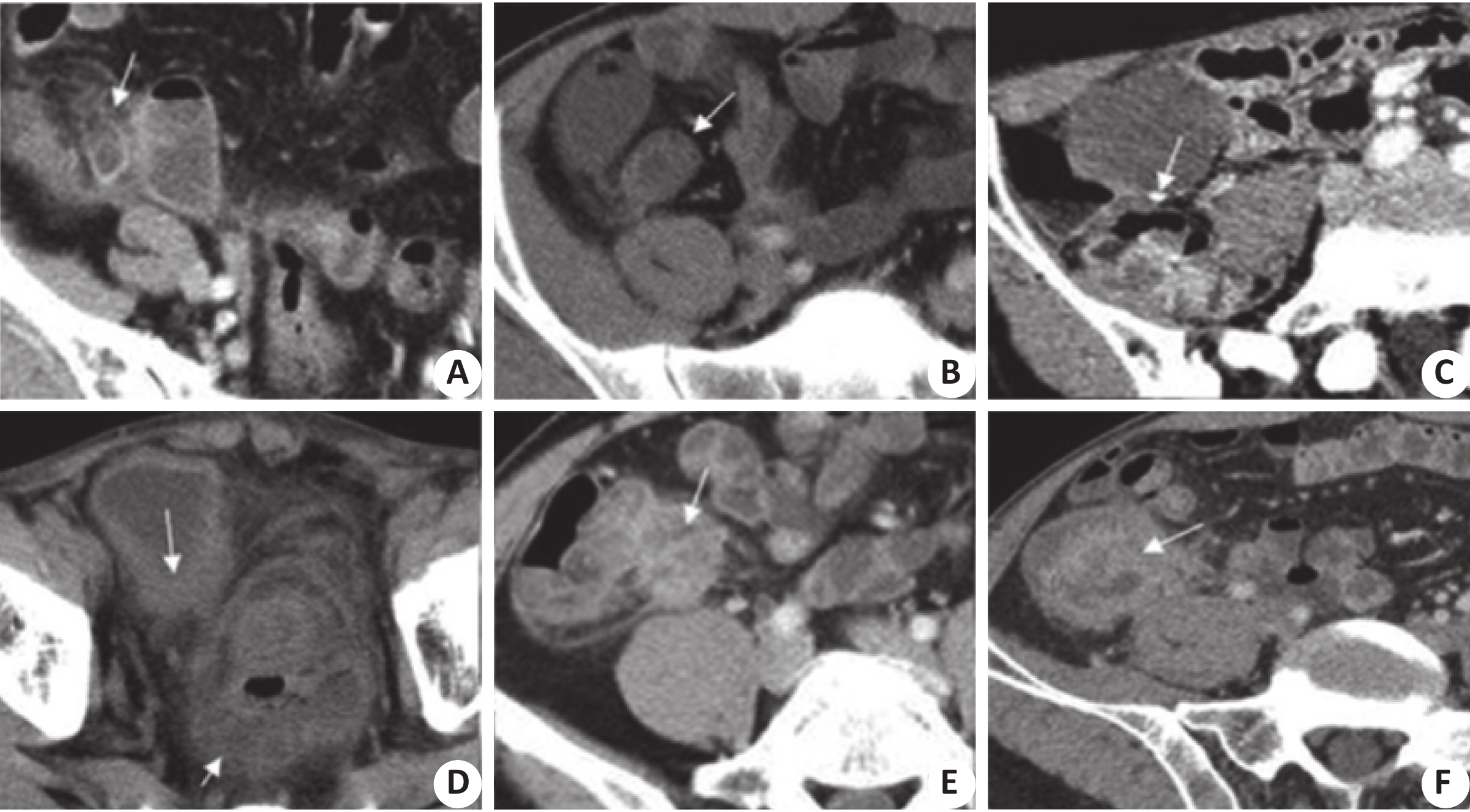
图 1 阑尾粘液囊肿患者CT表现
A:患者表现为阑尾粘液囊肿,壁薄光整,伴有阑尾周围炎征象;B:患者表现为阑尾粘液囊肿正常,阑尾远端囊性肿块,周围脂肪间隙清晰;C:患者表现为阑尾粘液囊腺瘤,囊肿5 cm,周围存在脂肪条纹征;D:患者表现为阑尾粘液囊腺瘤伴急性阑尾炎,阑尾远端呈囊实性肿块,囊壁明显强化,周围脂肪条纹征;E:患者表现为阑尾粘液囊腺癌,阑尾远端囊实性肿块,壁结节强化明显.
Figure 1. CT manifestations of appendicitis mucocyst

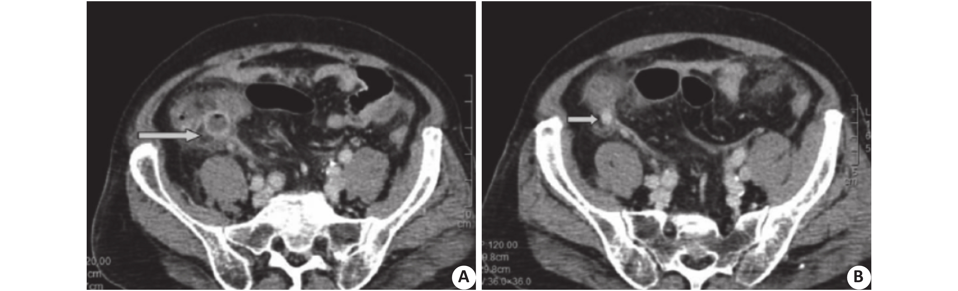
图 2 阑尾类癌患者CT表现
阑尾类癌合并急性阑尾炎CT示增粗的阑尾(箭头表示增粗肿胀的阑尾,周围大量粘液渗出,且呈明显结节状强化,直径约1 cm)。
Figure 2. CT manifestations of carcinoid of appendix
表 1 患者CT检查阑尾图像直径、阑尾壁厚度、钙化情况以及周围侵犯情况
Table 1. Appendix image diameter, appendix wall thickness, calcification and surrounding invasion of patients in CT detection
组别 直径(cm) 阑尾壁厚度(%) 是否钙化(%) 周围侵犯情况(%) 囊腺瘤组(n=50) 1~3 52 42 62 类癌组(n=20) 3~10 68 46 76 t 2.395 2.141 2.139 2.054 P 0.005 0.001 0.012 0.001  下载: 导出CSV
表 2 患者CT检查阑尾图像阑尾壁增厚、壁异常强化及粪石发生率
Table 2. Appendiceal wall thickening, wall abnormal strengthening and the incidence of faecal stone examined by CT
组别 阑尾壁增厚(%) 壁异常强化(%) 粪石发生率(%) 囊腺瘤组(n=50) 52 42 62 类癌组(n=20) 74 52 66 t 2.167 3.139 1.284 P 0.001 0.012 0.001
下载: 导出CSV
表 3 CT鉴别诊断检出前阑尾粘液囊腺瘤和类癌的结果
Table 3. CT results of differential diagnosis of anterior appendicitis mucinous cystadenoma and carcinoid carcinoma
组别 检出率(%) 准确率(%) 囊腺瘤组(n=50) 90.56 85.32 类癌组(n=20) 95.68 90.26 t 8.265 6.387 P 0.006 0.009
下载: 导出CSV
表 4 CT评估阑尾粘液囊腺瘤和类癌的异质性检测
Table 4. Heterogeneity of CT assessment of adenoma and carcinoid carcinoma of the appendicular mucus capsule
组别 敏感度 特异度 阳性比 阴性比 诊断比值比 囊腺瘤组(n=50) 0.83 0.81 4.21 0.19 23.65 类癌组(n=20) 0.95 0.86 5.56 0.23 56.12 t 7.145 8.236 6.114 7.265 8.221 P 0.002 0.001 0.012 0.025 0.002
下载: 导出CSV
表 5 影响CT评估阑尾粘液囊腺瘤和类癌诊断价值的相关因素分析
Table 5. Analysis of relevant factors affecting CT assessment of diagnostic value of appendicitis mucinous cystadenoma and carcinoid
相关因素 囊腺瘤体积 病程 年龄 发病范围 囊腺瘤体积 3.265 1.456 3.214 3.113 病程 1.256 1.254 3.265 3.244 年龄 1.256 1.225 5.689 7.012 发病范围 1.632 2.564 2.336 1.236 F 2.564 7.214 10.112 9.264 P 0.012 0.006 0.006 0.002
下载: 导出CSV
-
[1] Legué LM, Gj C, Ihjt DH, et al. Huysentruyt.review:pathology and its clinical relevance of mucinous appendiceal neoplasms and PseudomyxomaPeritonei[J]. Clin Colorectal Cancer, 2019, 18(1): 1-7. doi: 10.1016/j.clcc.2018.11.007 [2] Nutu OA, Marcacuzcoquinto AA, Manriquemunicio A, et al. Mucinous appendiceal neoplasms:incidence,diagnosis and surgical treatment[J]. Cirugía Española (English Edition), 2017, 95(6): 321-7. doi: 10.1016/j.cireng.2017.07.011 [3] Quadri R, Vasan V, Hester C, et al. Comprehensive review of typical and atypical pathology of the appendix on CT:cases with clinical implications[J]. Clin Imaging, 2019, 53(2): 65-77. [4] Menassel B, Duclos A, Passot G, et al. Preoperative CT and MRI prediction of non-resectability in patients treated for pseudomyxomaperitonei from mucinous appendicealneoplasms[J]. Europ J Surg Oncol, 2016, 42(4): 558-66. doi: 10.1016/j.ejso.2016.01.005 [5] Lord AC, Shihab O, Chandrakumaran K, et al. Recurrence and outcome after complete tumour removal and hyperthermicintraperitoneal chemotherapy in 512 patients with pseudomyxomaperitonei from perforated appendiceal mucinous tumors[J]. Europ J Surg Oncol, 2015, 41(3): 396-9. doi: 10.1016/j.ejso.2014.08.476 [6] 韩太林, 赵慧萍, 曾蒙苏, 等. 阑尾黏液性肿瘤的CT和MRI诊断及良恶性鉴别[J]. 放射学实践, 2014, 29(7): 808-13. [7] 潘江峰, 马周鹏, 厉学民, 等. 阑尾粘液性囊腺癌的CT表现及临床分析[J]. 中华普通外科杂志, 2016, 31(4): 350-1. doi: 10.3760/cma.j.issn.1007-631X.2016.04.029 [8] Noh BJ, Kim YW, Park YK. A rare,low-grade appendiceal mucinous neoplasm(Pseudomyxomaperitonei)with ossification:a case report with morphoproteomic analysis of bone formation[J]. Pathol Res Pract, 2016, 212(11): 1085-8. doi: 10.1016/j.prp.2016.09.015 [9] 陈 明, 查云飞, 王艳艳. 阑尾黏液性囊腺瘤CT表现及文献复习[J]. 实用放射学杂志, 2016, 32(1): 147-9. doi: 10.3969/j.issn.1002-1671.2016.01.039 [10] 张 娣, 王红霞, 张培功, 等. 阑尾黏液性肿瘤影像学表现及病理相关性研究[J]. 实用放射学杂志, 2017, 33(1): 69-71. doi: 10.3969/j.issn.1002-1671.2017.01.019 [11] Sk BB, Jasuja P. Appendicealmucocele-a rare case report[J]. Int J Surg Case Rep, 2019, 58(3): 21-5. [12] Komo T, Kohashi T, Hihara J, et al. Intestinal obstruction caused by low-grade appendiceal mucinous neoplasm:a case report and review of the literature[J]. Int J Surg Case Rep, 2018, 51(6): 37-40. [13] 李彩琴, 王振光, 王希林, 等. 多层螺旋CT在急性阑尾炎诊断中的应用价值[J]. 实用放射学杂志, 2016, 32(7): 1059-62. doi: 10.3969/j.issn.1002-1671.2016.07.017 [14] 李玉广, 石新兰, 贾 静, 等. 阑尾低级别粘液性肿瘤的病理特征[J]. 世界最新医学信息文摘, 2016, 16(87): 242-3. [15] Misdraji J. Mucinous epithelial neoplasms of the appendix and pseudomyxoma peritonei[J]. Mod Pathol, 2015, 28(Suppl 1): S67-79. [16] Rouchaud A, Glas L, Gayet M, et al. Appendiceal mucinous cystadenoma[J]. Diagn Interv Imaging, 2014, 95(1): 113-6. doi: 10.1016/j.diii.2013.07.015 [17] 陈瑞云, 张 琳, 孙国峰, 等. 腹腔镜手术治疗阑尾粘液性肿瘤的临床体会[J]. 腹腔镜外科杂志, 2017, 22(8): 612-4. [18] 陈 玲, 张 超, 童梦玲, 等. 多层螺旋CT及薄层重建技术在阑尾病变中的诊断价值[J]. 世界最新医学信息文摘, 2017, 17(71): 137-9. [19] 彭贵平, 梁 敏, 李智惠. 13例阑尾粘液囊肿的超声表现及临床分析[J]. 现代诊断与治疗, 2018, 29(1): 65-6. doi: 10.3969/j.issn.1001-8174.2018.01.032 [20] 林莉萍, 蔡春仙. 阑尾粘液囊肿的影像表现[J]. 现代诊断与治疗, 2016, 27(21): 4094-5. doi: 10.3969/j.issn.1001-8174.2016.21.061 [21] 张 娣, 王红霞, 张培功, 等. 阑尾粘液性肿瘤的影像学表现及病理相关性研究[J]. 实用放射学杂志, 2017, 33(1): 102-6. [22] 李高峰, 黄耀华, 杨贤卫. 阑尾粘液性囊腺瘤1例并文献复习[J]. 罕少疾病杂志, 2017, 24(6): 71-2. doi: 10.3969/j.issn.1009-3257.2017.06.029 [23] 李逢芳, 张 娣, 陈慧铀, 等. MSCT对阑尾囊性囊腺瘤的诊断价值[J]. 中国医疗设备, 2016, 31(2): 73-6. doi: 10.3969/j.issn.1674-1633.2016.02.018 [24] 刘 瑞, 米 英, 高芬霞. 阑尾粘液性囊腺瘤误诊为卵巢肿瘤1例[J]. 临床医药文献电子杂志, 2017, 4(29): 5717-8. [25] 龚碧云, 丁旭恩, 陈苍松, 等. 阑尾粘液性肿瘤的多层螺旋CT表现及诊断价值[J]. 中外医疗, 2019, 23(18): 189-91. -

 点击查看大图
计量
- 文章访问数: 865
- HTML全文浏览量: 427
- PDF下载量: 10
- 被引次数: 0