


FMRI study of motor, sensory and speech-related brain areas in healthy middle-aged and elderly women after the first intervention of international standard scalp acupuncture
-
摘要:
目的运用静息态脑功能成像(fcMRI)技术,探讨在健康中老年女性人群中施行头针组穴针刺后脑区激活的情况。 方法本研究于2017年1~5月招募11例已自愿签署知情同意书的健康中老年女性志愿者,年龄60~63岁(56.18±3.82岁),于顶中线、左侧顶颞前斜线和顶颞后斜线施行头针刺激。针刺前、后采用西门子3.0 T超导MRI获取受试者的fcMRI数据,并基于Matlab 2012a平台,使用DPABI、SPM12和Rest1.8工具包进行数据分析和作图,以低频振幅比率和局部一致性(ReHo)作为结局指标。 结果最终10例受试者接受国际标准头针干预后脑局部活动变化如下:以右侧小脑后叶、小脑角、悬雍垂和山坡为主的脑区低频振幅比率降低(T= −6.576 1),而以左侧角回、楔前叶和BA7为主的脑区低频振幅比率增强(T= 6.219 3)。以左梭形回(T= −5.609 5)为主,以左侧脑桥(T= −6.431 6)为主,以颞上回和海马旁回(T= −6.442 4)为主,以右侧距状裂周围皮层(T= −5.520 1)为主和以右侧梭形回(T= −6.477 6)为主的脑区ReHo降低,而双侧楔前叶和BA7、角回为主(T= 4.822 4、5.606 4),左颞中回和颞上回为主(T= 6.745 8),左顶下小叶和中央后回为主(T= 8.089 1),左边缘叶、扣带回、辅助运动区、中央前回、中央后回(T= 6.714 1)为主的脑区ReHo增强。 结论健康中老年女性脑功能在国际标准头针首次干预后,与感觉、肢体运动、言语功能相关的脑区出现特异性改变。 -
关键词:
- 静息态脑功能成像技术 /
- 国际标准头针 /
- 经穴特异性
Abstract:ObjectiveTo investigate the activation of brain regions after international standard scalp acupuncture on healthy middle-aged and elderly women using resting state brain functional imaging (Rs-fMRI). MethodsWe recruited 11 healthy middle-aged and elderly female volunteers who had voluntarily signed informed consent from January 2017 to May 2017, with the age from 60 to 63 years old (average 56.18±3.82). They had voluntarily signed informed consent were recruited to receive scalp acupuncture stimulation at MS5, the left MS6, and the left MS7. Rs-fMRI data of subjects were obtained by Siemens 3.0 t superconducting MRI before and after acupuncture. It was based on Matlab 2012a platform, using DPABI、SPM12 and REST1.8 software for data analysis and mapping with fractional amplitude low-frequency fluctuation (fALFF) and regional homogeneity (ReHo) as an outcome indicator. ResultsFinally, the changes of local brain activity after the international standard scalp acupuncture intervention in ten subjects were as follows: fALFF decreased at the right posterior cerebellar lobe, cerebellopontine angle, uvula and declive (T= −6.576 1), and increased at the left angular gyrus, precuneus and BA7 (T= 6.219 3). ReHo decreased at mainly the left spindle gyrus (T= −5.609 5), and the land the left pontine (T= −6.431 6), and mainly the superior temporal gyrus (BA38) and para hippocampus (T= −6.442 4), and decreased at Cortical cortex (T=−5.520 1) and right fusiform gyrus (T= −6.477 6) as well, but increased at the bilateral anterior wedge, BA7, and angular gyrus (T=4.822 4 and 5.606 4), the left middle temporal superior and superior temporal gyrus (BA21, BA22) are increased (T= 6.745 8), and increased at mainly the left top parietal lobe and central posterior gyrus (T= 8.089 1) are increased, and the left marginal lobe, cingulate gyrus (BA24), auxiliary sports area, central anterior gyrus (BA4), central posterior gyrus (BA3) (T= 6.714 1) are increased too. ConclusionBrain function in healthy middle-aged and elderly women shows specific changes in brain areas related to sensory, motor and speech functions after the first intervention with the international standard scalp acupuncture. -
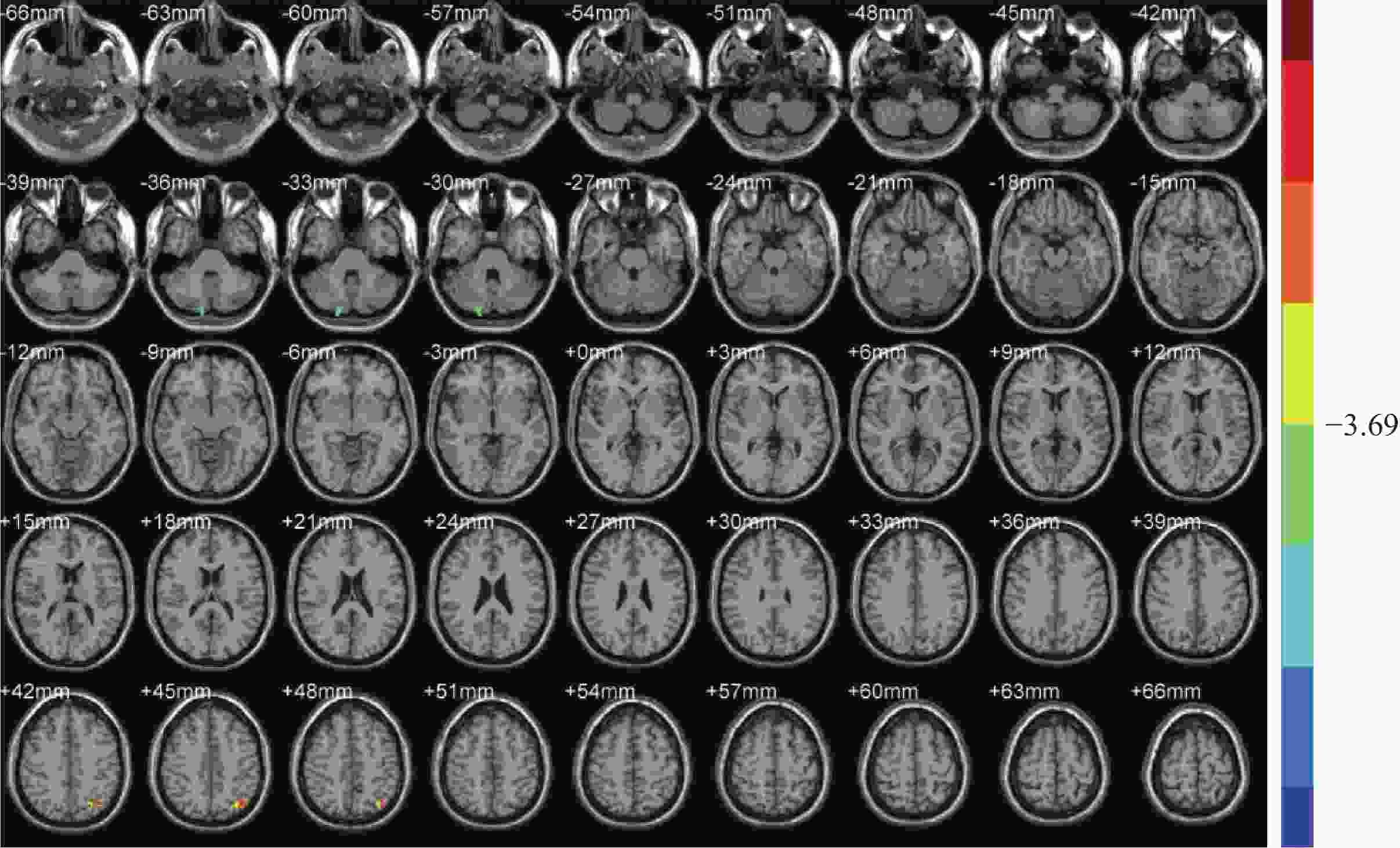
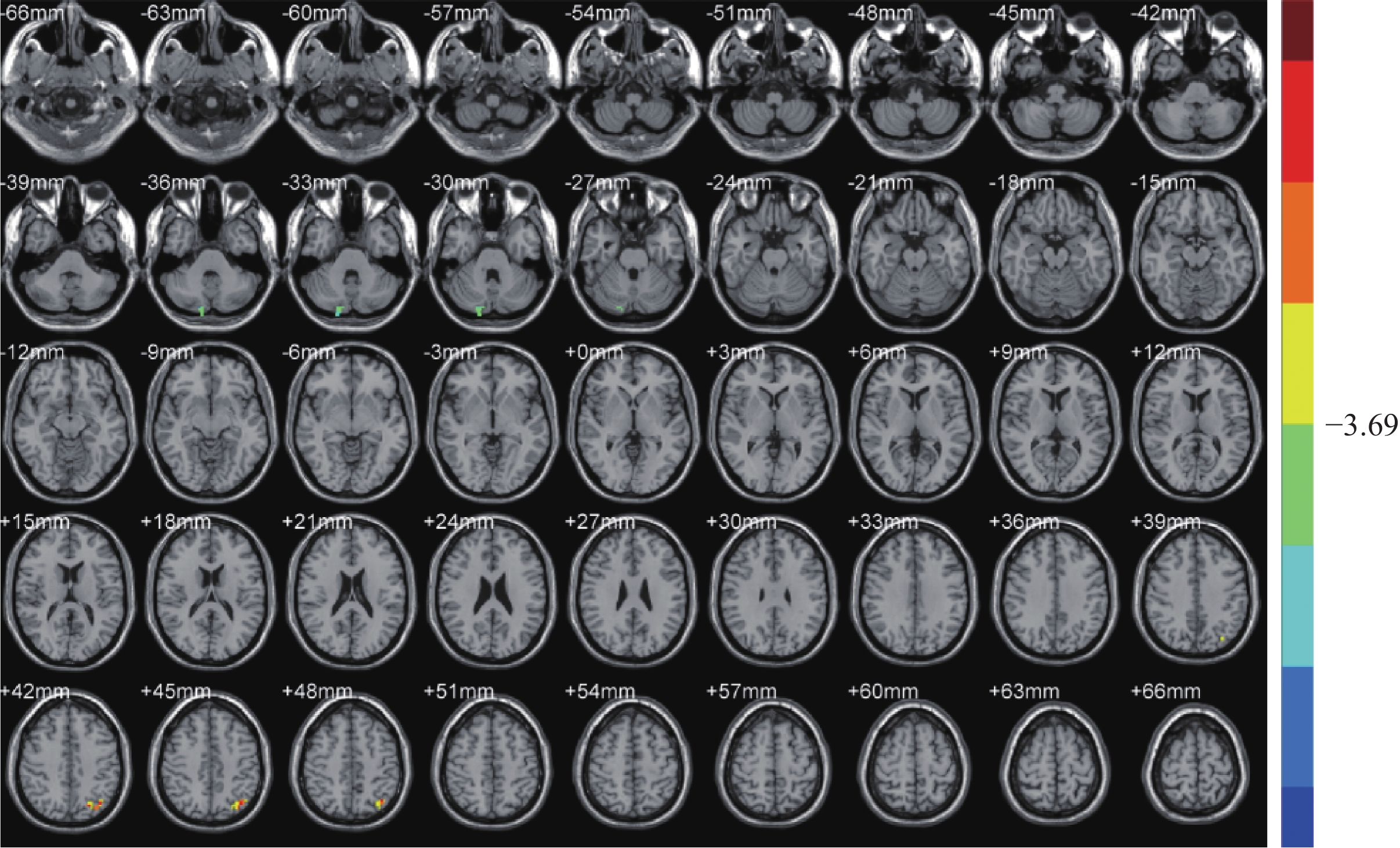
图 1 国际标准头针首次干预后fALFF变化的脑区
Figure 1. Brain regions of fALFF after first intervention of international standard scalpel
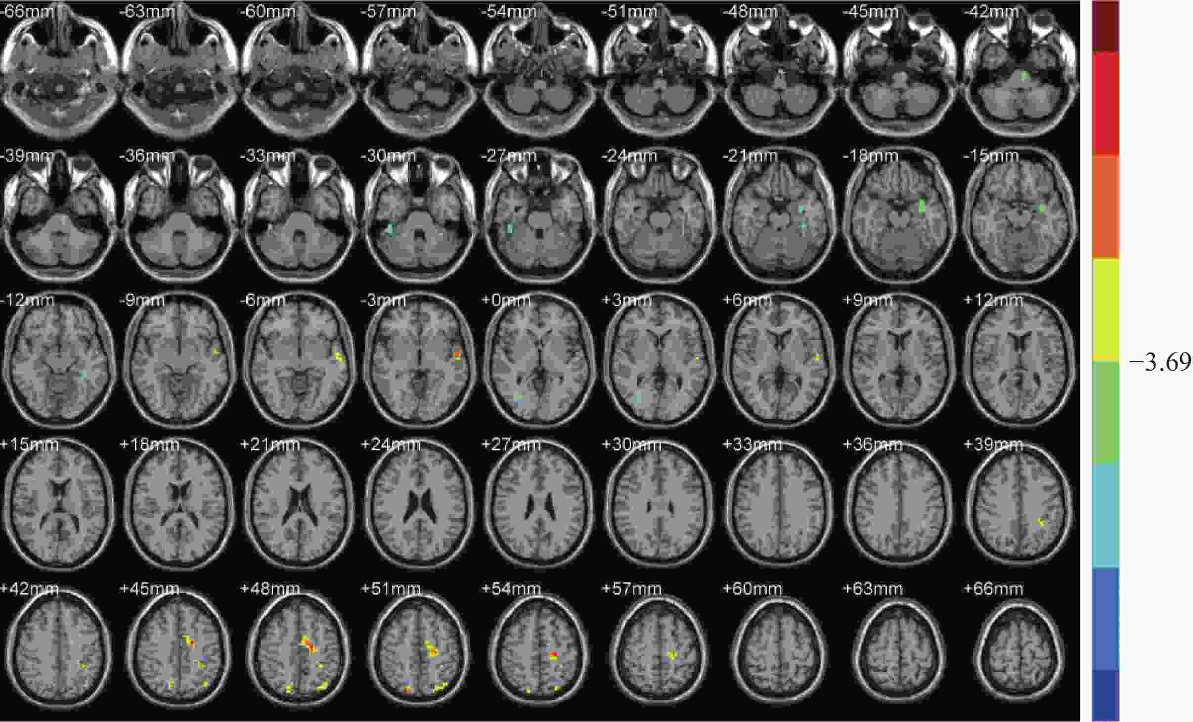
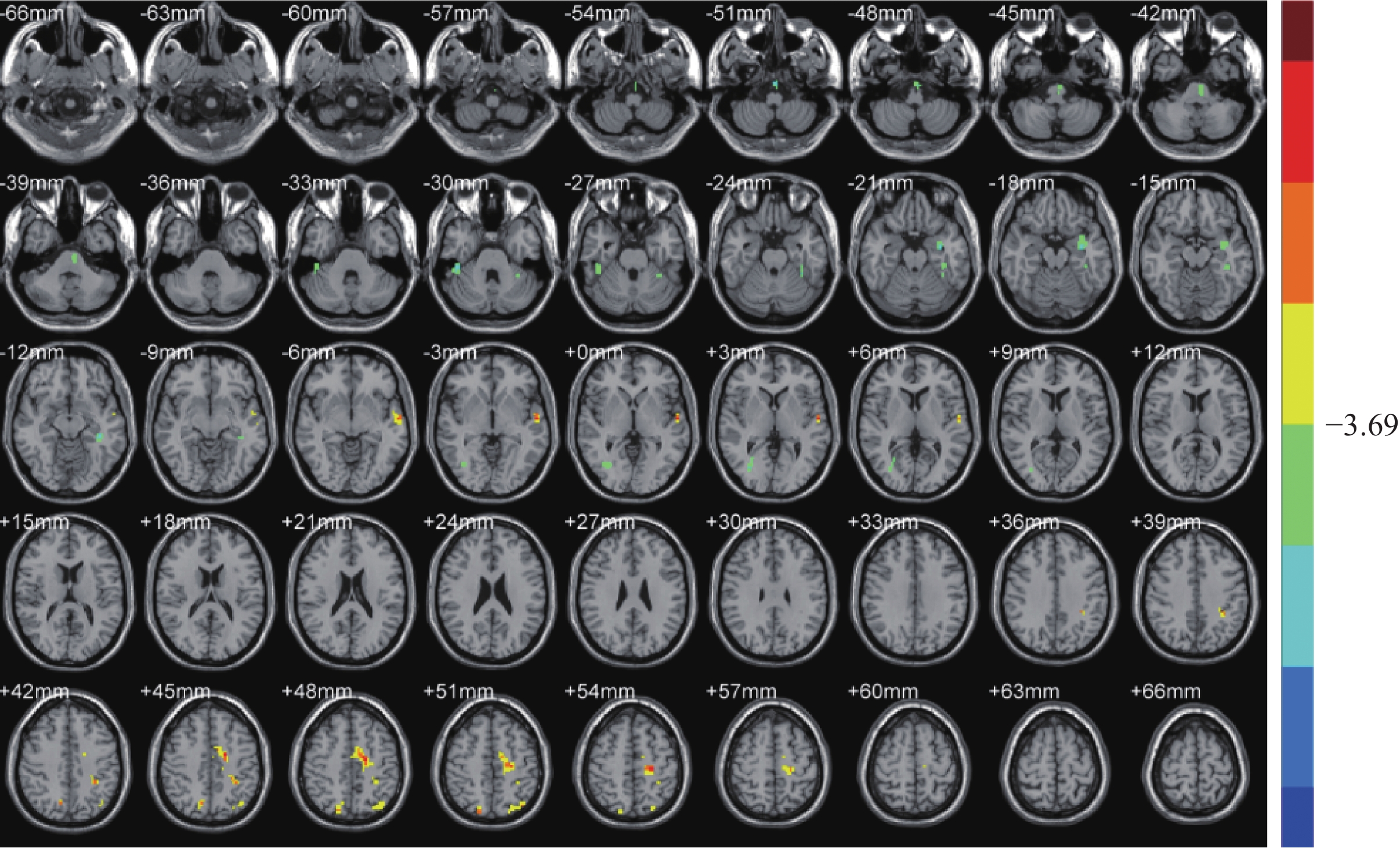
图 2 国际标准头针首次干预后ReHo变化的脑区
Figure 2. Brain regions and the location of ReHo after first intervention of international standard scalpel
表 1 国际标准头针首次干预后fALFF和ReHo变化的脑区及其定位
Table 1. Brain regions and the location of fALFF and ReHo after first intervention of international standard scalpel
参数 脑区效应 脑区 MiNi坐标 激活强度(T) X Y Z fALFF 降低 右小脑后叶、小脑中脚、悬雍垂和山坡 15 −84 −36 −6.576 1 增强 左角回、楔前叶、BA7 −36 −75 45 6.219 3 ReHo 降低 右距状裂周围皮层 33 −72 3 −5.520 1 降低 右梭形回 48 −33 −30 −6.477 6 降低 左梭形回 −39 −33 −12 −5.609 5 降低 左脑桥 −3 −15 −51 −6.431 6 降低 左颞上回、海马旁回 −36 −3 −18 −6.442 4 增强 左颞中回和颞上回 −57 −9 −3 6.745 8 增强 左顶下小叶和中央后回 −33 −39 39 8.089 1 增强 左边缘叶、扣带回、辅助运动区、
中央前回、中央后回−18 −12 48 6.714 1 增强 左角回、BA7、楔前叶 −39 −72 51 4.822 4 增强 右楔前叶、BA7 18 −78 48 5.606 4 X、Y、Z代表脑空间定位轴,分别表示在MiNi标准化空间坐标系中左右,前后和上下方位的坐标位置.  下载: 导出CSV
-
[1] 柳默涵, 王述菊, 马 骏, 等. 头针治疗帕金森病的Meta分析[J]. 时珍国医国药, 2019, 30(8): 2011-4. [2] 朱增越, 高 旸. 头针与常规疗法治疗失眠疗效比较的随机对照试验的系统评价与Meta分析[J]. 上海针灸杂志, 2018, 37(06): 713-9. [3] 但小龙. 针刺治疗脑卒中后失语的系统评价[D]. 成都: 成都中医药大学, 2015. [4] Russell AP, Jeanette AM, Thomas EN[著]. 功能磁共振成像数据分析手册[M]. 马国林等译. 北京: 人民军医出版社, 2017. [5] 姜思竹. 基于静息态fMRI和DTI探讨头针治疗脑卒中偏瘫的机制研究[D]. 北京: 北京中医药大学, 2017. [6] Chung WY, Liu SY, Gao JC, et al. Modulatory effect of international standard scalp acupuncture on brain activation in the elderly as revealed by resting-state fMRI[J]. Neur Regenerat Res, 2017, 14(12): 2126-31. [7] 李佳子. 基于分数低频振幅算法的大脑功能性别差异的静息态功能磁共振研究[D]. 合肥: 安徽医科大学, 2017. [8] 针灸技术操作规范第2部分: 头针[Z].2008, GB/T 21709.2-2008. [9] 薛红莉, 薛 贵. 阅读能力个体差异的神经机制研究进展[J]. 当代语言学, 2016, 18(4): 549-67. [10] 刘梦秋, 刘 婕, 刘 影. 采用静息态功能磁共振研究先天性重度感音神经性耳聋患儿相关脑区的ALFF值[J]. 磁共振成像, 2019, 10(9): 650-4. [11] 韩红艳, 王柯柯, 王文献, 等. 针刺间使穴前后健康人脑内静息态fALFF时变效应分析[J]. 中国中医基础医学杂志, 2019, 25(7): 972-5, 998. [12] Margulies DS, Vincent JL, Kelly C, et al. Precuneus shares intrinsic functional architecture in humans and monkeys[J]. Proc Natl Acad Sci USA, 2009, 106(47): 20069-74. doi: 10.1073/pnas.0905314106 [13] 徐君海. 静息状态下脑网络及其与注意的关系[D]. 济南: 山东大学, 2014. [14] 张傲霜. 男性精神分裂症患者源记忆的神经心理学和脑磁图研究[D].南京: 南京医科大学, 2009. [15] 张吉昌. 独立成分分析在静息态功能磁共振成像中定位语言网络的研究[D]. 石家庄: 河北工业大学, 2016. [16] 付晓璇. 基于静息态下功能磁共振的语言网络功能连接的研究[D]. 石家庄: 河北工业大学, 2016. [17] Singh-Curry V, Husain M. The functional role of the inferior parietal lobe in the dorsal and ventral stream dichotomy[J]. Neuropsychologia, 2009, 47(12): 1434-48. [18] 余 寒, 龙珊珊, 蒋 硕, 等. 氟西汀联合重复经颅磁刺激低频刺激辅助运动区治疗强迫障碍的研究[J]. 四川医学, 2019, 40(8): 811-4. [19] 贾建平, 陈生弟. 神经病学[M]. 8版. 北京: 人民卫生出版社, 2018. [20] Fitzpatrick LE, Crowe SF. Cognitive and emotional deficits in chronic alcoholics:a role for the cerebellum[J]. Cerebellum, 2013, 12(4): 520-33. doi: 10.1007/s12311-013-0461-3 [21] 姜树军, 孙 勍, 陈宏义, 等. 巴拉尼协会血流动力性直立性头晕/眩晕诊断标准解读[J]. 北京医学, 2019, 41(9): 832-4. [22] 韩中胜, 乔健天. 小脑: 它的组件式神经元环路是如何进行运动学习和经典式条件反射活动的[J]. 生理科学进展, 2008, 27(1): 15-20. [23] 隆秀灵, 夏振元, 戴 旖, 等. 健康人群不同年龄组小脑脚及小脑上脚交叉的DTI相关参数特征分析[J]. 中国临床新医学, 2019, 12(6): 642-5. doi: 10.3969/j.issn.1674-3806.2019.06.16 [24] Christopher B, Diane M, Li FF. Differential connectivity within the parahippocampal place area[J]. NeuroImage, 2013, 75(3): 228-37. [25] Brunyé TT, Moran JM, Holmes A, et al. Non-invasive brainstimulation targeting the right fusiform gyrus selectively increases working memory for faces[J]. Brain Cogn, 2017, 113(2): 32-9. [26] Jonas J, Rossion B, Brissart H, et al. Beyond the core face- processing network: Intracerebral stimulation of a face-selective area in the right anterior fusiform gyrus elicits transient prosopagnosia[J]. Cortex, 2015, 72(2): 140-55. -

 点击查看大图
计量
- 文章访问数: 605
- HTML全文浏览量: 295
- PDF下载量: 12
- 被引次数: 0