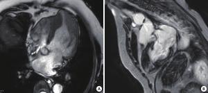
Application value of magnetic resonance imaging in patients with apical hypertrophy cardiomyopathy
-
摘要:
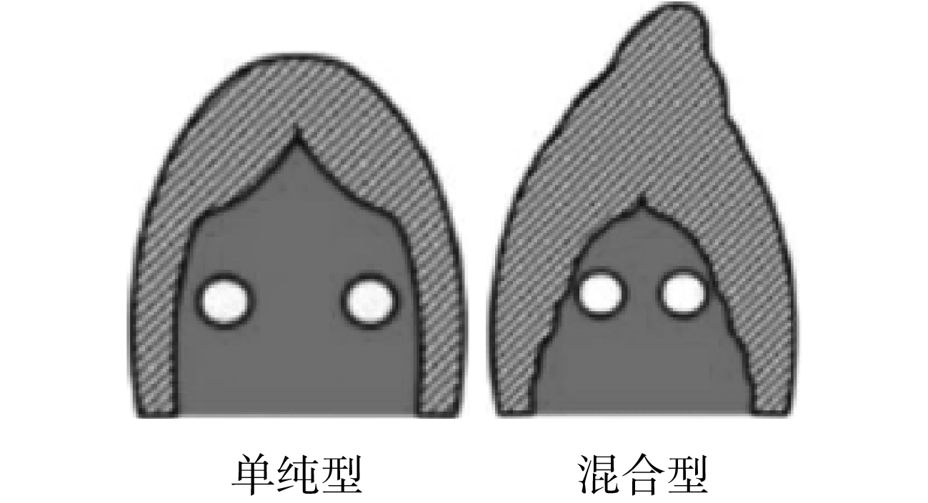
目的 评价心脏磁共振在心尖肥厚型心肌病诊断方面的临床应用价值。 方法 回顾性分析2017~2018年在我院就诊的9例心尖肥厚型心肌病患者,所有患者均行心脏磁共振检查,观察心肌受累节段,测量心肌厚度、心室心房容积、左心室射血分数,评价心肌首过灌注及延迟强化特点。 结果 9例患者心脏磁共振表现为左心室心尖部心肌肥厚,符合心尖肥厚型心肌病诊断标准。分析磁共振图像特点,将患者分为单纯型和混合型。其中单纯型心尖肥厚型心肌病6例,混合型心尖肥厚型心肌病3例。3例可见心尖室壁瘤。8例出现延迟强化,表现为肥厚的心肌内斑片样、点灶样强化。其中5例单纯型延迟强化范围局限在心尖部,3例混合型延迟强化范围累及左室中部、甚至乳头肌。 结论 心脏磁共振一站式成像对于诊断心尖肥厚型心肌病有独特作用,磁共振对比剂延迟强化,提示心肌纤维化,对临床治疗和预后有指导意义。 Abstract:Objective To evaluate the clinical value of cardiac magnetic resonance (CMR) in the diagnosis of apical hypertrophic cardiomyopathy (AHCM). Methods Nine patients with AHCM from 2017 to 2018 in our hospital were successfully underwent magnetic resonance scanning. Myocardial thickness, ventricular atrial volume, left ventricular ejection fraction were measured. Myocardial involvement segments, the characteristics of myocardial first perfusion and delayed enhancement were evaluated. Results CMR showed apical hypertrophic in all the patients. These patients were divided into two groups: pure AHCH and mixed AHCH. Among them, 6 patients were pure AHCM, 3 patients were mixed AHCM. 3 patients had apical aneurysm. All patients underwent myocardial contrast enhancement scanning, 8 patients presented late gadolinium enhancement (LGE) which showed plaques and focal enhancement. 5 pure AHCH’s LGE were located in the apex. 3 mixed AHCH’s LGE were involved the central left ventricle and even the papillary muscles. Conclusions CMR is best diagnostic modality for AHCM. LGE can characterize areas of myocardial fibrosis, which plays an important role in the natural history of AHCM. -
Key words:
- magnetic resonance imaging /
- hypertrophy cardiomyopathy /
- diagnosis
-
表 1 两组AHCM患者比较
分组 例数 心肌厚度(mm) 肥厚节段分布(n) 室壁瘤(n) 延迟强化(例) 均匀 不均匀 单纯型 6 15~20 4 2 1 5 混合型 3 16~30 1 2 2 3 -
[1] Jan MF, Todaro MC, Oreto L, et al. Apical hypertrophic cardiomyopathy: Present status[J]. Int J Cardiol, 2016, 222(8): 745-59 [2] Yan LR, Wang ZM, Xu ZM, et al. Two hundred eight patients with apical hypertrophic cardiomyopathy in China: clinical feature, prognosis, and comparison of pure and mixed forms[J]. Clin Cardiol, 2012, 35(2): 101-6 doi: 10.1002/clc.20995 [3] 冉玲平, 黄 璐, 赵培君, 等. RSNA2016心脏MRI/CT[J]. 放射学实践, 2017, 32(1): 3-7 [4] Maron MS. Clinical utility of cardiovascular magnetic resonance in hypertrophic cardiomyopathy[J]. J Cardiovasc Magn Reson, 2012, 14(1): 13-33 doi: 10.1186/1532-429X-14-13 [5] Kim EK, Lee SC, Hwang JW, et al. Differences in apical and non-apical types of hypertrophic cardiomyopathy: a prospective analysis of clinical, echocardiographic, and cardiac magnetic resonance findings and outcome from 350 patients[J]. Eur Heart J Cardiovasc Imaging, 2016, 17(6): 678-86 doi: 10.1093/ehjci/jev192 [6] Kebed KY, Al Adham RI, Bishu K, et al. Evaluation of apical subtype of hypertrophic cardiomyopathy using cardiac magnetic resonance imaging with Gadolinium enhancement[J]. Am J Cardiol, 2014, 114(5): 777-82 doi: 10.1016/j.amjcard.2014.05.067 [7] 马晓海, 赵 蕾, 葛海龙, 等. 非对称性肥厚型心肌病与心尖肥厚型心肌病心脏磁共振成像特点分析[J]. 中国全科医学, 2015, 18(18): 2166-9 doi: 10.3969/j.issn.1007-9572.2015.18.011 [8] 袁思殊, 李志伟, 夏黎明. 心尖肥厚型心肌病的MRI与超声心动图对比研究[J]. 磁共振成像, 2015, 6(3): 187-93 doi: 10.3969/j.issn.1674-8034.2015.03.006 [9] 刘 洪, 余建群, 彭礼清. 磁共振延迟强化在肥厚型心肌病中的临床应用价值研究[J]. 放射学实践, 2017, 32(12): 1271-6 [10] 刘东婷, 马晓海, 赵 蕾, 等. 钆布醇在磁共振延迟增强成像诊断肥厚型心肌病中的应用[J]. 中国医学影像学杂志, 2016, 24(5): 337-41 doi: 10.3969/j.issn.1005-5185.2016.05.005 [11] Kherada N, Vinardell JM, Mihos CG. Apical hypertrophic cardiomyopathy with left ventricular apical aneurysm: Importance of multi-modality imaging[J]. Echocardiography, 2017, 34(9): 1392-5 doi: 10.1111/echo.13581 [12] Fattori R, Biagini E, Lorenzini MA, et al. Significance of magnetic resonance imaging in apical hypertrophic cardiomyopathy[J]. Am J Cardiol, 2010, 105(11): 1592-6 doi: 10.1016/j.amjcard.2010.01.020 [13] Rosario, Parisi, Francesca, et al. Multimodality imaging in apical hypertrophic cardiomyopathy[J]. World J Cardiol, 2014, 6(9): 916-23 doi: 10.4330/wjc.v6.i9.916 [14] Doctorian T, Mosley WJ, Do B. Apical hypertrophic cardiomyopathy: case report and literature review[J]. Am J Case Rep, 2017, 18(6): 525-8 [15] Alvarez P, Tang WH. Recent advances in understanding and managing cardiomyopathy[J]. Res, 2017, 9(7): 1659-64 [16] Chan RH, Maron BJ, Olivotto I, et al. Prognostic value of quantitative contrast-enhanced cardiovascular magnetic resonance for the evaluation of sudden death risk in patients with hypertrophic cardiomyopathy[J]. Circulation, 2014, 130(6): 484-95 doi: 10.1161/CIRCULATIONAHA.113.007094 [17] Shah M. Hypertrophic cardiomyopathy[J]. Cardiol Young, 2017, 27(S1): S25-30 doi: 10.1017/S1047951116002195 [18] Gersh BJ, Maron BJ, Bonow RO, et al. 2011 ACCF/AHA guideline for the diagnosis and treatment of hypertrophic cardiomyopathy: a report of the American College of Cardiology Foundation/American Heart Association Task Force on Practice Guidelines[J]. Circulation, 2011, 124(24): e783-831 [19] Rowin EJ, Maron BJ, Haas TS, et al. Hypertrophic cardiomyopathy with left ventricular apical aneurysm: implications for risk stratification and management t[J]. J Am Coll Cardio, 2017, 69(7): 761-73 doi: 10.1016/j.jacc.2016.11.063 [20] Dominguez F, Sanz J, Garcia P, et al. Follow-up and prognosis of HCM[J]. Glob Cardiol Sci Pract, 2018, 8(3): 33-8 [21] Reichek N. Imaging cardiac morphology in hypertrophic cardiomyopathy: recent advances[J]. Curr Opin Cardiol, 2015, 30(5): 461-7 doi: 10.1097/HCO.0000000000000209 -


 下载:
下载:


