


Imaging study on the orientation of acetabular prosthesis based on orthographic radiographs of hip joint
-
摘要:
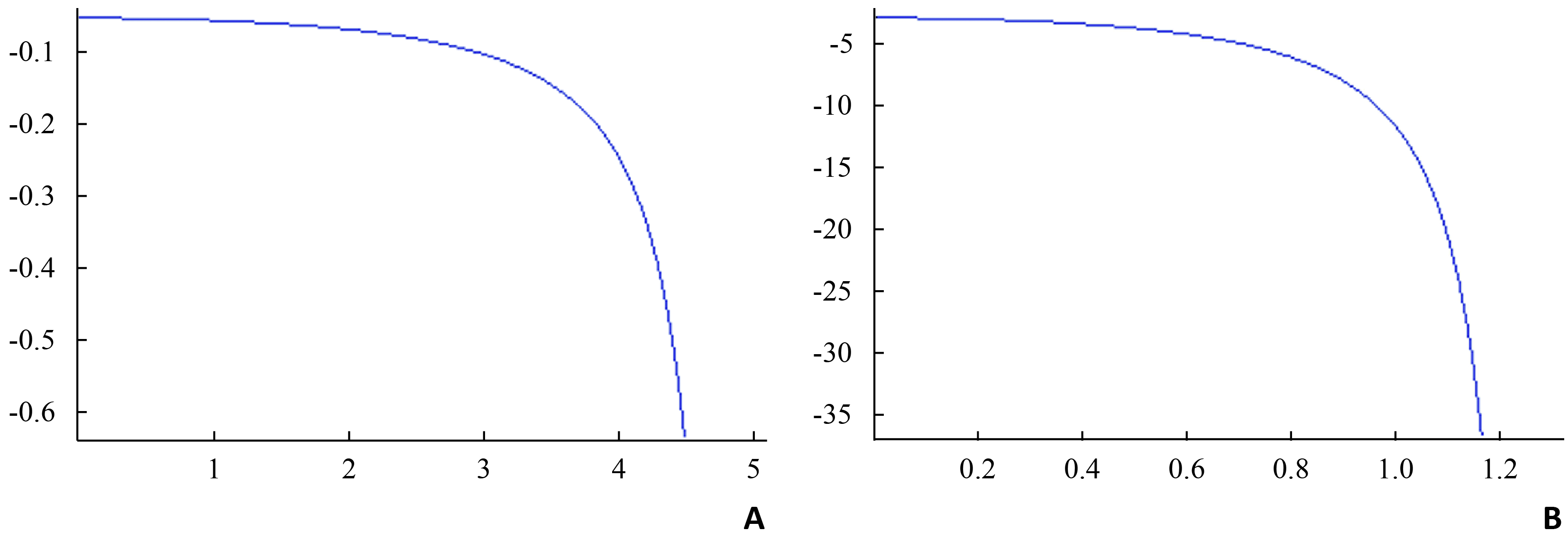
目的 系统回顾以往基于髋关节正位X线片测量髋臼假体方位的方法,提出更加可靠的测量工具。 方法 利用Matlab软件建立基于髋关节正位X线片拟合臼杯开口椭圆的新方法,并测量34例全髋关节置换术后臼杯的前倾角和倾斜角。由4名骨科医生完成信度实验,评价该测量方法的观察者间及观察者内信度。 结果 椭圆二次导数函数图可知,椭圆切线斜率的变化率在长短轴附近最小,距离长短轴端点越近,所选拟合点对拟合椭圆形态的影响也最小。观察者内信度表明:前倾角和倾斜角的组内相关系数ICC分别为0.916(95% CI:0.858~0.954)和0.948(95% CI:0.910~0.972)。观察者间信度结果为:前倾角和倾斜角的组间相关系数ICC分别为0.893(95% CI:0.821~0.941)和0.937(95% CI:0.892~0.966)。 结论 基于椭圆导数理论,本研究提出了一种基于髋关节正位X线片测量髋臼假体方位的新方法,可以用于临床评价全髋关节置换术后的假体位置。 Abstract:Objective To analyze the current measurement methods for determining the indication and antevesion of acetabular cup, and then a new radiographic measurement tool was developed and verified. Methods Based on the formula of ellipse, the second derivative of ellipse was set up and plotted using the Mathematica software. We analyzed the plot of the second derivative of ellipse and determine the relationship between the slope of ellipse and different points. The best method to select the points of the fitted ellipse was explored. Based on the previous analyses, a new radiographic measurement tool was developed using Matlab software and to calculate the antevesions and indications of 34 acetabular cups after total hip arthrosplasty. The reliability of the current established measurement approach was tested by the intra-class correlation coefficient (ICC). Results The plot of the second derivative of ellipse revealed that the slope of ellipse varied relatively little near the end of the major and minor axises of ellipse. Thus, the selected fitted points near the end of the major and minor axises could influence little on the shape of fitted ellipse. The reliability studies showed that high intraobserver reliability (n=34) was observed in the same observer’s measurements with an ICC between 0.972 and 0.996. A high agreement (n=34) was observed between different observers with an ICC between 0.973 and 0.996. The mean squares within trials ranged from 0.022 to 0.482 with all P values more than 0.05. Conclusion Based on the principle of the derivative of ellipse, a new radiographic measurement tool for determining the indication and antevesion of acetabular cup is developed using Matlab software. -
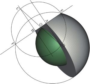
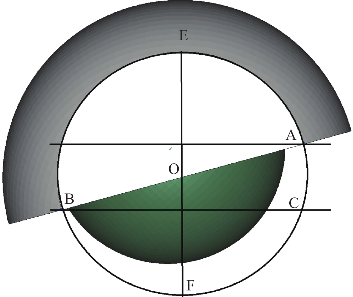
图 4 半球为模拟出的臼杯,球为模拟出的股骨头假体
作出臼杯所在的完整,EF为垂直于长轴切割臼杯半球的平面,AB分别为被切割后臼杯半球上的两个顶点,以O为中心,OE为半径作圆,AD和BC分别为垂直于EF的直线;角OCB即为放射学前倾角.
-
[1] Murray DW. The definition and measurement of acetabular orientation[J]. J Bone Joint Surg Br, 1993, 75(2): 228-32 [2] Langton DJ, Sprowson AP, Mahadeva D, et al. Cup anteversion in hip resurfacing: validation of EBRA and the presentation of a simple clinical grading system[J]. J Arthroplasty, 2010, 25(4): 607-13 doi: 10.1016/j.arth.2009.08.020 [3] Lewinnek GE, Lewis JL, Tarr R, et al. Dislocations after total hip-replacement arthroplasties[J]. J Bone Joint Surg Am, 1978, 60(2): 217-20 doi: 10.2106/00004623-197860020-00014 [4] Widmer KH. A simplified method to determine acetabular cup anteversion from plain radiographs[J]. J Arthroplasty, 2004, 19(3): 387-90 doi: 10.1016/j.arth.2003.10.016 [5] Pradhan R. Planar anteversion of the acetabular cup as determined from plain anteroposterior radiographs[J]. J Bone Joint Surg Br, 1999, 81(3): 431-5 doi: 10.1302/0301-620X.81B3.9067 [6] Ackland MK, Bourne WB, Uhthoff HK. Anteversion of the acetabular cup. Measurement of angle after total hip replacement[J]. J Bone Joint Surg Br, 1986, 68(3): 409-13 [7] Hassan DM, Johnston GH, Dust WN, et al. Radiographic calculation of anteversion in acetabular prostheses[J]. J Arthroplasty, 1995, 10(3): 369-72 doi: 10.1016/S0883-5403(05)80187-1 [8] Liaw CK, Hou SM, Yang RS, et al. A new tool for measuring cup orientation in total hip arthroplasties from plain radiographs[J]. Clin Orthop Relat Res, 2006, 451(1): 134-9 [9] Fabeck L, Farrokh D, Tolley M, et al. A method to measure acetabular cup anteversion after total hip replacement[J]. Acta Orthop Belg, 1999, 65(4): 485-91 [10] Bachhal V, Jindal N, Saini G, et al. A new method of measuring acetabular cup anteversion on simulated radiographs[J]. Int Orthop, 2012, 36(9): 1813-8 doi: 10.1007/s00264-012-1583-9 [11] Hart AJ, Dandachli W, Schlueter-Brust K, et al. Large ball metal on metal hips obscure cup angle measurement on plain radiographs[J]. Hip Int, 2009, 19(4): 323-9 doi: 10.1177/112070000901900405 [12] Olivecrona H, Weidenhielm L, Olivecrona L, et al. A new CT method for measuring cup orientation after total hip arthroplasty: a study of 10 patients[J]. Acta Orthop Scand, 2004, 75(2): 252-260 [13] Ghelman B, Kepler CK, Lyman S, et al. CT outperforms radiography for determination of acetabular cup version after THA[J]. Clin Orthop Relat Res, 2009, 467(6): 2362-70 [14] Kalteis T, Handel M, Herold T, et al. Position of the acetabular cup - accuracy of radiographic calculation compared to CT-based measurement[J]. Eur J Radiol, 2006, 58(2): 294-300 doi: 10.1016/j.ejrad.2005.10.003 [15] Mclaren RH. Prosthetic hip angulation[J]. Radiology, 1973, 107(3): 705-6 doi: 10.1148/107.3.705 [16] Pettersson H, Gentz CF, Lindberg HO, et al. Radiologic evaluation of the position of the acetabular component of the total hip prosthesis[J]. Acta Radiol Diagn (Stockh), 1982, 23(3A): 259-63 doi: 10.1177/028418518202303A15 [17] Marx A, von Knoch M, Pförtner J, et al. Misinterpretation of cup anteversion in total hip arthroplasty using planar radiography[J]. Arch Orthop Trauma Surg, 2006, 126(7): 487-92 doi: 10.1007/s00402-006-0163-0 [18] Haenle M, Mittelmeier W, Barbano R, et al. Accuracy and reliability of different methods to evaluate the acetabular cup version from plain radiographs[J]. Surg Radiol Anat, 2010, 32(8): 725-30 doi: 10.1007/s00276-010-0682-9 [19] Nho JH, Lee YK, Kim HJ, et al. Reliability and validity of measuring version of the acetabular component[J]. J Bone Joint Surg Br, 2012, 94(1): 32-36 [20] Nomura T, Naito M, Nakamura Y, et al. An analysis of the best method for evaluating anteversion of the acetabular component after total hip replacement on plain radiographs[J]. Bone Joint J, 2014, 96(2): 597-603 [21] Kosiyatrakul A, Luenam S, Chotanaphuti T. Measurement of acetabular cup anteversion with the circle theorem[J]. J Med Assoc Thai, 2009, 92(Suppl 6): S128-33 [22] Lu M, Zhou YX, Du H, et al. Reliability and validity of measuring acetabular component orientation by plain anteroposterior radiographs[J]. Clin Orthop Relat Res, 2013, 471(9): 2987-94 doi: 10.1007/s11999-013-3021-8 [23] Liaw CK, Wu TY, Hou SM, et al. Computerized ellipse method for measuring acetabular version after total hip replacement--a precision study using synthetic and real radiographs[J]. Comput Aided Surg, 2013, 18(5/6): 195-200 [24] Walter SD, Eliasziw M, Donner A. Sample size and optimal designs for reliability studies[J]. Stat Med, 1998, 17(1): 101-10 doi: 10.1002/(ISSN)1097-0258 [25] Weir JP. Quantifying test-retest reliability using the intraclass correlation coefficient and the SEM[J]. J Strength Cond Res, 2005, 19(1): 231-40 -

 下载:
下载:

 点击查看大图
图(4)
计量
- 文章访问数: 1944
- HTML全文浏览量: 656
- PDF下载量: 3
- 被引次数: 0