


Value of calcification in CT diagnosis of benign and malignant thyroid nodules
-
摘要:
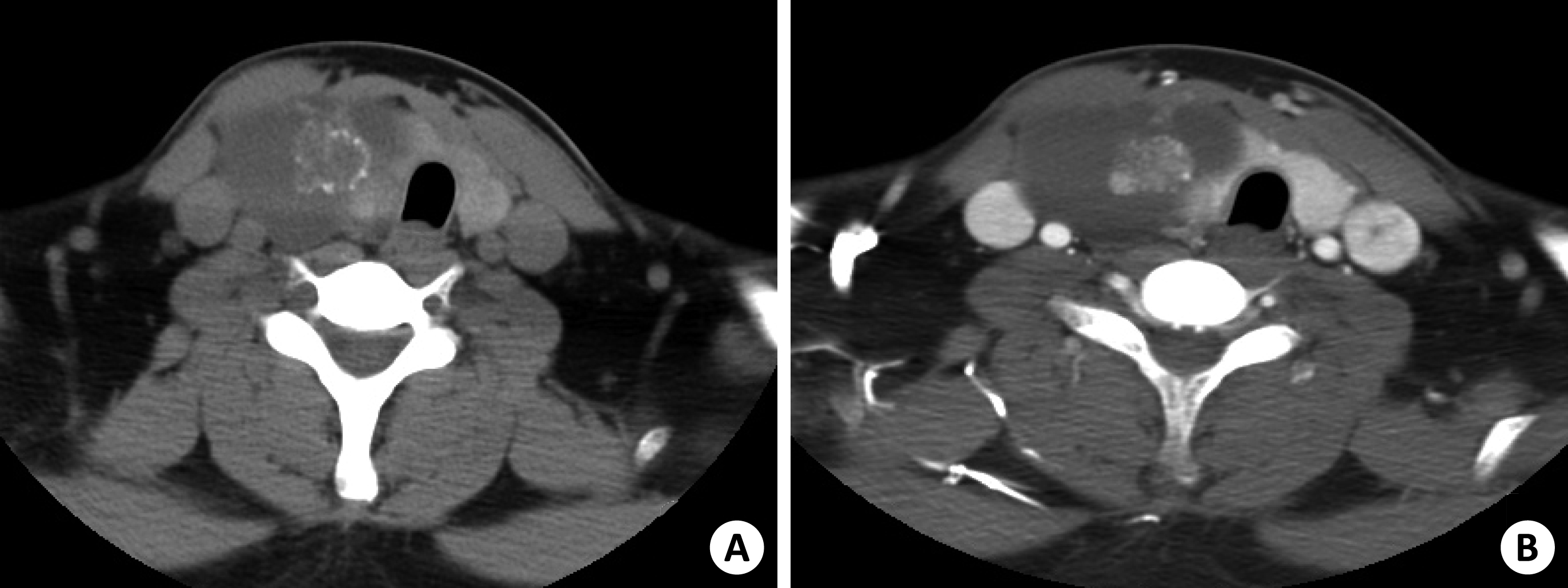
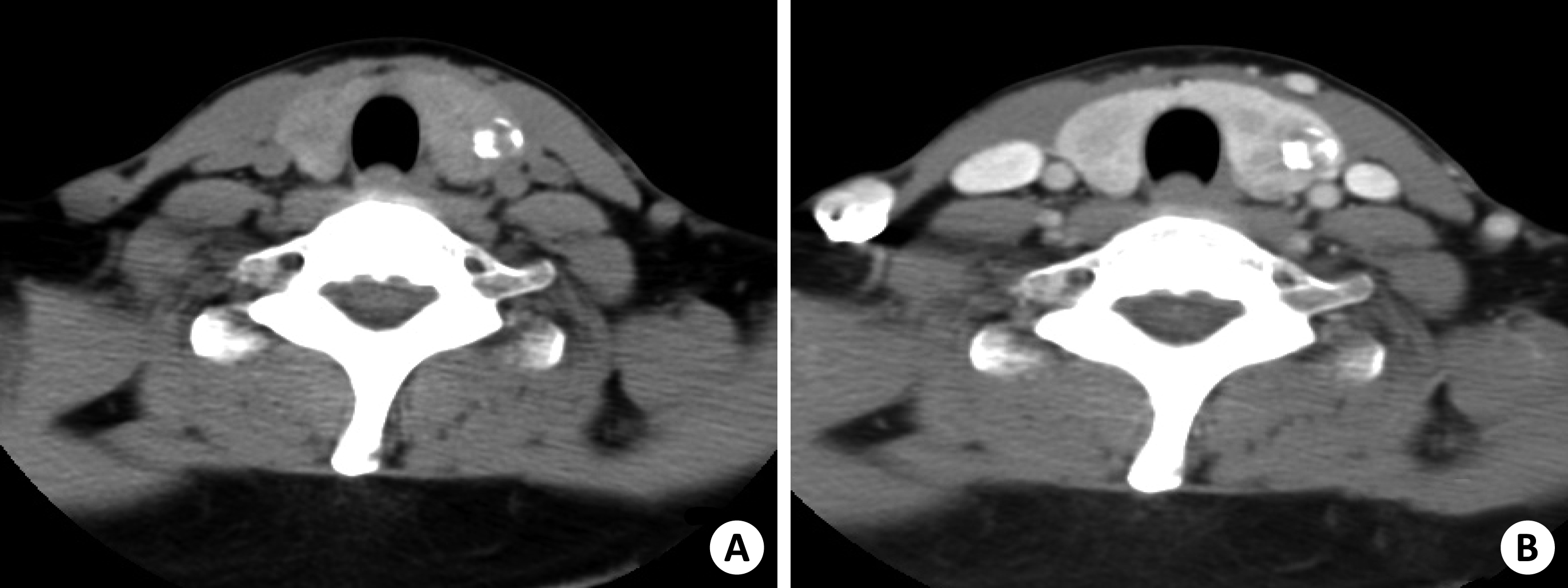
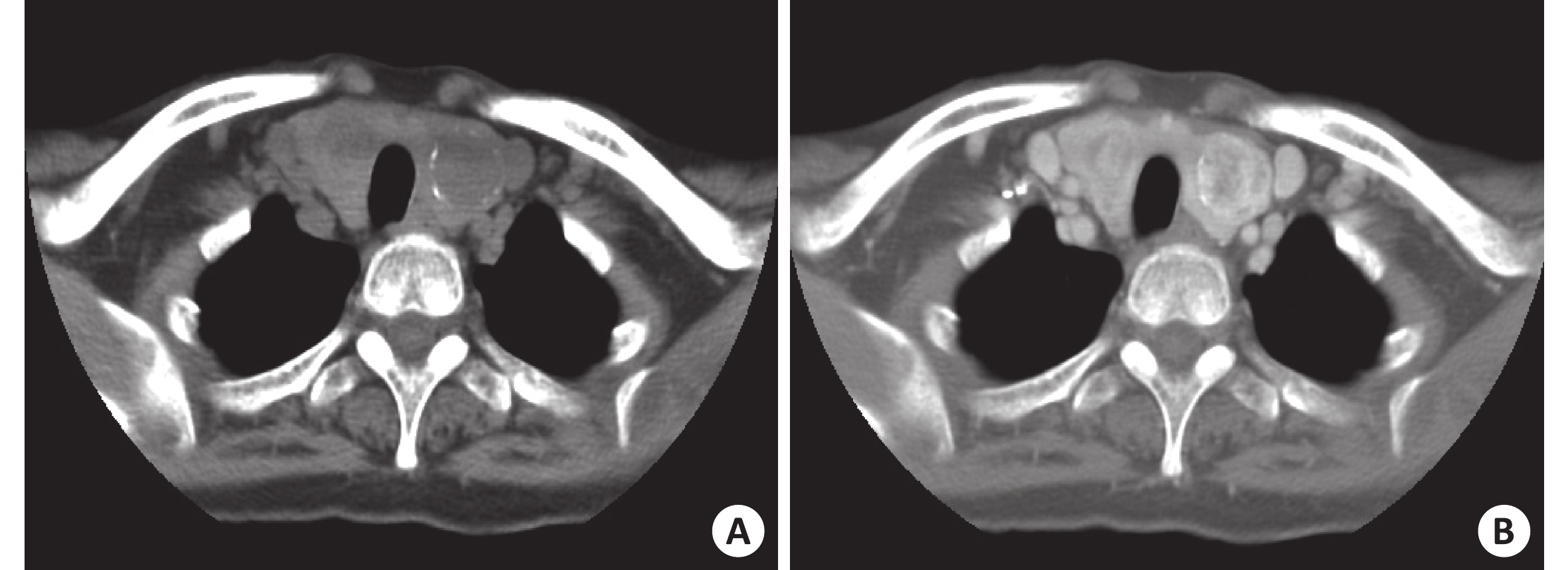
目的 分析甲状腺结节钙化的CT特点,探讨甲状腺结节的钙化特征对其良恶性诊断与鉴别诊断中的价值。 方法 回顾性分析259例277个经病理证实的甲状腺钙化结节的CT特点,重点观察结节钙化的大小、边缘、位置及数量。 结果 纳入259例患者,年龄16~84岁(54.73±12.43岁),男性66例,女性193例。总277个钙化结节,良性钙化结节205个,占总钙化结节74.01%,恶性钙化结节72个,占总钙化结节25.99%。边缘毛糙、微小钙化(直径<2 mm)的恶性率明显高于边缘光滑、粗大结节样的钙化组(χ2=13.669~13.950,P<0.001)。边缘毛糙、细颗粒微钙化对恶性结节诊断的敏感度分别为30.56%、59.72%,特异度分别为88.29%、65.36%,准确度分别为73.29%、63.90%,阳性预测值分别为47.83%、37.72%,阴性预测值分别为78.35%、82.21%。良恶性结节平均年龄差异具有统计学意义(t=4.452,P<0.001)。良恶性钙化结节在位置、数量、形状及性别之间差异无统计学意义(P>0.05)。 结论 MSCT检查甲状腺结节钙化特点对鉴别甲状腺良恶性病变有重要指导意义,微钙化、钙化结节边缘毛糙提示恶性结节,临床医生对这类结节应提高警惕并应进一步细针穿刺检查。 Abstract:Objective To investigate the diagnostic value of X-ray computed tomography detection for calcification in benign and malignant thyroid nodules. Methods A total of 277 thyroid calcification nodules confirmed either by surgery or histopathological examination in 259 cases were evaluated.The size, margin and quantity of nodular calcification were observed. Results In 277 calcified nodules,there were 205 benign calcified nodules (74.01%) and 72 malignant calcified nodules (25.99%) in 66 males and 193 females.The average age was 54.73 ±12.43 years. Overall, the malignant rate in patients with microcalcification (<2 mm in diameter) and margin calcification was significantly higher than the smooth edge in coarse calcification group (χ2=13.669~13.950, P<0.001). The accuracy, sensitivity, specificity, positive predictive value (PPV)and the negative predictive value (NPV) of microcalcification and marginal rough calcified nodules to diagnosis of malignant nodules were 73.29%, 63.90%, 30.56% and 59.72%, 88.29%, and 65.36%, 47.83% and 37.72%, 78.35% and 82.21%, respectively.In terms of age, the mean age of benign and malignant nodules was significantly different (t=4.452, P<0.001). There was no significant difference in location, quantity, shape and sex between benign and malignant calcified nodules (P>0.05). Conclusion The calcification characteristics of thyroid nodules by MSCT have important guiding significance in differentiating benign and malignant thyroid lesions. Microcalcification and rough calcified nodules with rough edges suggest malignant nodules. Clinicians should be alert to such nodules and further FNA examination should be performed in these patients. -
Key words:
- thyroid nodule /
- thyroid carcinoma /
- calcification /
- tomography /
- X-ray computed
-
表 1 良恶性结节钙化征的比较
结节类型 数量(个) 钙化数量 钙化边缘 钙化大小 钙化位置 钙化形状 单发 多发 光滑 毛糙 细 粗 混合 内部 边缘 混合 结节 蛋壳状 环形 良性 205 84 121 181 24 35 134 36 51 99 55 140 25 14 恶性 72 27 45 50 22 20 29 23 27 34 11 39 4 9 χ2 - 0.268 13.669 13.950 6.012 4.959 P - 0.605 <0.001 0.001 0.05 0.084  下载: 导出CSV
-
[1] 韩志江, 项晶晶, 包凌云, 等. 甲状腺钙化性病变的超声和CT联合诊断[J]. 国际医学放射学杂志, 2016, 38(4): 416-21 [2] Lu Z, Mu Y, Zhu H, et al. Clinical value of using ultrasound to assess calcification patterns in thyroid nodules[J]. World J Surg, 2011, 35(1): 122-7 [3] Wu CW, Dionigi G, Lee KW, et al. Calcifications in thyroid nodules identified on preoperative computed tomography: patterns and clinical significance[J]. Surgery, 2012, 151(3): 464-70 [4] Wu G, Zhou ZW, Li TY, et al. Do hyperechoic thyroid nodules on B-ultrasound represent calcification[J]. J Intern Med Res, 2013, 41(3): 848-54 doi: 10.1177/0300060513480083 [5] 周 健, 赖旭峰, 韩志江, 等. 多种CT征象对甲状腺良,恶性结节的预测价值[J]. 中华全科医师杂志, 2018, 16(1): 44-9 doi: 10.3760/cma.j.issn.1671-7368.2018.01.010 [6] Bai YH, Zhou GY, Nakamura M, et al. Survival impact of psammoma body, stromal calcification, and bone formation in papillary thyroid carcinoma[J]. Mod Pathol, 2009, 22(7): 887-94 doi: 10.1038/modpathol.2009.38 [7] Hegedüs L. Thyroid nodule[M]. [S.l.]: [s.n.], 2018. [8] Haugen BR, Alexander EK, Bible KC, et al. 2015 American thyroid association management guidelines for adult patients with thyroid nodules and differentiated thyroid cancer: the American thyroid association guidelines task force on thyroid nodules and differentiated thyroid cancer[J]. Thyroid, 2016, 26(1): 1-133 doi: 10.1089/thy.2015.0020 [9] Wang Z, Zhang H, Zhang P, et al. Diagnostic value of ultrasound-detected calcification in thyroid nodules[J]. Ann Acad Med Singapore, 2014, 43(2): 102-6 [10] Kim BK, Choi YS, Kwon HJ, et al. Relationship between patterns of calcification in thyroid nodules and histopathologic findings[J]. Endocr J, 2013, 60(2): 155-60 [11] Shi C, Li S, Shi T, et al. Correlation between thyroid nodule calcification morphology on ultrasound and thyroid carcinoma[J]. J Int Med Res, 2012, 40(1): 350-7 doi: 10.1177/147323001204000136 [12] Han ZJ, Shu YY, Lai XF, et al. Value of computed tomography in determining the Nature of papillary thyroid microcarcinomas: evaluation of the computed tomographic characteristics[J]. Clin Imaging, 2013, 37(4): 664-8 doi: 10.1016/j.clinimag.2012.12.005 [13] Na DG, Kim DS, Kim SJ, et al. Thyroid nodules with isolated macrocalcification:malignancy risk and diagnostic efficacy of fine-needle aspiration and core needle biopsy[J]. Ultrasonography, 2016, 35(3): 212-9 doi: 10.14366/usg.15074 [14] Lee J, Lee SY, Cha SH, et al. Fine-Needle aspiration of thyroid nodules with macrocalcification[J]. Thyroid, 2013, 23(9): 1106-12 doi: 10.1089/thy.2012.0406 [15] Kobayashi K, Fujimoto T, Ota H, et al. Calcifications in thyroid tumors on ultrasonography:calcification types and relationship with histopathological type[J]. Ultrasound Int Open, 2018, 4(2): E45-51 doi: 10.1055/a-0591-6070 [16] Arpaci D, Ozdemir D, Cuhaci N, et al. Evaluation of cytopathological findings in thyroid nodules with macrocalcification: macrocalcification is not innocent as it seems[J]. Arq Bras Endocrinol Metabol, 2014, 58(9): 939-45 doi: 10.1590/0004-2730000003602 [17] Lacout A, Chevenet C, Thariat J, et al. Thyroid calcifications: a pictorial essay[J]. J Clin Ultrasound, 2016, 44(4): 245-51 [18] 戴中强, 许镇城, 陈文陆, 等. 甲状腺结节内钙化灶良恶性病变的CT鉴别诊断[J]. 齐鲁医学杂志, 2016, 30(2): 142-4 [19] 朱玲英, 王恩雨, 巴 蕾, 等. CT鉴别甲状腺微小结节良恶性的价值[J]. 实用放射学杂志, 2013, 29(10): 1566-9 doi: 10.3969/j.issn.1002-1671.2013.10.004 [20] 刘 伟, 杨 军, 张 毅, 等. 钙化征在CT鉴别甲状腺良、恶性病变中的价值[J]. 中华放射学杂志, 2010, 44(2): 147-51 doi: 10.3760/cma.j.issn.1005-1201.2010.02.010 [21] 骆洪浩, 彭玉兰, 赵海娜. 不同类型的钙化对甲状腺良恶性肿瘤的诊断价值[J]. 中国医学计算机成像杂志, 2016, 21(2): 162-6 [22] 顾立军, 陆 杨, 王垚青, 等. 钙化在甲状腺结节MSCT诊断中的临床价值[J]. 放射学实践, 2014, 29(3): 279-82 [23] Li JW, Chang C, Chen M, et al. Is ultrasonography more sensitive than computed tomography for identifying calcifications in thyroid nodules[J]. J Ultrasound Med, 2016, 35(10): 2183-90 [24] 李如强, 许 敏, 袁戈恒. 甲状腺良恶性结节钙化超声特点分析[J]. 医学综述, 2018, 24(14): 2896-8 doi: 10.3969/j.issn.1006-2084.2018.14.038 [25] Liu W, Dong XY, Zhu CS, et al. Association between computed tomography-detected calcification and thyroid carcinoma[J]. Neoplasma, 2015, 62(4): 641-5 doi: 10.4149/neo_2015_077 -

 点击查看大图
图(3) / 表(1)
计量
- 文章访问数: 2186
- HTML全文浏览量: 754
- PDF下载量: 7
- 被引次数: 0