


Relationship between MRI redundant nerve roots and lumbar spinal canal stenosis
-
摘要:
目的 探讨腰椎管狭窄与马尾神经冗余征的关系。 方法 回顾性分析2015年3月~2017年12月160例腰椎管狭窄的患者信息,将其分为冗余组和非冗余组。测量狭窄处椎管前后径、狭窄层面硬膜囊面积、马尾神经冗余相对长度等数据;记录性别、年龄、腰椎管最狭窄的位置、狭窄处突起是否锐利、是否多节段狭窄等参数。统计上述变量是否与马尾神经冗余征及马尾神经冗余相对长度相关。 结果 不同马尾神经冗余征的性别、年龄、腰椎管最狭窄的位置、狭窄层面硬膜囊面积、狭窄处突起锐利、多节段狭窄等变量的差异均有统计学意义(P<0.05);除腰椎管多节段狭窄(P<0.01),不同性别、狭窄处是否突起锐利、狭窄处椎管前后径的马尾神经冗余相对长度差异无统计学意义(P>0.05)。 结论 马尾神经冗余是一种与腰椎管狭窄相关的MRI征象,其中性别、年龄、腰椎管多节段狭窄、狭窄层面硬膜囊面积、腰椎管最狭窄的位置、狭窄处锐利突起等均是其危险因素。 Abstract:Objective To explore the relationship between various factors of lumbar spinal canal stenosis and redundant nerve roots. Methods A total of 160 patients with lumbar spinal stenosis from March 2015 to December 2017 were included. The patients were divided into redundant group and non redundant group.The front to rear with lumbar stenosis, scar area of narrow, relatively long redundant caudal nerve etc data were measured. Gender, age, the narrowest position of lumbar spinal canal, the protuberance, and multinode stenosis were recorded. The correlation between the above variables with redundant nerve roots and relatively long redundant caudal nerve were analyzed. Result Gender, age, the narrowest position of lumbar spinal canal, Scar area of narrow, sharp protuberance on narrow area, multinode stenosis were related with cauda equina redundant (P<0.05). Gender, sharp protuberance on narrow area, diameter of front to rear with lumbar stenosis were not related with relatively long redundant caudal nerve, except for multisegmental stenosis (P<0.01). Conclusion Redundant nerve roots is a MRI feature associated with lumbar spinal canal stenosis. Gender, age, the narrowest position of lumbar spinal canal, scar area of narrow, sharp protuberance on narrow area, multinode stenosis are risk factors. -
Key words:
- lumbar stenosis /
- redundant nerve roots /
- MRI
-
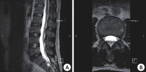
图 3 25岁男性志愿者
A: MRI矢状位示正常椎管及其椎管内分布排列的马尾神经根, 腰1至腰5椎体上缘马尾神经均靠背侧整束行走, 形态自然; B: MRI轴位示马尾神经分布在椎管后1/3份, 粗细均匀, 排列规整.
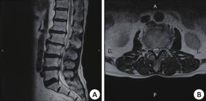
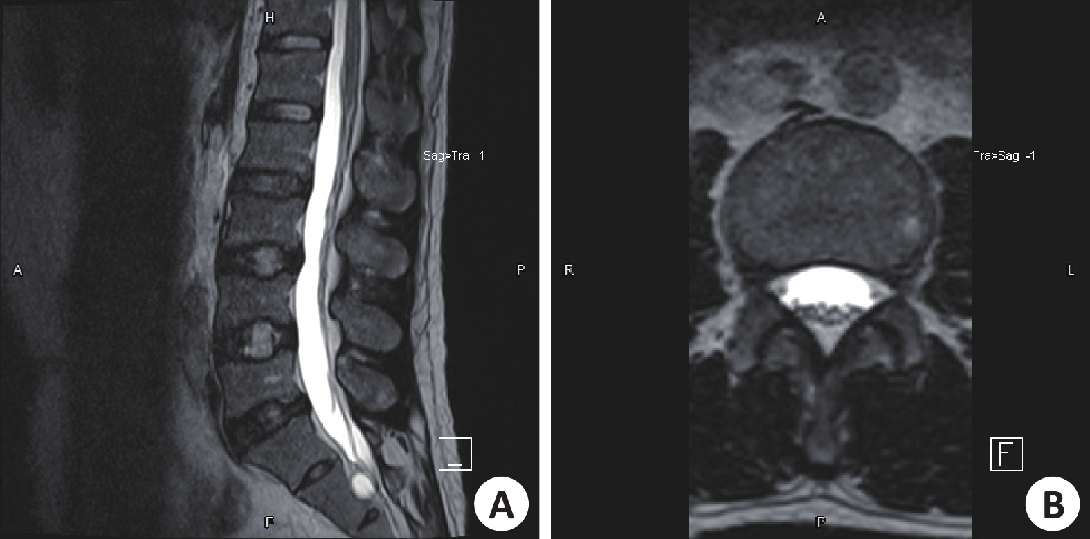
图 1 女,76岁,腰腿疼多年伴偶发神经源性跛行
A: 图MRI矢状位示腰椎体上缘至腰4/5椎间盘区间, 可见腰椎管多节段狭窄及椎管内马尾神经迂曲冗长; B: 图MRI轴位示增粗的马尾神经在狭窄椎管内的异常分布排列.
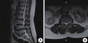
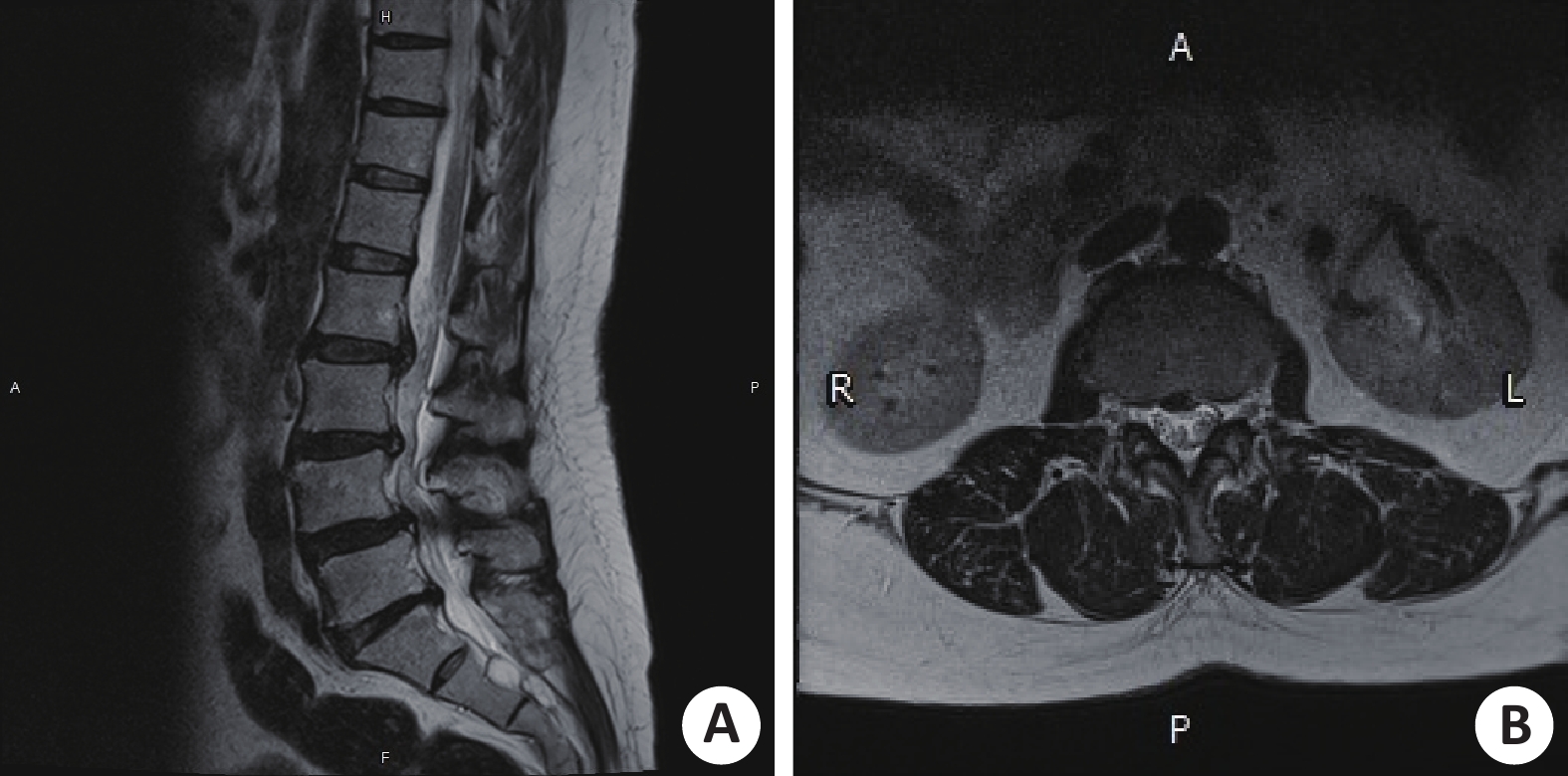
图 2 女,54岁,双下肢交替出现疼痛无力麻木多年
A: 图MRI矢状位示腰1椎体上缘至腰4/5水平段, 显示腰椎间盘多发突出及腰椎管多节段狭窄, 狭窄椎管内的马尾神经迂曲冗长; B: 图MRI轴位示马尾神经正常结构消失, 形态增粗, 分布成团, 排列紊乱.
表 1 冗余组和非冗余组多参数的统计分析
分类 冗余组(n=78) 非冗余组(n=82) χ2/t P 性别(n, %) χ2=6.320 0.016 女 40(51.3%) 26(31.7%) 男 38(48.7%) 56(68.3%) 年龄(岁, Mean±SD) 69±9.888 54.66±16.617 t=6.592 0.000 腰椎管狭窄的位置(n, %) χ2=19.639 0.000 腰1/2 0(0%) 0(0%) 腰2/3 3(3.8%) 4(4.87 %) 腰3/4 28(35.9%) 6(7.31%) 腰4/5 45(57.7%) 68(82.9%) 腰5/骶1 2(2.5%) 4(4.87%) 狭窄层面硬膜囊面积(mm2, Mean±SD) 57.95±27.724 80.29±29.166 t=4.962 0.000 突出是否锐利(n, %) χ2=23.761 0.000 是 42(53.8%) 14(17.1%) 否 36(46.2%) 68(82.9%) 是否多节段狭窄(n, %) χ2=19.850 0.000 是 54(69.2%) 19(23.2%) 否 24(30.8%) 63(76.8%)  下载: 导出CSV
表 2 冗余马尾神经相对长度与相关参数的统计分析
分类 例数 冗余马尾神经
相对长度
(cm, Mean±SD)t P 性别 0.990 0.325 男 38 1.94±0.69 女 40 2.07±0.42 狭窄处突起是否锐利 0.360 0.720 是 42 2.02±0.56 否 36 1.98±0.59 腰椎管是否多节段狭窄 13.020 0.000 是 54 2.30±0.20 否 24 1.42±0.40 狭窄处椎管前后径(mm) 0.870 0.387 >10 20 2.11±0.35 ≤10 58 1.99±0.58
下载: 导出CSV
-
[1] 柏树令, 应大君. 系统解剖学[M]. 北京: 人民卫生出版社, 2005. [2] 张 宝. 马尾神经的影像解刨学研究[D]. 吉林: 吉林大学, 硕士, 2013. [3] 赵 兴, 范顺武. 若干未受重视的腰椎管狭窄的MRI表征[J]. 中华骨科杂志, 2016, 36(22): 1405-9 doi: 10.3760/cma.j.issn.0253-2352.2016.22.001 [4] Nogueira BM, Savarese LG, Herrero C, et al. Redundant nerve Roots of the cauda equina:review of the literature[J]. Radiol Bras, 2012, 45(3): 155-9 doi: 10.1590/S0100-39842012000300007 [5] Lurie J, Tomkins-Lane C. Management of lumbar spinal stenosis[J]. BMJ, 2016, 352(23): 6234-9 [6] Lee SY, Kim TH, Oh JK, et al. Lumbar stenosis: a recent update by review of literature[J]. Asian Spine J, 2015, 9(5): 818-28 doi: 10.4184/asj.2015.9.5.818 [7] Shamji MF, Mroz T, Hsu W, et al. Management of degenerative lumbar spinal stenosis in the elderly[J]. Neurosurgery, 2015, 77(Suppl 4): S68-74 [8] Menon VK, Raniga SB, Al Busaidi AQ. MRI morphology of surgically treated lumbar canal stenosis: a retrospective study[J]. J Spinal Disord Tech, 2015, 28(1): 12-8 doi: 10.1097/BSD.0000000000000053 [9] Ozturk AK, Gokaslan ZL. Clinical significance of redundant nerve Roots of the cauda equina[J]. World Neurosurg, 2014, 82(6): e717-8 doi: 10.1016/j.wneu.2013.10.047 [10] Deyo RA, Gray DT, Kreuter W, et al. United States trends in lumbar fusion surgery for degenerative conditions[J]. Spine (Phila Pa 1976), 2005, 30(12): 1441-5 doi: 10.1097/01.brs.0000166503.37969.8a [11] Andreisek G, Imhof M, Wertli M, et al. A systematic review of semiquantitative and qualitative radiologic criteria for the diagnosis of lumbar spinal stenosis[J]. Am J Roentgenol, 2013, 201(5): W735-46 doi: 10.2214/AJR.12.10163 [12] 张 强. 腰椎管狭窄症患者中马尾神经冗余征的MR分析[D]. 浙江: 浙江大学, 硕士, 2016. [13] 张光辉, 刘旭林, 初英萍. 腰椎正中矢状径在椎间隙层面与椎体层面研究[J]. 实用放射学杂, 2006, 22(9): 1104-7 doi: 10.3969/j.issn.1002-1671.2006.09.023 [14] Verbiest H. A radicular syndrome from developmental narrowing of the lumbar vertebral canal[J]. J Bone Joint Surg Br, 1954, 36B(2): 230-7 [15] Min JH, Jang JS, Lee SH. Clinical significance of redundant nerve Roots of the cauda equina in lumbar spinal stenosis[J]. Clin Neurol Neurosurg, 2008, 110(1): 14-8 doi: 10.1016/j.clineuro.2007.08.005 [16] Ehni G, Moiel RH, Bragg TG. The "redundant" or "knotted" nerve root: a clue to spondylotic cauda equina radiculopathy. Case report[J]. J Neurosurg, 1970, 32(2): 252-4 doi: 10.3171/jns.1970.32.2.0252 [17] Fox JL. Redundant nerve Roots in the cauda equina. Case report[J]. J Neurosurg, 1969, 30(1): 74-5 doi: 10.3171/jns.1969.30.1.0074 [18] Gulati DR, Rout D. Myelographic block caused by redundant lumbar nerve root. Case report[J]. J Neurosurg, 1973, 38(4): 504-5 doi: 10.3171/jns.1973.38.4.0504 [19] Suzuki K, Ishida Y, Ohmori K, et al. Redundant nerve Roots of the cauda equina: clinical aspects and consideration of pathogenesis[J]. Neurosurgery, 1989, 24(4): 521-8 doi: 10.1227/00006123-198904000-00006 [20] Suzuki K, Takatsu T, Inoue H, et al. Redundant nerve Roots of the cauda equina caused by lumbar spinal canal stenosis[J]. Spine (Phila Pa 1976), 1992, 17(11): 1337-42 doi: 10.1097/00007632-199211000-00013 [21] 冯建刚, 韩永台, 王 飞, 等. 犬马尾神经分级压迫的MRI与组织学对比观察[J]. 中华放射学杂志, 2000, 34(3): 63-6 [22] Poureisa M, Daghighi MH, Eftekhari P, et al. Redundant nerve Roots of the cauda equina in lumbar spinal canal stenosis, an Mr study on 500 cases[J]. Eur Spine J, 2015, 24(10): 2315-20 doi: 10.1007/s00586-015-4059-y -

 点击查看大图
图(3) / 表(2)
计量
- 文章访问数: 1139
- HTML全文浏览量: 318
- PDF下载量: 6
- 被引次数: 0