| Citation: | Zhong WEI, Hua MAO. Predictive value of non-invasive parameter for the degree of esophageal varices in patients with post-hepatitis B cirrhosis[J]. Journal of Molecular Imaging, 2018, 41(2): 233-236. doi: 10.3969/j.issn.1674-4500.2018.02.22

|

| [1] |
Kim DH, Park JY. Prevention and management of variceal hemorrhage[J]. Int J Hepatol, 2013(3): 4346-9.
|
| [2] |
Buechter M, Kahraman A, Manka P, et al. Spleen and liver stiffness is positively correlated with the risk of esophageal variceal bleeding[J]. Digestion, 2016, 94(3): 138-44. doi: 10.1159/000450704
|
| [3] |
中华医学会肝病学分会, 中华医学会消化病学分会, 中华医学会内镜学分会. 肝硬化门静脉高压食管胃静脉曲张出血防治指南(2015)[J]. 中华胃肠内镜电子杂志, 2015, 2(4): 1-21. http://news.medlive.cn/liver/info-progress/show-85344_35.html
|
| [4] |
卢敏, 陈新杰, 黄纯炽, 等. Fibrotouch检测慢性乙型肝炎患者肝脏硬度指标的影响因素分析[J]. 实用医学杂志, 2014, 30(8): 1245-8. http://med.wanfangdata.com.cn/Paper/Detail/PeriodicalPaper_syyxzz201408020
|
| [5] |
方正亚, 张国顺, 刘斌, 等. 肝脏硬度指标对ALT正常慢性乙型肝炎患者肝纤维化的诊断价值[J]. 山东医药, 2017, 57(21): 55-7. doi: 10.3969/j.issn.1002-266X.2017.21.018
|
| [6] |
李建志. Fibroscan硬度值测量对乙肝相关肝纤维化的诊断及其与超声影像检查相关性的研究[D]. 济南: 山东大学, 2011.
|
| [7] |
杨学平. 瞬时弹性成像检测肝脾硬度预测食管静脉曲张的价值[J]. 中国超声医学杂志, 2017(02): 139-42. http://www.cqvip.com/QK/98512X/201603/668242137.html
|
| [8] |
Pár G, Trosits A, Pakodi F, et al. Transient elastography as a predictor of oesophageal varices in patients with liver cirrhosis[J]. Orv Hetil, 2014, 155(7): 270-6. doi: 10.1556/OH.2014.29824
|
| [9] |
蓝思荣, 张淼源, 周剑辉. 门静脉高压食管静脉曲张患者超声造影及彩色多普勒参数的检测及意义[J]. 广东医学院学报, 2015, 33(3): 343-5. http://www.cqvip.com/QK/90390A/201503/666532424.html
|
| [10] |
崔亚云, 王玲, 张超学, 等. 肝超声血流动力学参数评估门静脉高压中重度食管静脉曲张的应用价值[J]. 中华超声影像学杂志, 2013, 22(9): 788-91.
|
| [11] |
Sangma MA, Biswas N, Ahmed MU, et al. Doppler assessment of hepatic venous waves for evaluation of large varices in cirrhotic patient[J]. Mymensingh Med J, 2016, 25(4): 641-6.
|
| [12] |
赵丹, 毛华, 黄纯炽, 等. 乙型肝炎后肝硬化患者食管静脉曲张发生的无创性预测指标研究[J]. 上海交通大学学报:医学版, 2015, 35(3): 386-90. http://www.cnki.com.cn/Article/CJFDTotal-SHEY201503020.htm
|
| [13] |
凤辉, 龚镭, 唐学军, 等. 无创血清学肝纤维化评分系统对肝硬化食管胃底静脉曲张的预测价值[J]. 中华消化杂志, 2017, 37(8): 143-7. http://news.medlive.cn/liver/info-progress/show-55337_35.html
|
| [14] |
Bledar, Kraja, Iris, et al. Predictors of esophageal varices and first variceal bleeding in liver cirrhosis patients[J]. World J Gastroenterol, 2017, 23(26): 4806-14. doi: 10.3748/wjg.v23.i26.4806
|
| [15] |
中华医学会肝病学分会, 中华医学会感染病学分会. 慢性乙型肝炎防治指南(2015年版) [J]. 中华实验和临床感染病杂志:电子版, 2015, 9(5): 570-89. http://guide.medlive.cn/guideline/9744
|
| [16] |
令孤恩强. 消化道静脉曲张及出血的内镜诊断和治疗规范试行方案[C]//第十届国际治疗内镜及消化病学术会议论文集, 苏州, 2010.
|
| [17] |
Massimo, Bolognesi, Marco, et al. Clinical role of non-invasive assessment of portal hypertension[J]. World J Gastroenterol, 2017, 23(01): 1-10. doi: 10.3748/wjg.v23.i1.1
|
| [18] |
Ke, Pu, Jing JH, et al. Diagnostic accuracy of transient elastography (Fibro Scan) in detection of esophageal varices in patients with cirrhosis: A meta-analysis[J]. World J Gastroenterol, 2017, 23(02): 345-56. doi: 10.3748/wjg.v23.i2.345
|
| [19] |
肝脏硬度评估小组. 瞬时弹性成像技术诊断肝纤维化专家意见[J]. 中华肝脏病杂志, 2013, 21(6): 420-4. http://guide.medlive.cn/guideline/4823
|
| [20] |
Kitson MT, Kemp WW, Iser DM, et al. Utility of transient elastography in the non-invasive evaluation of cystic fibrosis liver disease[J]. Liver Int, 2013, 33(5): 698-705. doi: 10.1111/liv.12113
|
| [21] |
Gao L, Meng F, Cheng J, et al. Prediction of oesophageal varices in patients with primary biliary cirrhosis by non-invasive markers[J]. Arch Med Sci, 2017, 13(2): 370-6. http://www.termedia.pl/Journal/-19/pdf-29239-10?filename=prediction%20of%20oesophageal.pdf
|
| [22] |
Sebastiani G, Tempesta D, Fattovich G, et al. Prediction of oesophageal varices in hepatic cirrhosis by simple serum non-invasive markers: Results of a multicenter, large-scale study[J]. J Hepatol, 2010, 53(4): 630-8. doi: 10.1016/j.jhep.2010.04.019
|
| [23] |
刘芳, 李庭红, 韩涛, 等. 瞬时弹性成像在肝硬化门静脉高压中的临床评价[J]. 中华肝脏病杂志, 2013, 21(11): 840-4. doi: 10.3760/cma.j.issn.1007-3418.2013.11.010
|
| [24] |
徐瀚清, 唐煜文, 杜志娜. 联合检测PVW、SSM、LSM对肝硬化发生胃底静脉曲张出血风险的预测价值[J]. 肝脏, 2017, 22(06): 548-50. doi: 10.3969/j.issn.1008-1704.2017.06.022
|
| [25] |
李勤涛, 蒋力, 张珂, 等. 无创模型预测肝炎肝硬化患者食管静脉曲张[J]. 中华肝脏病杂志, 2015, 23(5): 339-42. http://d.wanfangdata.com.cn/Periodical_zhgzbzz201505004.aspx
|
| [26] |
王晓彤, 韩涛, 李雅玥. 无创血清学模型对酒精性肝硬化食管静脉曲张的预测价值[J]. 山东医药, 2016, 56(8): 4-6. http://med.wanfangdata.com.cn/Paper/Detail/PeriodicalPaper_shandyy201608002
|
| [27] |
Castera L, Pinzani M, Bosch J. Non invasive evaluation of portal hypertension using transient elastography[J]. J Hepatol, 2012, 56(3): 696-703. doi: 10.1016/j.jhep.2011.07.005
|
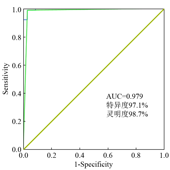
Figures(1) / Tables(2)
Supported by: Beijing Renhe Information Technology Co. Ltd support: info@rhhz.net


 DownLoad: