| Citation: | Chengde ZHU. Clinical value and significance of ultrasonography in the diagnosis of fetal umbilical vasa previa[J]. Journal of Molecular Imaging, 2018, 41(2): 178-180. doi: 10.3969/j.issn.1674-4500.2018.02.11

|

| [1] |
宣兰萍, 张韩珉. 22例胎膜血管前置的产前超声诊断分析[J]. 中国计划生育学杂志, 2016, 24(06): 409-12.
|
| [2] |
Suzuki S, Kato M. Clinical significance of pregnancies complicated by velamentous umbilical cord insertion associated with other umbilical cord/placental abnormalities[J]. J Clin Med Res, 2015, 7(11): 853-6. doi: 10.14740/jocmr2310w
|
| [3] |
Ruiter L, Kok N, Limpens J, et al. Systematic review of accuracy of ultrasound in the diagnosis of vasa previa[J]. Ultrasound Obstet Gynecol, 2015, 45(5): 516-22. doi: 10.1002/uog.14752
|
| [4] |
Sullivan EA, Javid N, Duncombe G, et al. Vasa previa diagnosis, clinical practice, and outcomes in Australia[J]. Obstet Gynecol, 2017, 130(3): 591-8. doi: 10.1097/AOG.0000000000002198
|
| [5] |
佟玲玲, 刘 莉, 敖 禹, 等. 球盘状胎盘前置血管破裂并发胎盘早剥1例分析[J]. 中国实验诊断学, 2016, 20(11): 1946-7.
|
| [6] |
刘希婧, 白 一, 龚云辉, 等. 前置血管5例临床特征分析[J]. 实用妇产科杂志, 2014, 30(05): 380-2. http://med.wanfangdata.com.cn/Paper/Detail/PeriodicalPaper_syfckzz201405021
|
| [7] |
Melcer Y, Maymon R, Pekar-Zlotin M, et al. The mid-gestation triple test profile among women diagnosed with vasa previa[J]. J Matern Fetal Neonatal Med, 2018, 31(11): 1402-6. doi: 10.1080/14767058.2017.1317343
|
| [8] |
肖 兵, 熊 庆. 前置血管的孕期处置[J]. 实用妇产科杂志, 2014, 30(08): 572-4. http://med.wanfangdata.com.cn/Paper/Detail/PeriodicalPaper_syfckzz201408006
|
| [9] |
陈凤英, 张大伟, 刘正平, 等. 凶险型前置胎盘伴胎盘植入的产前MRI平扫影像学表现及诊断价值[J]. 中国临床医学影像杂志, 2016, 27(5): 359-62.
|
| [10] |
Nohuz E, Boulay E, Gallot D, et al. Can we perform a prenatal diagnosis of vasa previa to improve its obstetrical and neonatal outcomes[J]. J Gynecol Obstetr Human Reproduct, 2017, 46(4): 373-7. doi: 10.1016/j.jogoh.2017.02.009
|
| [11] |
Kapoor S, Thomas JT, Petersen SG, et al. Is the third trimester repeat ultrasound scan for placental localisation needed if the placenta is low lying but clear of the os at the mid-trimester morphology scan[J]. Aust N Z J Obstet Gynaecol, 2014, 54(5): 428-32. doi: 10.1111/ajo.2014.54.issue-5
|
| [12] |
Belmonte L, Fuente AM, Soler RM. Vasa praevia diagnosis during transvaginal measurement of cervical length as preventing preterm delivery in the second quarter[J]. Ginecol Obstet Mex, 2016, 84(3): 186-93.
|
| [13] |
van Steenis A, Zhao DP, Steggerda SJ, et al. Double fatal outcome after ruptured vasa previa in monochorionic twins: case report and review of the literature[J]. J Matern Fetal Neonatal Med, 2016, 29(15): 2523-6.
|
| [14] |
韦 静, 居红芳. 胎盘前置血管产妇临床早期诊断对新生儿预后的影响[J]. 浙江临床医学, 2015, 14(9): 1539-40. http://med.wanfangdata.com.cn/Paper/Detail/PeriodicalPaper_zjlcyx201509040
|
| [15] |
Matsubara S, Kuwata T, Takahashi H, et al. Vasa previa: Another ultrasound sign and caution at cesarean section[J]. J Matern Fetal Neonatal Med, 2016, 29(7): 1139-40. doi: 10.3109/14767058.2015.1038233
|
| [16] |
陈 乐, 王 银, 江燕萍, 等. 产前超声诊断重复胎盘并脐带帆状附着1例[J]. 现代妇产科进展, 2016, 25(07): 558-61.
|
| [17] |
王 铭, 栗河舟, 刘 云, 等. 超声中孕期筛查胎盘脐带入口位置异常的临床评价[J]. 实用医院临床杂志, 2016, 13(01): 96-8. doi: 10.3969/j.issn.1672-6170.2016.01.034
|
| [18] |
王晓波, 陈 忠, 邓连桂, 等. 帆状胎盘的彩色多普勒超声征象及其临床意义[J]. 中国超声医学杂志, 2015, 31(12): 1108-11. http://med.wanfangdata.com.cn/Paper/Detail/PeriodicalPaper_zgcsyxzz201512021
|
| [19] |
顾莉莉, 姜 凡, 解 翔, 等. 产前超声在诊断血管前置中的临床应用价值[J]. 安徽医学, 2016, 37(01): 50-2. doi: 10.3969/j.issn.1000-0399.2016.01.015
|
| [20] |
廖凤琴, 郑 慧, 穆仲平. 产前超声诊断血管前置病例分析[J]. 中国超声医学杂志, 2016, 32(09): 850-3. doi: 10.3969/j.issn.1002-0101.2016.09.030
|
| [21] |
李雪艳, 于 松, 吴青青. 帆状胎盘的危险因素及围产儿结局[J]. 中国医学科学院学报, 2015, 37(03): 355-7. doi: 10.3881/j.issn.1000-503X.2015.03.022
|
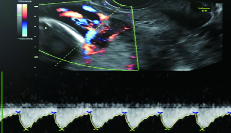
Figures(1)
Supported by: Beijing Renhe Information Technology Co. Ltd support: info@rhhz.net


 DownLoad: