| Citation: | Guangyin CHEN, Yuexi CHEN. Value of MSCT in diagnosis of nodular goiter with coexistent thyroid carcinoma[J]. Journal of Molecular Imaging, 2018, 41(2): 175-177. doi: 10.3969/j.issn.1674-4500.2018.02.10

|

| [1] |
Edino ST, Mohammed AZ, Ochicha O, et al. Thyroid cancers in nodular goiters in Kano, Nigeria[J]. Niger J Clin Pract, 2010, 13(3): 298-300. https://www.ajol.info/index.php/njcp/article/download/59739/48020
|
| [2] |
吴 唯, 钱立元, 吴君辉, 等. 结节性甲状腺肿合并甲状腺癌的诊断和治疗[J]. 中国普通外科杂志, 2014, 23(05): 596-600.
|
| [3] |
Hermusa R, Huymansd A. Treatment of benign nodular thyroid disease[J]. N Engl J Med, 1998, 338(20): 1438-47. doi: 10.1056/NEJM199805143382007
|
| [4] |
李 鹏, 玄国庆. CT在乳头状甲状腺结节微小癌和微小结节性甲状腺肿鉴别诊断中的价值分析[J]. 中国CT和MRI杂志, 2015, 13(03): 30-2. doi: 10.3969/j.issn.1672-5131.2015.03.10
|
| [5] |
Wu CW, Dionigi G, Lee KW, et al. Calcifications in thyroid nodules identified on preoperative computed tomography: patterns and clinical significance[J]. Surgery, 2012, 151(3): 464-70. doi: 10.1016/j.surg.2011.07.032
|
| [6] |
郭仪仙. 结节性甲状腺肿与甲状腺腺瘤的诊断与治疗[J]. 中国医药指南, 2013, 11(13): 383-5. doi: 10.3969/j.issn.1671-8194.2013.13.309
|
| [7] |
梁欢庆, 翟健坤. 结节性甲状腺肿合并甲状腺癌动态增强CT[J]. 放射学实践, 2010, 25(07): 743-5. doi: 10.3969/j.issn.1000-0313.2010.07.010
|
| [8] |
桂广华, 韩 萍, 吴发银, 等. 64层螺旋CT灌注成像对甲状腺病变的应用价值[J]. 临床放射学杂志, 2013, 32(01): 52-5.
|
| [9] |
吕英志, 柳剑英, 廖松林. 结节性甲状腺肿与甲状腺癌关系的探讨[J]. 中华普通外科杂志, 2004, 19(5): 298-300. http://www.wanfangdata.com.cn/details/detail.do?_type=perio&id=zhptwk200405015
|
| [10] |
盖宝东, 郑泽霖, 张德恒, 等. 169例结节性甲状腺肿与甲状腺癌并存的诊治体会[J]. 中国普外基础与临床杂志, 2004, 11(6): 496-7. http://med.wanfangdata.com.cn/Paper/Detail?id=PeriodicalPaper_zgpwjcylczz200406009
|
| [11] |
Arora N, Scognamiglio T, Zhu BX, et al. Do benign thyroid nodules have malignant potential? An evidence-based review[J]. World J Surg, 2008, 32(7): 1237-46. doi: 10.1007/s00268-008-9484-1
|
| [12] |
Luo J, Mcmanus C, Chen H, et al. Are there predictors of malignancy in patients with multinodular goiter[J]. J Surg Res, 2012, 174(2): 207-10. doi: 10.1016/j.jss.2011.11.1035
|
| [13] |
Negro R, Piana S, Ferrari M, et al. Assessing the risk of false-negative fine-needle aspiration cytology and of incidental cancer in nodular goiter[J]. Endocr Pract, 2013, 19(3): 444-50. doi: 10.4158/EP12271.OR
|
| [14] |
米泰宇, 刘开坤. 结节性甲状腺肿合并甲状腺癌的临床分析[J]. 中国普通外科杂志, 2011, 20(09): 979-83. http://med.wanfangdata.com.cn/Paper/Detail/PeriodicalPaper_sjzxyy-e201524047
|
| [15] |
许媛媛, 裘浙林, 陈秋燕, 等. 结节性甲状腺肿合并甲状腺癌的超声诊断临床分析[J]. 医学影像学杂志, 2012, 22(05): 836-8. doi: 10.3969/j.issn.1006-9011.2012.05.051
|
| [16] |
王赛云. 超声与CT检查在甲状腺疾病诊断中的临床价值分析[J]. 医学影像学杂志, 2013, 23(4): 608-10. http://www.cqvip.com/QK/91034X/201304/45595091.html
|
| [17] |
李泉水, 邓水平, 陈胜华, 等. 超声对甲状腺癌漏误诊原因的探讨[J]. 中国超声医学杂志, 2012, 28(10): 890-2. doi: 10.3969/j.issn.1002-0101.2012.10.011
|
| [18] |
韩本谊, 顾立军, 赵亚娥, 等. 甲状腺癌多排螺旋CT诊断及鉴别诊断[J]. 中国医学影像学杂志, 2011, 19(10): 749-52. doi: 10.3969/j.issn.1005-5185.2011.10.008
|
| [19] |
陈传新, 胡春洪, 马 岩, 等. 乳头状甲状腺癌的CT表现与病理对照分析[J]. 中国CT和MRI杂志, 2015, 13(08): 30-2. doi: 10.3969/j.issn.1672-5131.2015.08.010
|
| [20] |
谢榜昆, 关玉宝, 袁小平, 等. 甲状腺癌的CT表现与病理相关性研究[J]. 癌症, 2003, 22(2): 192-7. https://www.wenkuxiazai.com/doc/3eb9a9a6bceb19e8b8f6ba95.html
|
| [21] |
白瑞霞, 顾 浩, 王守玉. 甲状腺癌的CT表现与病理学对照分析[J]. 当代医学, 2009, 15(18): 87-90. doi: 10.3969/j.issn.1009-4393.2009.18.067
|
| [22] |
徐列印, 邱维加, 何敏丽, 等. 结节性甲状腺肿合并甲状腺癌的CT诊断[J]. 临床放射学杂志, 2015, 34(01): 28-31. http://med.wanfangdata.com.cn/Paper/Detail/PeriodicalPaper_lcfsxzz201501009
|
| [23] |
韩志江, 陈文辉, 周 健, 等. 微小甲状腺癌的CT特点[J]. 中华放射学杂志, 2012, 46(2): 135-8. http://med.wanfangdata.com.cn/Paper/Detail/PeriodicalPaper_gyzyxyxb201302103
|
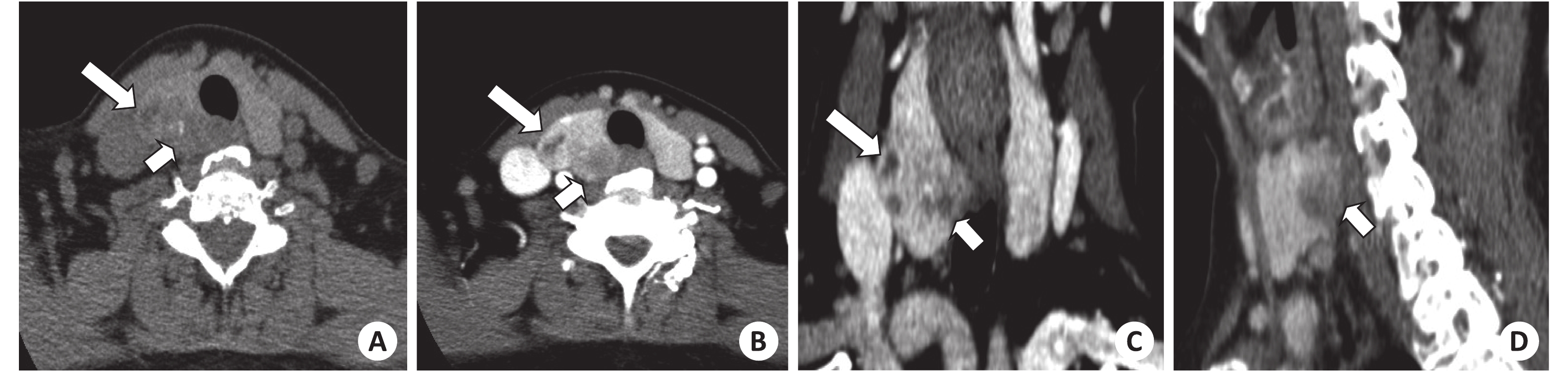
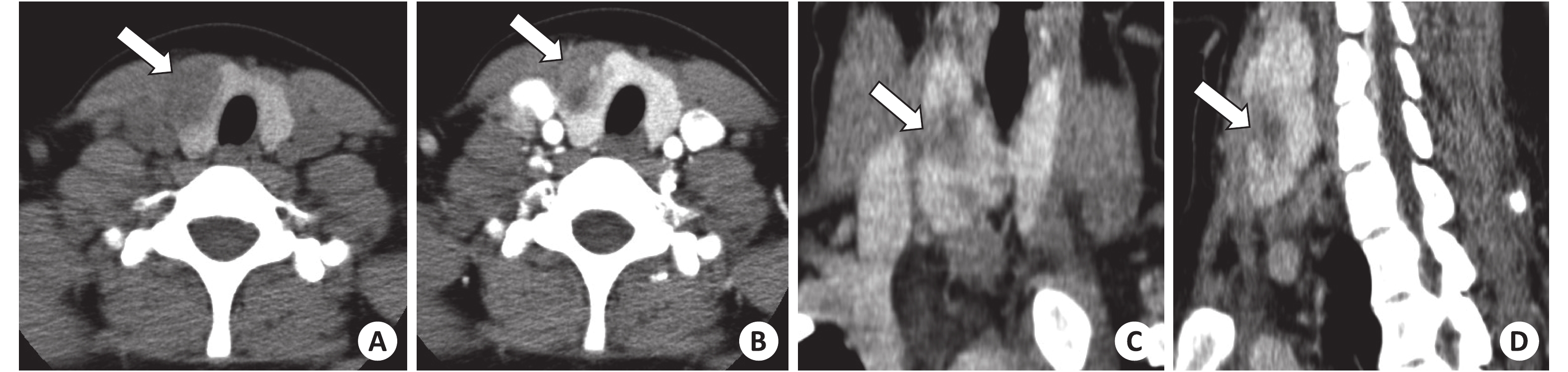
Figures(2)
Supported by: Beijing Renhe Information Technology Co. Ltd support: info@rhhz.net


 DownLoad: