


Correlation of serum inflammatory factors and MRI features of knee osteoarthritis
-
摘要:
目的 探讨血清炎症因子与膝骨关节炎(KOA)MRI影像学特征的相关性。 方法 选取2020年1月~2021年5月于我院收治的膝骨关节炎患者120例作为研究组,另选取同期健康体检人员90例作为对照组。根据是否有滑膜炎将研究组分为Ⅰ组(滑膜炎,n=71)和Ⅱ组(无滑膜炎,n=49)。比较所有患者MRI影像学表现和血清炎症因子[肿瘤坏死因子-α(TNF-α)、白细胞介素-1(IL-1)、白细胞介素-6(IL-6)和白细胞介素-1β(IL-1β)]水平,并对KOA患者MRI影像学特征与血清炎症因子相关性进行分析。 结果 Ⅰ组所有患者均存在软骨病变,且66.20%为2级软骨病变,而Ⅱ组患者有5例无软骨病变,病变患者59.18%为1级。Ⅰ组软骨厚度低于Ⅱ组(P < 0.05)。血清中IL-1β、IL-6和TNF-α表达水平对照组 < Ⅱ组 < Ⅰ组(P < 0.05),IL-10表达水平Ⅱ组 < Ⅰ组 < 对照组(P < 0.05);Ⅰ组血清炎性细胞因子中IL-1β、IL-6和TNF-α与MRI软骨分级呈正相关关系(r=0.387、0.289、0.426,P < 0.05),IL-10无相关性(P > 0.05);Ⅱ组血清炎性细胞因子中仅IL-1β与MRI软骨分级呈正相关关系(r=0.509,P < 0.05),IL-6、IL-10和TNF-α均无相关性(P > 0.05);KOA合并滑膜炎患者MRI滑膜厚度与血清炎性细胞因子中IL-1β、IL-6和TNF-α呈正相关(r=0.497、0.425、0.506,P < 0.05);MRI软骨厚度与血清炎性细胞因子中IL-1β和TNF-α呈负相关(r=-0.503、-0.313,P < 0.05)。 结论 KOA患者MRI可见明显异常,与血清炎性因子有一定的相关性,膝关节影像学特征与血清炎性因子水平结合可对KOA临床诊断及治疗提供指导价值。 Abstract:Objective To investigate the correlation between serum inflammatory factors and MRI features of knee osteoarthritis (KOA). Methods A total of 120 patients with knee osteoarthritis admitted to our hospital from January 2020 to May 2021 were selected as the research group, and another 90 healthy persons during the same period were selected as the control group. Patients in research group were divided into group Ⅰ (synovitis) and group Ⅱ (no synovitis) according to whether there was synovitis. MRI findings and serum levels of inflammatory factors [tumor necrosis factor-α (TNF-α), interleukin-1 (IL-1), interleukin-6 (IL-6) and interleukin-1β (IL-1β)] in all patients were compared. The correlation between MRI imaging features and serum inflammatory factors in KOA patients was analyzed. Results All patients in group Ⅰ had chondropathy, and 66.20% had grade 2 chondropathy, while 5 patients in group Ⅱ had no chondropathy, 59.18% had grade 1 chondropathy. The thickness of cartilage in group Ⅰ was lower than that in group Ⅱ (P < 0.05). The expression levels of IL-1β, IL-6 and TNF-α in serum were lower than those in control group < group Ⅱ < group Ⅰ (P < 0.05). The expression levels of IL-10 were lower than those in group Ⅱ < group Ⅰ < control group (P < 0.05). Il-1β, IL-6 and TNF-α were positively correlated with MRI cartilage grading in group Ⅰ (r= 0.387, 0.289, 0.426, P < 0.05), while IL-10 was not correlated with MRI cartilage grading (P > 0.05). Only IL-1β was positively correlated with MRI cartilage grading in group Ⅱ (r=0.509, P < 0.05), while IL-6, IL-10 and TNF-α were not correlated (P > 0.05). MRI synovial thickness was positively correlated with IL-1β, IL-6 and TNF-α in serum inflammatory cytokines in patients with KOA combined with synovitis (r=0.497, 0.425, 0.506, P < 0.05). The thickness of MRI cartilage was negatively correlated with il-1β and TNF-α in serum inflammatory cytokines (r=-0.503, -0.313, P < 0.05). Conclusion MRI abnormalities in patients with KOA are significantly correlated with serum inflammatory factors. The combination of knee imaging features and serum inflammatory factors can provide guidance for the clinical diagnosis and treatment of KOA. -

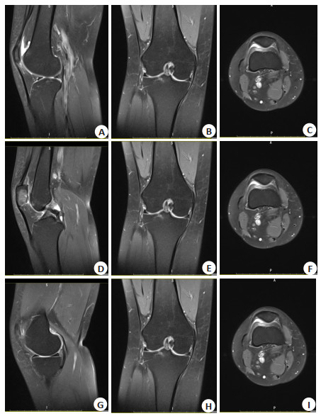
图 1 右膝内外侧半月板MRI平扫
A、D、G:右膝内外侧半月板矢状面平扫图像; B、E、H:右膝内外侧半月板冠状面平扫图像; C、F、I:右膝内外侧横断面平扫图像.
Figure 1. MRI plain scan of medial and lateral meniscus of right knee.

图 2 右膝前后韧带MRI平扫
A、D、G:右膝前后韧带矢状面平扫图像; B、E、H:右膝前后韧带冠状面平扫图像; C、F、I:右膝前后韧带横断面平扫图像.
Figure 2. MRI plain scan of anterior and posterior ligaments of the right knee.
表 1 两组一般资料比较
Table 1. Comparison of general data of two groups
组別 性別(n) 年龄(岁) 膝关节综合评分(分, Mean±SD) 男 女 最小 最大 Mean±SD 对照组(n=90) 40 50 41 73 59.49±6.31 8.19±0.89 研究组(n=120) 55 65 48 75 60.97±6.78 8.07±0.85 χ2/t 0.159 1.612 0.992 P 0.687 0.108 0.322  下载: 导出CSV
表 2 KOA患者膝关节MRI检查结果对比
Table 2. Comparison of MRI results of knee joint in patients with KOA
组別 软骨病变程度[n(%)] 软骨厚度(mm, Mean±SD) 0级 1级 2级 3级 4级 Ⅰ组(n=71) 0(0.00) 2(2.82) 47(66.20) 15(21.13) 7(9.86) 0.44±0.06 Ⅱ组(n=49) 5(10.20) 29(59.18) 8(16.33) 4(8.16) 3(6.12) 0.98±0.11 χ2/t 62.219 34.610 P < 0.001 < 0.001 Ⅰ组:滑膜炎; Ⅱ组:无滑膜炎.
下载: 导出CSV
表 3 三组血清炎性因子水平对比
Table 3. Comparison of serum inflammatory factors in three groups(pg/mL, Mean±SD)
组别 IL-1β IL-6 IL-10 TNF-α 对照组(n=90) 43.66±4.54 85.69±8.70 7.56±0.83 21.49±2.36 Ⅰ组(n=71) 116.36±12.01 146.07±15.85 7.08±0.74 53.66±5.45 Ⅱ组(n=49) 56.08±6.01 98.84±10.12 6.41±0.70 28.57±3.04 F 1698.575 536.373 35.479 1473.475 P < 0.001 < 0.001 < 0.001 < 0.001
下载: 导出CSV
表 4 KOA患者MRI软骨分级与血清炎性细胞因子相关性
Table 4. Correlation between MRI cartilage grading and serum inflammatory cytokines in patients with KOA
指标 Ⅰ组 Ⅱ组 r P r P IL-1β 0.387 < 0.001 0.509 < 0.001 IL-6 0.289 < 0.001 0.150 0.125 IL-10 0.108 0.756 0.114 0.347 TNF-α 0.426 < 0.001 0.193 0.346
下载: 导出CSV
表 5 KOA合并滑膜炎患者MRI软骨厚度、滑膜厚度与血清炎性细胞因子相关性
Table 5. Correlation between MRI cartilage thickness, synovial thickness and serum inflammatory cytokines in patients with KOA complicated with synovitis
指标 滑膜厚度 软骨厚度 r P r P IL-1β 0.497 < 0.001 -0.503 < 0.001 IL-6 0.425 < 0.001 0.057 0.521 IL-10 0.138 0.165 0.078 0.742 TNF-α 0.506 < 0.001 -0.313 0.006
下载: 导出CSV
-
[1] 关春辉, 周占锋, 李沛. 益肾荣筋汤辅助胫骨高位截骨治疗膝骨关节炎疗效及对患者关节液内细胞因子表达水平的影响[J]. 陕西中医, 2019, 40(5): 616-9. doi: 10.3969/j.issn.1000-7369.2019.05.021 [2] Booker SQ, Sibille KT, Terry EL, et al. Psychological predictors of perceived age and chronic pain impact in individuals with and without knee osteoarthritis[J]. Clin J Pain, 2020, 36(8): 569-77. doi: 10.1097/AJP.0000000000000842 [3] 季亚香, 邢伟. 骨关节炎患者膝关节内区形态学的超声评估[J]. 临床超声医学杂志, 2019, 21(3): 236-7. doi: 10.3969/j.issn.1008-6978.2019.03.030 [4] Hiyama Y, Takahashi R, Tanaka T, et al. Quantitative ultrasound of the heel in women with knee osteoarthritis[J]. J Clin Densitom, 2021, 24(4): 557-62. doi: 10.1016/j.jocd.2021.01.001 [5] 朱映红, 曹晓清, 蒋双兰, 等. 超声检查在早期膝骨性关节炎的诊断价值探讨[J]. 实用医院临床杂志, 2019, 16(6): 99-101. doi: 10.3969/j.issn.1672-6170.2019.06.031 [6] Giordano R, Petersen KK, Andersen HH, et al. Serum inflammatory markers in patients with knee osteoarthritis: a proteomic approach [J]. Clin J Pain, 2020, 36(4): 229-37. doi: 10.1097/AJP.0000000000000804 [7] 刘建华, 赵海勇, 温芳, 等. 炎性细胞因子在膝骨关节炎中的表达及与高敏C反应蛋白和红细胞沉降率的相关性[J]. 天津医药, 2020, 48 (1): 55-8. https://www.cnki.com.cn/Article/CJFDTOTAL-TJYZ202001013.htm [8] 中华医学会骨科学分会关节外科学组. 骨关节炎诊疗指南(2018年版) [J]. 中华骨科杂志, 2018, 38(12): 705-15. doi: 10.3760/cma.j.issn.0253-2352.2018.12.001 [9] 王岩, 董本超, 王颖, 等. 膝骨关节炎动物模型的研究进展[J]. 生物医学工程与临床, 2019, 23(3): 365-71. https://www.cnki.com.cn/Article/CJFDTOTAL-SGLC201903032.htm [10] Nikolic G, Nedeljkovic B, Trajkovic G, et al. Pain, physical function, radiographic features, and quality of life in knee osteoarthritis agricultural workers living in rural population[J]. Pain Res Manag, 2019, 2019: 7684762. [11] Camacho-Encina M, Balboa-Barreiro V, Rego-Perez I, et al. Discovery of an autoantibody signature for the early diagnosis of knee osteoarthritis: data from the Osteoarthritis Initiative[J]. Ann Rheum Dis, 2019, 78(12): 1699-705. doi: 10.1136/annrheumdis-2019-215325 [12] 吴炅, 周正球, 周定华, 等. 骨增定痛汤治疗膝骨性关节炎的临床疗效及对关节功能、骨代谢、血清炎症因子水平的影响[J]. 河北中医药学报, 2019, 34(4): 10-4. https://www.cnki.com.cn/Article/CJFDTOTAL-HZYX201904004.htm [13] 易南星, 梁倩倩, 张伟强, 等. 骨性关节炎相关滑膜炎症研究进展[J]. 昆明医科大学学报, 2019, 40(3): 136-9. doi: 10.3969/j.issn.1003-4706.2019.03.028 [14] 孔颖, 王国栋, 孟纯阳. 膝关节骨性关节炎血清炎性因子与血管生成因子的关系[J]. 中国矫形外科杂志, 2019, 27(10): 916-20. https://www.cnki.com.cn/Article/CJFDTOTAL-ZJXS201910015.htm [15] 李志刚, 文戈, 杜立新, 等. T2*mapping辅助MRI技术对差异性膝关节骨性关节炎软骨损伤程度的诊断价值[J]. 分子影像学杂志, 2020, 43(2): 242-6. doi: 10.12122/j.issn.1674-4500.2020.02.13 [16] Sun G, Ba CL, Gao R, et al. Association of IL- 6, IL-8, MMP- 13 gene polymorphisms with knee osteoarthritis susceptibility in the Chinese Han population [J]. Biosci Rep, 2019, 39(2): BSR20181346. doi: 10.1042/BSR20181346 [17] Schwarz S, Mrosewski I, Silawal S, et al. The interrelation of osteoarthritis and diabetes mellitus: considering the potential role of interleukin-10 and in vitro models for further analysis[J]. Inflamm Res, 2018, 67(4): 285-300. doi: 10.1007/s00011-017-1121-8 [18] Bemeur C, Desjardins P, Butterworth RF. 64 interleukin- 1β and tumor necrosis factor-Α[J]. Cytokine, 2008, 43(3): 251-2. [19] 陈宁杰, 赵卉, 郝风云, 等. 晚期膝骨关节炎的滑膜炎MRI厚度分级与滑膜病理相关性[J]. 中华关节外科杂志: 电子版, 2018, 12(5): 653- 61. doi: 10.3877/cma.j.issn.1674-134X.2018.05.010 [20] 陈元川, 庞坚, 石印玉, 等. 膝骨关节炎膝痛分布及其与影像学特征的关系研究[J]. 中国中医骨伤科杂志, 2018, 26(5): 39-43, 48. https://www.cnki.com.cn/Article/CJFDTOTAL-ZGZG201805010.htm [21] 李志刚, 文戈, 杜立新, 等. T2*mapping辅助MRI技术对差异性膝关节骨性关节炎软骨损伤程度的诊断价值[J]. 分子影像学杂志, 2020, 43(2): 242-6. doi: 10.12122/j.issn.1674-4500.2020.02.13 -


 点击查看大图
计量
- 文章访问数: 164
- HTML全文浏览量: 141
- PDF下载量: 5
- 被引次数: 0