


Application of three-dimensional reconstruction of head and neck with neck extension in thyroid surgery
-
摘要:
目的 探究在颈伸位下三维重建头颈结构在甲状腺手术中的应用价值。 方法 选择20例已经由病理结果证实的甲状腺疾病患者在颈伸位下的平扫CT图像,应用三维重建软件3DVWorks,对甲状腺、甲状腺结节、甲状软骨、下颌骨、锁骨、皮肤等组织进行三维重建,测量多个空间角度及径线以评估疾病及治疗方案。 结果 通过在头颈三维重建模型中对下段气管所在的不同平面与甲状腺手术相关解剖结构的角度与距离测量,综合评估后在甲状腺结节 < 3 cm的11例患者中有3例患者不适合行经口腔镜术式,可行其他腔镜等术式;3例患者不适合行经口、经胸乳腔镜术式及经腋窝等腔镜手术,仅适合开放手术;1例患者因结节分期较高,不推荐行腔镜手术;另外4例患者各类手术均可施行。通过构建胸廓入口及以此平面准确分割巨大甲状腺体积,对6例含有结节 > 3 cm的巨大甲状腺进行预评估后发现均未达经颈胸联合切除甲状腺的指标要求,6例患者行传统单纯经颈切口即可完整切除甲状腺。对有残留甲状腺的3例患者,预评估显示3例残余甲状腺均可准确定位并能指导手术切除。 结论 通过在颈伸位下三维重建头颈结构并建立评估指标,对腔镜手术方式选择、巨大甲状腺肿诊治、残余甲状腺术中定位有独特优势,有助于形成良好的手术策略,更有益于医患交流。 Abstract:Objective To evaluate the clinical application of three-dimensional reconstruction of head and neck with neck extension in thyroid surgery. Methods The unenhanced CT images which were scan with neck extension of 20 patients proved to be thyroid disease by pathology were collected. The thyroid gland, thyroid nodules, thyroid cartilage, mandible, clavicle, and skin were reconstructed with the software of 3DVWorks. Measuring different angles and diameters for disease evaluation and treatment base on the reconstruction model. Results In the 3D reconstruction model of head and neck, intersectoral angles and distances between different tracheal planes and anatomic structures of endoscopic thyroidectomy were measured. Based on these 3D data, 3 of 11 patients with thyroid nodule < 3 cm were not suitable for endoscopic thyroidectomy via a transoral approach. Meantime, 3 patient was not suitable for the thoracic and breast approach or axillo-breast approach and 1 patient was only suitable for open surgery. The other 4 of 11 patients were suitable for all surgical approaches. 6 patients with thyroid nodule > 3 cm were not diagnosed with a substernal goiter by the model, which could be accessible via a transcervical approach but not a transsternal operative approach. The model showed residual thyroid after surgery in 3 cases could be located accurately and directed the operation. Conclusion Three-dimensional reconstruction of head and neck with neck extension has unique advantages in choice of operative approach, diagnosis, and treatment of substernal goiter and intraoperative localization of residual thyroid tissue, which help to make a better surgical decision and benefit physician-patient communication. -
Key words:
- thyroid diseases /
- tomography /
- imaging, three-dimensional /
- surgical procedures
-

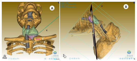
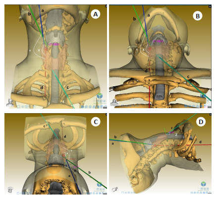
图 1 患者女性,22岁,术前颈伸位下多维度的术式评估三维模型
A:前面观; B:足侧面观; C:头侧面观; D:右侧面观. 条棒a为经口腔镜径路,条棒b为甲状腺结节内侧缘与甲状软骨板底边连线,条棒c为经胸乳腔镜径路,条棒d指示下段气管平行的平面。角度α为棒a与棒b的交角.
Figure 1. Female, 22 years old, a 3D model of operative methods evaluation with head and neck extension.
-
[1] Durante C, Grani G, Lamartina L, et al. The diagnosis and management of thyroid nodules[J]. JAMA, 2018, 319(9): 914. doi: 10.1001/jama.2018.0898 [2] 彭李青, 陈金品, 李燕梅, 等. 螺旋CT三维重建与仿真喉镜在甲状腺疾病中的临床应用[J]. 中国临床医学影像杂志, 2005, 16(3): 132-3, 152. doi: 10.3969/j.issn.1008-1062.2005.03.004 [3] 蔡念, 喻海波, 杨景哥, 等. 甲状腺三维重建在腔镜甲状腺切除术中的应用[J]. 腹腔镜外科杂志, 2012, 17(6): 408-11. doi: 10.3969/j.issn.1009-6612.2012.06.004 [4] Franca C, Levin-Plotnik D, Sehgal V, et al. Use of three-dimensional spiral computed tomography imaging for staging and surgical planning of head and neck cancer[J]. J Digit Imaging, 2000, 13(2 suppl 1): 24-32. [5] 翁梅, 陆云, 王琦, 等. 甲状腺手术体位护理的研究进展[J]. 护理研究, 2014, 28(15): 1803-5. https://www.cnki.com.cn/Article/CJFDTOTAL-SXHZ201415006.htm [6] 孙晓铮, 马静. 甲状腺手术体位的研究及护理进展[J]. 护士进修杂志, 2015, 30(17): 1574-6. https://www.cnki.com.cn/Article/CJFDTOTAL-FSJX201517013.htm [7] 方思月. CT增强联合三维重建技术评估分化型甲状腺癌T分期的价值[J]. 医学影像学杂志, 2015, 25(6): 981-4. https://www.cnki.com.cn/Article/CJFDTOTAL-XYXZ201506013.htm [8] 吴国洋, 傅锦波. 经口入路腔镜下甲状腺切除手术技术要点[J]. 临床外科杂志, 2015, 23(7): 497-9. doi: 10.3969/j.issn.1005-6483.2015.07.007 [9] Jiang WJ, Yan PJ, Zhao CL, et al. Comparison of total endoscopic thyroidectomy with conventional open thyroidectomy for treatment of papillary thyroid cancer: a systematic review and meta-analysis[J]. Surg Endosc, 2020, 34(5): 1891-903. doi: 10.1007/s00464-019-07283-y [10] Wang C, Sun P, Li J, et al. Strategies of laparoscopic thyroidectomy for treatment of substernal goiter via areola approach[J]. Surg Endosc, 2016, 30(11): 4721-30. doi: 10.1007/s00464-016-4814-0 [11] Hanson MA, Shaha AR, Wu JX. Surgical approach to the substernal goiter[J]. Best Pract Res Clin Endocrinol Metab, 2019, 33(4): 101312. doi: 10.1016/j.beem.2019.101312 [12] 何晓娜, 张小鹏, 王斌儒, 等. 甲状腺结节性疾病超声诊疗的研究进展[J]. 医学综述, 2018, 24(12): 2467-75. doi: 10.3969/j.issn.1006-2084.2018.12.033 [13] Guo J, Qian J, Yuan YF. Computed tomography measurements as a standard of exophthalmos? two-dimensional versus three-dimensional techniques[J]. Curr Eye Res, 2018, 43(5): 647-53. doi: 10.1080/02713683.2018.1431285 [14] 傅栋, 靳安民, 田京, 等. 颈段脊柱前路手术相关结构的三维可视化[J]. 南方医科大学学报, 2010, 30(4): 888-90, 894. https://www.cnki.com.cn/Article/CJFDTOTAL-DYJD201004063.htm [15] Eisenberg H, Pallotta J, Sacks B, et al. Parathyroid localization, three-dimensional modeling, and percutaneous ablation techniques[J]. Endocrinol Metab Clin North Am, 1989, 18(3): 659-700. doi: 10.1016/S0889-8529(18)30359-1 [16] Toyota K, Uchida H, Ozasa H, et al. Preoperative airway evaluation using multi-slice three-dimensional computed tomography for a patient with severe tracheal Stenosis[J]. Br J Anaesth, 2004, 93(6): 865-7. doi: 10.1093/bja/aeh283 [17] Binar M, Serindere M, Bozlar U, et al. Determining the thyroid gland volume causing tracheal compression: a semiautomated 3D CT volumetry study[J]. Medicina, 2019, 55(5): 143. doi: 10.3390/medicina55050143 [18] 王存川, 陈志强, 李进义. 内镜治疗甲状腺疾病[J]. 腹腔镜外科杂志, 2011, 16(8): 567-71. doi: 10.3969/j.issn.1009-6612.2011.08.003 [19] 陆锦俊, 龚卫东, 支巧明. 腔镜技术在甲状腺疾病外科治疗中的运用[J]. 中华腔镜外科杂志: 电子版, 2011, 4(4): 304-6. https://www.cnki.com.cn/Article/CJFDTOTAL-ZQJW201104018.htm [20] Winzek CF, Hartrampf LCM, Kampschulte M, et al. Unilateral and bilateral agenesis of the upper thyroid horns—A morphometric analysis of the larynx[J]. Forensic Sci Int, 2019, 301: 225-30. doi: 10.1016/j.forsciint.2019.05.038 [21] Shiozawa T, Epe P, Herlan S, et al. Clinically relevant variations of the superior thyroid Cornu[J]. Surg Radiol Anat, 2017, 39(3): 299-306. doi: 10.1007/s00276-016-1735-5 [22] Jennings A. Evaluation of substernal goiters using computed tomography and MR imaging[J]. Endocrinol Metab Clin North Am, 2001, 30(2): 401-14, ix. doi: 10.1016/S0889-8529(05)70192-4 [23] Allo MD, Thompson NW. Rationale for the operative management of substernal goiters[J]. Surgery, 1983, 94(6): 969-77. [24] McKenzie GA, Rook W. Is it possible to predict the need for sternotomy in patients undergoing thyroidectomy with retrosternal extension?[J]. Interact Cardiovasc Thorac Surg, 2014, 19(1): 139-43. doi: 10.1093/icvts/ivu094 [25] Qureishi A, Garas G, Tolley N, et al. Can pre-operative computed tomography predict the need for a thoracic approach for removal of retrosternal goitre?[J]. Int J Surg, 2013, 11(3): 203-8. doi: 10.1016/j.ijsu.2013.01.006 [26] Huins CT, Georgalas C, Mehrzad H, et al. A new classification system for retrosternal goitre based on a systematic review of its complications and management[J]. Int J Surg, 2008, 6(1): 71-6. doi: 10.1016/j.ijsu.2007.02.003 [27] Mercante G, Gabrielli E, Pedroni C, et al. CT cross-sectional imaging classification system for substernal goiter based on risk factors for an extracervical surgical approach[J]. Head Neck, 2011, 33(6): 792-9. doi: 10.1002/hed.21539 [28] Harris RS. The effect of extension of the head and neck upon the infrahyoid respiratory passage and the supraclavicular portion of the human Trachea[J]. Thorax, 1959, 14: 176-80. doi: 10.1136/thx.14.2.176 [29] 张卫国, 刘佳萍, 贾秀琴, 等. 胸部CT扫描中颈部伸展体位应用价值的探讨[J]. 中华放射医学与防护杂志, 2019(12): 951-4. doi: 10.3760/cma.j.issn.0254-5098.2019.12.014 [30] Cho SJ, Suh CH, Baek JH, et al. Diagnostic performance of CT in detection of metastatic cervical lymph nodes in patients with thyroid cancer: a systematic review and meta-analysis[J]. Eur Radiol, 2019, 29(9): 4635-47. doi: 10.1007/s00330-019-06036-8 -

 下载:
下载:
 点击查看大图
图(2)
计量
- 文章访问数: 266
- HTML全文浏览量: 142
- PDF下载量: 15
- 被引次数: 0