


Value of conventional ultrasound, ultrasound elastography and percutaneous contrast-enhanced ultrasound in sentinel lymph node diagnosis of breast cancer
-
摘要:
目的 探讨常规二维超声、超声弹性成像技术和经皮超声造影(CEUS)对乳腺癌患者同侧腋窝前哨淋巴结的诊断价值。 方法 对158例乳腺癌患者进行回顾性分析,术前均行常规二维超声、超声弹性成像、CEUS及粗针穿刺,以病理穿刺结果为金标准,与病理结果一致性进行Kappa检验,以病理诊断前哨淋巴结转移为因变量,以常规二维超声、超声弹性成像和CEUS为自变量,建立Logistic回归模型,并用ROC曲线评价常规二维超声、超声弹性成像和CEUS对乳腺癌前哨淋巴结的诊断价值。 结果 经病理诊断共检出206个淋巴结,其中转移性76个,非转移性130个。常规二维超声检查对乳腺癌前哨淋巴结转移的敏感度为69.7%,特异性为94.6%,准确度为85.4%;应用超声弹性成像技术检查的敏感度为84.0%,特异性为96.9%,准确度为92.2%;CEUS检查的敏感度为88.3%,特异性为98.4%,准确度为94.7%;三者联合检查的敏感度为93.4%,特异性为99.2%,准确度为97.1%;与病理结果一致性进行比较,弹性成像、CEUS的一致性均高于常规二维超声,三者联合诊断的一致性最好(Kappa= 0.683、0.828、0.884、0.937,P < 0.05)。ROC曲线显示:常规二维超声、超声弹性成像、CEUS和三者联合诊断乳腺癌前哨淋巴结转移的曲线下面积为0.668、0.712、0.738、0.756。 结论 超声弹性成像技术、CEUS对乳腺癌前哨淋巴结的诊断价值均高于常规二维超声,且三者联合诊断的敏感度、特异性、准确性更高,对术前乳腺癌患者同侧转移性前哨淋巴结的诊断具有重要价值。 Abstract:Objective To investigate the diagnostic value of conventional two-dimensional ultrasound, ultrasound elastography and percutaneous contrast ultrasound (CEUS) in ipsilateral axillary sentinel lymph node in breast cancer patients. Methods We retrospectively analyzed 158 cases of breast cancer patients. The patients undewent preoperative lines of conventional two-dimensional ultrasound, ultrasound elasticity imaging, CEUS and coarse needle puncture, with pathological biopsy results as the gold standard, and Kappa test for consistency with pathological results. Logistic regression model was established with pathological diagnosis of sentinel lymph node metastasis as dependent variable and routine two-dimensional ultrasound, ultrasound elastography and CEUS as independent variables. ROC curve was used to evaluate the diagnostic value of conventional two-dimensional ultrasound, ultrasound elastography and CEUS in sentinel lymph nodes of breast cancer. Results A total of 206 lymph nodes were detected by pathological diagnosis in 158 patients with breast cancer, including 76 metastatic nodes and 130 non-metastatic nodes. The sensitivity, specificity and accuracy of conventional two-dimensional ultrasonography for sentinel lymph node metastasis of breast cancer were 69.7%, 94.6% and 85.4%, respectively. The sensitivity, specificity and accuracy of ultrasound elastography were 84.0%, 96.9% and 92.2%, respectively. The sensitivity, specificity and accuracy of CEUS were 88.3%, 98.4% and 94.7% respectively. The sensitivity, specificity and accuracy were 93.4%, 99.2% and 97.1% respectively. The consistency of elastography and CEUS is higher than that of conventional two-dimensional ultrasound, the consistency of combined diagnosis of the three is the best (Kappa=0.683, 0.828, 0.884, 0.937, P < 0.05). ROC curve showed that the AUC of conventional two-dimensional ultrasound, ultrasound elastic imaging, CEUS and their combination in the diagnosis of sentinel lymph node metastasis of breast cancer were 0.668, 0.712, 0.738 and 0.756. Conclusion The diagnostic value of ultrasound elastic imaging and CEUS for sentinel lymph nodes of breast cancer is higher than conventional two-dimensional ultrasound. The combined diagnosis of the three has higher sensitivity, specificity and accuracy, which is of great value for the preoperative diagnosis of ipsilateral metastatic sentinel lymph nodes in breast cancer patients. -

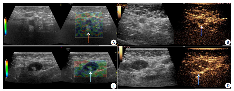
图 1 乳腺癌前哨淋巴结CEUS高峰期特征分型
A:Ⅰ型,完全均匀增强型; B:Ⅱ型,周边及髓质均匀增强型; C:Ⅲ型,周边和(或)髓质不均匀增强型; D:Ⅳ型,无增强型.
Figure 1. Sentinel lymph node of breast cancer with CEUS peak characteristic classification.

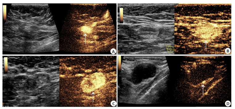
图 2 乳腺癌前哨淋巴结UE及CEUS图像
A:UE上淋巴结中央少许呈绿色,周边大部分呈蓝色,评为4分; B:此淋巴结在CEUS表现为Ⅳ型,无增强型; 病理结果为转移性淋巴结; C:UE上淋巴结蓝色与绿色占比几乎相等,评为3分; D:此淋巴结在CEUS表现为Ⅲ型,髓质不均匀增强; 病理结果为转移性淋巴结.
Figure 2. Images of sentinel lymph nodes with breast cancer by elastography and CEUS.

图 3 常规二维超声、UE、CEUS及联合检查对乳腺癌前哨淋巴结的ROC曲线
Figure 3. ROC curves of sentinel lymph nodes of breast cancer by conventional two-dimensional ultrasound, elastography, CEUS and combined examination.
表 1 病理检测、常规二维超声、UE、CEUS的诊断结果比较
Table 1. Comparison of diagnostic results of pathological examination, conventional two-dimensional ultrasound, elastography and CEUS (n)
类别 手术病理 合计 转移性前哨淋巴结 非转移性前哨淋巴结 常规二维超声 转移 53 6 59 非转移 23 123 147 总计 76 130 206 UE 转移 63 4 67 非转移 12 127 139 总计 75 131 206 CEUS 转移 68 2 70 非转移 9 127 136 总计 77 129 206 联合检查 转移 71 1 72 非转移 5 129 134 总计 76 130 206 UE:超声弹性成像; CEUS:超声造影.  下载: 导出CSV
-
[1] Siegel RL, Miller KD, Jemal A. Cancer statistics, 2019[J]. CA A Cancer J Clin, 2019, 69(1): 7-34. doi: 10.3322/caac.21551 [2] 孙可欣, 郑荣寿, 张思维, 等. 2015年中国分地区恶性肿瘤发病和死亡分析[J]. 中国肿瘤, 2019, 28(1): 1-11. https://www.cnki.com.cn/Article/CJFDTOTAL-ZHLU201901001.htm [3] Shah AN, Metzger O, Bartlett CH, et al. Hormone receptor-positive/ human epidermal growth receptor 2-negative metastatic breast cancer in young women: emerging data in the era of molecularly targeted agents[J]. Oncologist, 2020, 25(6): e900-e908. doi: 10.1634/theoncologist.2019-0729 [4] Xiao YJ, Xia JJ, Li LP, et al. Associations between dietary patterns and the risk of breast cancer: a systematic review and meta-analysis of observational studies[J]. Breast Cancer Res, 2019, 21(1): 16. doi: 10.1186/s13058-019-1096-1 [5] Zuo TT, Zheng RS, Zeng HM, et al. Female breast cancer incidence and mortality in China, 2013[J]. Thorac Cancer, 2017, 8(3): 214-8. doi: 10.1111/1759-7714.12426 [6] 中国抗癌协会乳腺癌专业委员会. 中国抗癌协会乳腺癌诊治指南与规范(2019年版[)J]. 中国癌症杂志, 2019, 29(8): 609-80. https://www.cnki.com.cn/Article/CJFDTOTAL-ZGAZ202110015.htm [7] Kang J, Chang JH, Kim SM, et al. Real-time sentinel lymph node biopsy guidance using combined ultrasound, photoacoustic, fluorescence imaging: in vivo proof-of-principle and validation with nodal obstruction[J]. Sci Rep, 2017, 7: 45008. doi: 10.1038/srep45008 [8] Pepels MJ, Vestjens JHMJ, de Boer M, et al. Safety of avoiding routine use of axillary dissection in early stage breast cancer: a systematic review[J]. Breast Cancer Res Treat, 2011, 125(2): 301-13. doi: 10.1007/s10549-010-1210-7 [9] 吴意赟, 蔡婷, 许华宁, 等. 联合经皮和经静脉超声造影对乳腺癌前哨淋巴结的诊断价值[J]. 中华医学超声杂志: 电子版, 2020, 17(12): 1168-72. doi: 10.3877/cma.j.issn.1672-6448.2020.12.004 [10] 左梦, 张海宇, 巴黎, 等. 超声造影联合声触诊组织成像定量技术对乳腺癌前哨淋巴结转移的评估[J]. 中华医学超声杂志: 电子版, 2021, 18(2): 171-6. doi: 10.3877/cma.j.issn.1672-6448.2021.02.009 [11] 韩霞. 2D-US联合UE技术对乳腺癌腋淋巴结转移患者诊断效能的影响[J]. 现代医用影像学, 2018, 27(4): 1410-1. https://www.cnki.com.cn/Article/CJFDTOTAL-XDYY201804168.htm [12] 李慧芳, 刘景萍, 郑薇薇, 等. 高频彩色多普勒超声及弹性成像在乳腺癌腋淋巴结转移中的应用价值[J]. 山西医药杂志, 2016, 45(5): 510-3. https://www.cnki.com.cn/Article/CJFDTOTAL-SXYY201605004.htm [13] 郭碧萍, 李艳宁, 徐丽芳, 等. 超声弹性成像联合常规超声评分对乳腺癌腋窝淋巴结转移的诊断价值[J]. 广西医科大学学报, 2017, 34(6): 832-4. https://www.cnki.com.cn/Article/CJFDTOTAL-GXYD201706008.htm [14] 王少芬, 李君庆. 常规超声联合超声造影在乳腺癌转移性前哨淋巴结中的诊断价值[J]. 临床医学研究与实践, 2021, 6(35): 114-6. https://www.cnki.com.cn/Article/CJFDTOTAL-YLYS202135036.htm [15] 纪岩磊, 韩真, 马恒敏, 等. 超声弹性成像对乳腺癌腋窝淋巴结诊断价值的探讨[J]. 中华肿瘤防治杂志, 2016, 23(16): 1081-4. doi: 10.3969/j.issn.1673-5269.2016.16.007 [16] Sever AR, Mills P, Weeks J, et al. Preoperative needle biopsy of sentinel lymph nodes using intradermal microbubbles and contrast-enhanced ultrasound in patients with breast cancer[J]. AJR Am J Roentgenol, 2012, 199(2): 465-70. doi: 10.2214/AJR.11.7702 [17] Shimazu K, Ito T, Uji K, et al. Identification of sentinel lymph nodes by contrast-enhanced ultrasonography with Sonazoid in patients with breast cancer: a feasibility study in three hospitals[J]. Cancer Med, 2017, 6(8): 1915-22. doi: 10.1002/cam4.1142 [18] Zhao Q, Sun JW, Zhou H, et al. Pre-operative conventional ultrasound and sonoelastography evaluation for predicting axillary lymph node metastasis in patients with malignant breast lesions[J]. Ultrasound Med Biol, 2018, 44(12): 2587-95. doi: 10.1016/j.ultrasmedbio.2018.07.017 [19] Guo RR, Lu GL, Qin BJ, et al. Ultrasound imaging technologies for breast cancer detection and management: a review[J]. Ultrasound Med Biol, 2018, 44(1): 37-70. doi: 10.1016/j.ultrasmedbio.2017.09.012 [20] Wang JW, Guo ZX, Lin QG, et al. Ultrasound elastography as an imaging biomarker for detection of early tumor response to chemotherapy in a murine breast cancer model: a feasibility study[J]. Br J Radiol, 2018, 91(1085): 20170698. [21] Qiu T, Wang H, Song J, et al. Assessment of liver fibrosis by ultrasound elastography and contrast-enhanced ultrasound: a randomized prospective animal study[J]. Exp Anim, 2018, 67(2): 117-26. doi: 10.1538/expanim.17-0098 [22] Zheng XY, Huang YN, Wang Y, et al. Combination of different types of elastography in downgrading ultrasound Breast Imaging-Reporting and Data System category 4a breast lesions[J]. Breast Cancer Res Treat, 2019, 174(2): 423-32. doi: 10.1007/s10549-018-05072-0 [23] 胥桐, 郭丽苹, 方红, 等. 经皮超声造影对乳腺癌前哨淋巴结的定性评估[J]. 中国医学影像学杂志, 2020, 28(2): 86-9, 94. doi: 10.3969/j.issn.1005-5185.2020.02.002 [24] Zhao J, Zhang J, Zhu QL, et al. The value of contrast-enhanced ultrasound for sentinel lymph node identification and characterisation in pre-operative breast cancer patients: a prospective study[J]. Eur Radiol, 2018, 28(4): 1654-61. doi: 10.1007/s00330-017-5089-0 [25] 郭晓霞, 刘昱含, 李潜. 乳腺癌前哨淋巴结超声造影不均匀增强模式的表现分析[J]. 中华实用诊断与治疗杂志, 2019, 33(4): 386-8. https://www.cnki.com.cn/Article/CJFDTOTAL-HNZD201904023.htm [26] 梁舒媛, 罗渝昆, 费翔, 等. 高帧频超声造影在鉴别浅表淋巴结性质中的应用[J]. 中华医学超声杂志: 电子版, 2020, 17(9): 841-7. doi: 10.3877/cma.j.issn.1672-6448.2020.09.005 [27] Wang Y, Zhou WB, Li CY, et al. Variation of sentinel lymphatic channels (SLCs) and sentinel lymph nodes (SLNs) assessed by contrast-enhanced ultrasound (CEUS) in breast cancer patients[J]. World J Surg Oncol, 2017, 15(1): 127. doi: 10.1186/s12957-017-1195-3 -


 点击查看大图
计量
- 文章访问数: 223
- HTML全文浏览量: 145
- PDF下载量: 10
- 被引次数: 0