


MRI apparent diffusion coefficient histogram and serum CA125 help to evaluate the histopathological characteristics of endometrial adenocarcinoma
-
摘要:
目的 探究基于子宫内膜样腺癌表观扩散系数(ADC)直方图分析和糖类抗原125(CA125)与组织分化程度、微卫星、Ki-67的相关性。 方法 搜集68例首诊且经病理证实为子宫内膜样腺癌患者资料。所有患者均行超导核磁共振扫描,用FireVoxel软件在ADC图测得肿瘤感兴趣区的直方图参数。分析子宫内膜样腺癌的直方图参数和CA125与组织分化程度、微卫星、Ki-67的相关性。 结果 直方图参数、Ki-67差距在不同组织分化程度中均有统计学意义(P < 0.05)。直方图参数、Ki-67及CA125均值差距在微卫星稳定组与不稳定组没有统计学意义(P>0.05)。ROC曲线统计计算出P90值对应的曲线下面积最大(0.937)。直方图参数与组织分化程度呈低或中等程度负相关(P < 0.05);平均数、最大值、P10、P25、P50、P75、P90、P95与Ki-67呈低等程度负相关(P < 0.05)。 结论 ADC直方图参数和血清CA125有助于评估子宫内膜腺癌的病理组织学特征。 Abstract:Objective To explore the correlation between the whole focus apparent diffusion coefficient (ADC) histogram analysis and CA125 and the degree of differentiation, microsatellite and Ki-67 of endometrioid adenocarcinoma, respectively. Methods Sixty-eight patients with endometrial carcinoma confirmed by preoperative pathology. All patients underwent superconducting MRI scans. The ROIs were drawn by FireVoxel software and histogram analysis was carried out. The correlation between the ADC histogram parameters and CA125 and differentiation, microsatellite and Ki-67 of endometrioid adenocarcinoma were analyzed. Results The ADC histogram parameter and Ki-67 were statistically significant in different tissue differentiation degrees(P < 0.05). The ADC histogram parameters, Ki-67 and CA125 differences were not statistically significant between the stable and unstable microsatellite groups(P>0.05). ROC curve analysis showed that P90 achieved the highest AUC of 0.937. Histogram parameters were negatively correlated with the degree of tissue differentiation with low or middle degree. Mean, maximum, P10, P25, P50, P75, P90, P95 and Ki-67 were negatively correlated with low degrees. Conclusion ADC histogram parameters and serum CA125 help to evaluate the histopathological characteristics of endometrial adenocarcinoma. -
Key words:
- endometrioid adenocarcinoma /
- apparent diffusion coefficient /
- histogram /
- histological grade /
- microsatellite /
- CA125
-

图 1 患者女,49岁,子宫内膜腺癌(中分化),微卫星稳定(临床分期为三期)
A:T1WI;B:T2WI压脂;C:DWI;D:T1WI增强;E:ADC图;F:ROI勾画示意图。宫腔增大,其内见团块状软组织信号影,T1WI呈略低信号,T2WI压脂呈高信号,DWI呈明显高信号,ADC图呈明显低信号,增强扫描呈轻度不均匀强化,结合带消失.
Figure 1. A 49-year-old female patient with endometrial adenocarcinoma (moderately differentiated), microsatellite stable (clinical stage: stage III).

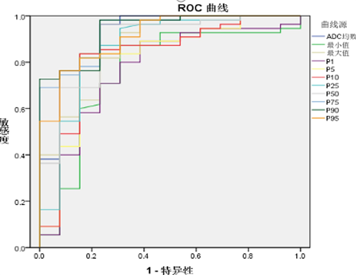
图 2 ADC直方图参数对不同分化程度子宫内膜腺癌的ROC曲线分析
Figure 2. ROC curve analysis of ADC histogram parameters for endometrial adenocarcinoma with different degrees of differentiation.
表 1 子宫内膜腺癌不同分化程度的ADC直方图参数、Ki-67、CA125的比较
Table 1. Comparison of ADC histogram parameters, Ki-67 and CA125 in endometrial adenocarcinoma with different degrees of differentiation(Mean±SD)
参数 高分化 中分化 低分化 F P 平均值(x10-3 mm2/s) 1.139±0.163 1.015±0.093 0.824±0.129 25.598 < 0.001 最小值(x10-3mm2/s) 0.667±0.215 0.625±0.185 0.438±0.324 5.608 0.006 最大值(x10-3mm2/s) 1.870±0.410 1.686±0.291 1.330±0.224 11.356 < 0.001 P(x10-3 mm2/s) 0.732±0.215 0.673±0.157 0.492±0.223 6.716 0.002 P5(x10-3 mm2/s) 0.834±0.169 0.753±0.107 0.604±0.164 10.854 < 0.001 P10(x10-3 mm2/s) 0.894±0.171 0.799±0.097 0.652±0.158 12.847 < 0.001 P25(x10-3 mm2/s) 0.990±0.177 0.880±0.102 0.717±0.152 15.616 < 0.001 P50(x10-3 mm2/s) 1.115±0.178 0.984±0.103 0.806±0.145 20.249 < 0.001 P75(x10-3 mm2/s) 1.270±0.184 1.126±0.100 0.912±0.125 27.565 < 0.001 P90(x10-3 mm2/s) 1.427±0.209 1.281±0.131 1.046±0.104 24.024 < 0.001 P95(x10-3 mm2/s) 1.535±0.263 1.383±0.151 1.132±0.130 17.672 < 0.001 Ki-67 28.890±18.789 49.640±18.556 70.000±14.142 23.137 < 0.001 CA125(U/mL) 16.990±14.886 20.249±17.498 29.355±18.522 2.243 0.096  下载: 导出CSV
表 2 子宫内膜腺癌不同分化程度的ADC直方图参数、Ki-67、CA125差异的两两比较
Table 2. Pairwise comparison of ADC histogram parameters, Ki-67, CA125 in endometrial adenocarcinoma with different degrees of differentiation
P 高分化vs中分化 高分化vs低分化 中分化vs低分化 P平均值 0.001 < 0.001 < 0.001 P最小值 0.449 0.002 0.009 P最大值 0.047 < 0.001 0.002 Pp1 0.270 0.001 0.007 PP5 0.044 < 0.001 0.004 Pp10 0.015 < 0.001 0.003 PP25 0.007 < 0.001 0.001 PP50 0.001 < 0.001 0.001 PP75 < 0.001 < 0.001 < 0.001 PP90 0.002 < 0.001 < 0.001 PP95 0.007 < 0.001 < 0.001 PKi67 < 0.001 < 0.001 0.001 PCA125 0.472 0.032 0.108
下载: 导出CSV
表 3 子宫内膜腺癌的微卫星稳定与不稳定组的ADC直方图参数、Ki-67、CA125比较
Table 3. Comparison of ADC histogram parameters, Ki-67 and CA125 between microsatellite stable and unstable groups in endometrial adenocarcinoma(Mean±SD)
参数 稳定组 不稳定组 统计值 P 平均值(x10-3 mm2/s) 1.031±0.182 1.016±0.146 0.319 0.751 最小值(x10-3 mm2/s) 0.609±0.210 0.597±0.256 0.188 0.851 最大值(x10-3 mm2/s) 1.648±0.338 1.823±0.485 -1.649 0.104 P1(x10-3 mm2/s) 0.673±0.200 0.630±0.245 0.716 0.476 P5(x10-3 mm2/s) 0.766±0.159 0.728±0.186 0.808 0.422 P10(x10-3 mm2/s) 0.818±0.161 0.779±0.180 0.853 0.397 P2(x10-3 mm2/s) 0.903±0.176 0.861±0.169 0.862 0.392 P50(x10-3 mm2/s) 1.009±0.191 0.981±0.156 0.543 0.589 P7(x10-3 mm2/s) 1.143±0.205 1.140±0.150 0.046 0.964 P90(x10-3 mm2/s) 1.287±0.209 1.316±0.192 -0.485 0.629 P9(x10-3 mm2/s) 1.376±0.232 1.453±0.284 -1.110 0.271 Ki-67 46.410±21.905 43.530±26.970 0.443 0.659 CA125(U/mL) 20.234±17.288 22.105±16.797 -0.389 0.698
下载: 导出CSV
表 4 ADC直方图参数对不同分化程度子宫内膜腺癌的ROC曲线分析
Table 4. ROC curve analysis of ADC histogram parameters for endometrial adenocarcinoma with different degrees of differentiation
ADC直方图参数 曲线下面积 95%CI P 平均值(x10-3mm2/s) 0.919 0.822~1.000 < 0.001 最小值(x10-3mm2/s) 0.760 0.596~0.924 0.004 最大值(x10-3mm2/s) 0.870 0.756~0.984 < 0.001 P1(x10-3mm2/s) 0.769 0.613~0.925 0.003 P5(x10-3mm2/s) 0.817 0.670~0.964 < 0.001 P10(x10-3mm2/s) 0.824 0.680~0.968 < 0.001 P25(x10-3mm2/s) 0.865 0.731~0.999 < 0.001 P50(x10-3mm2/s) 0.887 0.779~0.995 < 0.001 P75(x10-3mm2/s) 0.934 0.867~1.000 < 0.001 P90(x10-3mm2/s) 0.937 0.872~1.000 < 0.001 P95(x10-3mm2/s) 0.912 0.830~0.994 < 0.001
下载: 导出CSV
表 5 子宫内膜腺癌患者的病理组织分化程度、Ki-67与ADC直方图参数、CA125的相关性分析
Table 5. Correlation analysis of histological differentiation degree, Ki-67 and ADC histogram parameters and CA125 in patients with endometrial adenocarcinoma
参数 分化程度 Ki-67 r P r P 平均值(x10-3mm2/s) -0.657 < 0.001 -0.445 < 0.001 最小值(x10-3 mm2/s) -0.350 0.003 -0.124 0.313 最大值(x10-3mm2/s) -0.497 < 0.001 -0.458 < 0.001 P1(x10-3mm2/s) -0.389 < 0.001 -0.144 0.243 P5(x10-3mm2/s) -0.491 < 0.001 -0.263 0.030 P10(x10-3mm2/s) -0.527 < 0.001 -0.308 0.011 P25(x10-3mm2/s) -0.565 < 0.001 -0.354 0.003 P50(x10-3mm2/s) -0.617 < 0.001 -0.412 < 0.001 P75(x10-3mm2/s) -0.672 < 0.001 -0.469 < 0.001 P90(x10-3mm2/s) -0.644 < 0.001 -0.473 < 0.001 P95(x10-3mm2/s) -0.586 < 0.001 -0.483 < 0.001 CA125(U/mL) 0.250 0.039 0.134 0.276
下载: 导出CSV
-
[1] Siegel RL, Miller KD, Jemal A. Cancer statistics, 2019[J]. CA Cancer J Clin, 2019, 69(1): 7-34. doi: 10.3322/caac.21551 [2] 陈基明, 李周丽, 朱晴, 等. DWI和动态增强MRI定量参数诊断子宫内膜癌肌层浸润[J]. 中国医学影像技术, 2019, 35(2): 226-30. https://www.cnki.com.cn/Article/CJFDTOTAL-ZYXX201902024.htm [3] Ono T, Kishimoto K, Tajima S, et al. Apparent diffusion coefficient (ADC) values of serous, endometrioid, and clear cell carcinoma of the ovary: pathological correlation[J]. Acta Radiol, 2020, 61(7): 992-1000. doi: 10.1177/0284185119883392 [4] 周欣, 赵玉娇, 黄黎香, 等. 全容积ADC直方图鉴别Ⅰa期子宫内膜癌与子宫内膜息肉的价值[J]. 放射学实践, 2021, 36(12): 1538-42. https://www.cnki.com.cn/Article/CJFDTOTAL-FSXS202112015.htm [5] 生晓惠, 田浩, 赵金丽, 等. 基于ADC图的全域直方图鉴别子宫内膜癌和子宫黏膜下肌瘤的价值[J]. 临床放射学杂志, 2020, 39(11): 2322-5. https://www.cnki.com.cn/Article/CJFDTOTAL-LCFS202011040.htm [6] 张箭, 薛旭涛, 刘燕, 等. 肿瘤全域ADC直方图在鉴别子宫内膜癌组织级别中的应用[J]. 临床放射学杂志, 2019, 38(4): 678-83. https://www.cnki.com.cn/Article/CJFDTOTAL-LCFS201904026.htm [7] Cohn DE, Frankel WL, Resnick KE, et al. Improved survival with an intact DNA mismatch repair system in endometrial cancer[J]. Obstet Gynecol, 2006, 108(5): 1208-15. doi: 10.1097/01.AOG.0000239097.42987.0c [8] Li Y, Liu XY, Wang XQ, et al. Using amide proton transferweighted MRI to non-invasively differentiate mismatch repair deficient and proficient tumors in endometrioid endometrial adenocarcinoma[J]. Insights Imaging, 2021, 12(1): 182. doi: 10.1186/s13244-021-01126-y [9] 田士峰, 刘爱连, 刘静红, 等. 初探基于肿瘤全域ADC图的灰度共生矩阵纹理分析与子宫内膜癌Ki-67表达的相关性[J]. 磁共振成像, 2019, 10(11): 826-9. doi: 10.12015/issn.1674-8034.2019.11.006 [10] Nougaret S, Reinhold C, Alsharif SS, et al. Endometrial cancer: combined MR volumetry and diffusion-weighted imaging for assessment of myometrial and lymphovascular invasion and tumor grade[J]. Radiology, 2015, 276(3): 797-808. doi: 10.1148/radiol.15141212 [11] Yan B, Zhao TT, Liang XF, et al. Can the apparent diffusion coefficient differentiate the grade of endometrioid adenocarcinoma and the histological subtype of endometrial cancer?[J]. Acta Radiol, 2018, 59(3): 363-70. doi: 10.1177/0284185117716198 [12] 刘艳美, 王倩倩, 侯聪, 等. 单指数、双指数及拉伸指数模型扩散加权成像在卵巢肿瘤及子宫内膜癌中的应用进展[J]. 分子影像学杂志, 2021, 44(6): 1024-8. doi: 10.12122/j.issn.1674-4500.2021.06.28 [13] 周涛, 殷成, 王燕鸣, 等. 体素内非相干运动在子宫及子宫疾病中应用进展[J]. 分子影像学杂志, 2021, 44(4): 725-8. doi: 10.12122/j.issn.1674-4500.2021.04.30 [14] Surov A, Hamerla G, Meyer HJ, et al. Whole lesion histogram analysis of meningiomas derived from ADC values. Correlation with several cellularity parameters, proliferation index KI 67, nucleic content, and membrane permeability[J]. Magn Reson Imaging, 2018, 51: 158-62. doi: 10.1016/j.mri.2018.05.009 [15] 吴娟, 陆蓉, 林璐. 弥散成像与肾透明细胞癌分级及Ki67相关性分析[J]. 中国医学计算机成像杂志, 2018, 24(3): 219-23. doi: 10.3969/j.issn.1006-5741.2018.03.008 [16] Ramon Iglesias Gamarra J, Luz Tapias Diaz O. Image processing applied to medical science for the study of liver cancer using segmentation in magnetic resonance imaging[J]. Int J Inf Commun Sci, 2020, 5(1): 8. [17] 张瑜, 张静, 王乐, 等. 血清人附睾蛋白4及糖类抗原125对判断子宫内膜癌淋巴结转移的临床意义[J]. 实用医学杂志, 2020, 36(10): 1340-3, 1348. doi: 10.3969/j.issn.1006-5725.2020.10.014 [18] 刘秀玲, 贺全勤. 血清HE4、CA125、SCC-Ag联合检测对诊断子宫内膜癌的临床价值[J]. 实用癌症杂志, 2019, 34(4): 675-7. doi: 10.3969/j.issn.1001-5930.2019.04.045 [19] 张虎, 沈善昌, 李霞, 等. FIGO ⅠA期子宫内膜样腺癌病理组织分化程度与MRI定量、半定量灌注参数、Ki-67、CA12-5的相关性研究[J]. 临床放射学杂志, 2021, 40(8): 1610-5. https://www.cnki.com.cn/Article/CJFDTOTAL-LCFS202108034.htm [20] Wu XY, Snir O, Rottmann D, et al. Minimal microsatellite shift in microsatellite instability high endometrial cancer: a significant pitfall in diagnostic interpretation[J]. Mod Pathol, 2019, 32(5): 650-8. doi: 10.1038/s41379-018-0179-3 [21] Bhosale P, Ramalingam P, Ma JF, et al. Can reduced field-of-view diffusion sequence help assess microsatellite instability in FIGO stage 1 endometrial cancer?[J]. J Magn Reson Imaging, 2017, 45 (4): 1216-24. doi: 10.1002/jmri.25427 [22] 田士峰, 刘爱连, 陈丽华, 等. 磁敏感序列多定量参数预测子宫内膜癌微卫星不稳定状态[J]. 磁共振成像, 2020, 11(7): 493-6. https://www.cnki.com.cn/Article/CJFDTOTAL-CGZC202007006.htm [23] Ma XL, Ren XJ, Shen MH, et al. Volumetric ADC histogram analysis for preoperative evaluation of LVSI status in stage I endometrioid adenocarcinoma[J]. Eur Radiol, 2022, 32(1): 460-9. doi: 10.1007/s00330-021-07996-6 [24] 田士峰, 刘爱连, 陈丽华, 等. 酰胺质子转移成像和扩散峰度成像评估子宫内膜癌微卫星不稳定状态[J]. 中国临床医学影像杂志, 2022, 33 (5): 345-9. https://www.cnki.com.cn/Article/CJFDTOTAL-LYYX202205009.htm -

 点击查看大图

计量
- 文章访问数: 274
- HTML全文浏览量: 157
- PDF下载量: 9
- 被引次数: 0